Review
. 2012 Nov;250(1):180-98.
doi: 10.1111/imr.12005.

Structural insights into key sites of vulnerability on HIV-1 Env and influenza HA

Affiliations
Review

Structural insights into key sites of vulnerability on HIV-1 Env and influenza HA

Jean-Philippe Julien et al. Immunol Rev. 2012 Nov.

Abstract

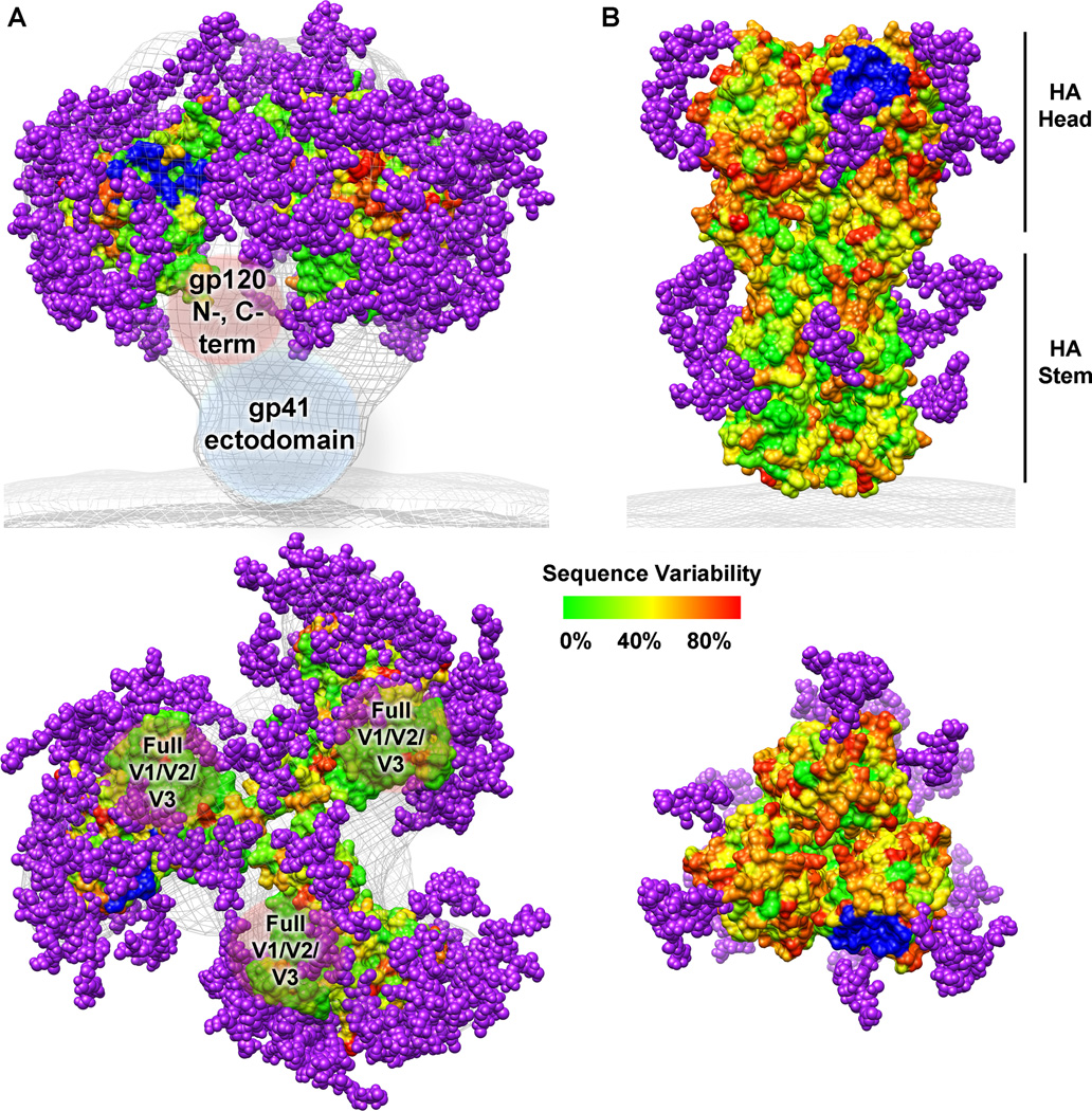
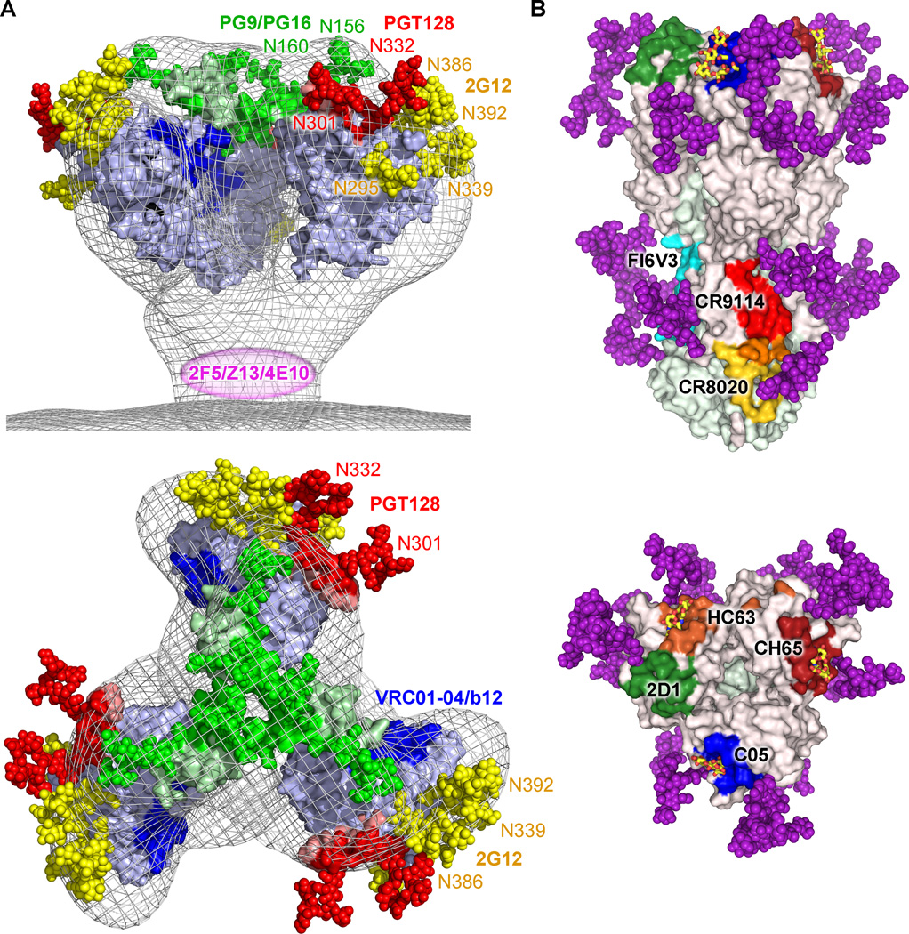
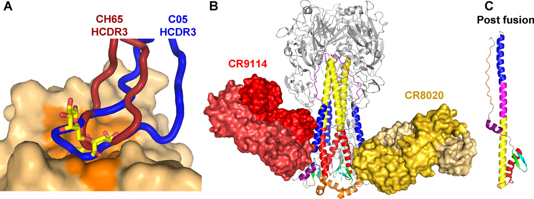
Human immunodeficiency virus-1 (HIV-1) envelope protein (Env) and influenza hemagglutinin (HA) are the surface glycoproteins responsible for viral entry into host cells, the first step in the virus life cycle necessary to initiate infection. These glycoproteins exhibit a high degree of sequence variability and glycosylation, which are used as strategies to escape host immune responses. Nonetheless, antibodies with broadly neutralizing activity against these viruses have been isolated that have managed to overcome these barriers. Here, we review recent advances in the structural characterization of these antibodies with their viral antigens that defines a few sites of vulnerability on these viral spikes. These broadly neutralizing antibodies tend to focus their recognition on the sites of similar function between the two viruses: the receptor-binding site and membrane fusion machinery. However, some sites of recognition are unique to the virus neutralized, such as the dense shield of oligomannose carbohydrates on HIV-1 Env. These observations are discussed in the context of structure-based design strategies to aid in vaccine design or development of antivirals.


Conflict of interest statement

The authors state to have no financial or personal relationships that could be viewed as a potential conflict of interest.

Figures

Fig. 1
Fig. 1. HIV-1 Env and influenza HA sequence variability and glycosylation
Sequence variability is represented on the molecular surface as varying colors described on the scale. Potential N-linked glycosylation sites from the consensus sequences are shown as purple spheres. The receptor binding site is colored in blue. (A) As no crystal structure of the full HIV-1 Env trimer is known, a model was generated from the electron microscopy reconstruction of the unliganded HIV-1 Env trimer (gray mesh, EMD ID 5019) (8), the gp120 core structure (PDB ID 3DNN) (8, 15), the gp120 mini-V3 loop (PDB ID 3TYG) (134), and the gp120 V1/V2 loops (PDB ID 3U4E) (17). Missing regions of gp120 (N- and C- termini, and the full V1/V2 and V3 loops) as well as the gp41 ectodomain are labeled inside brown and blue spherical shapes, respectively. (B) The influenza HA trimer structure was rendered using the coordinates from PDB ID 3GBN. This figure was prepared using Chimera (198).
Fig. 2
Fig. 2. HIV-1 Env and influenza HA sites of vulnerability
(A) Crucial oligomannose carbohydrates of the HIV-1 glycan coat are part of epitopes recognized by broadly neutralizing antibodies PGT128 (N301 and N332, red), PG9 (N156 and N160, green), and 2G12 (N295, N339, N386 and N392, yellow + N332, red). Other sites of vulnerability recognized by broadly neutralizing antibody include the CD4bs (blue) and the gp41 membrane proximal external region (MPER) (magenta). (B) Comparison of structurally characterized neutralizing antibody epitopes against influenza HA. For each antibody-HA complex, contacting residues were determined by Contacsym (199) and the equivalent positions are mapped onto the surface representation of A/Brevig Mission/1/1918 (H1N1) HA from PDB ID 3GBN: red, CR9114 (PDB ID 4FQI); yellow, CR8020 (PDB ID 3SDY); cyan, FI6V3 (PDB ID 3ZTN); blue, C05 (PDB ID 4FP8); dark red, CH65 (PDB ID 3SM5); green, 2D1 (PDB ID 3LZF); orange, HC63 (PDB ID 1KEN). Overlapping residues between the CR9114 and CR8020 footprints are colored in orange. The α2-6 sialic acid receptor analog from PDB ID 3UBE is shown with carbon in yellow, oxygen in red, and nitrogen in blue. Glycans are illustrated as in Fig. 1. This figure was prepared using Pymol.
Fig. 3
Fig. 3. Recognition of carbohydrate-containing HIV-1 Env epitopes by three broadly neutralizing antibodies
Protein components are rendered as secondary structure cartoons, whereas glycans are represented as sticks. The light chain and heavy chain of the antibodies are colored in orange and shades of blue, respectively. The glycans recognized by the antibodies are represented as cartoon models below the crystal structures with squares and circles representing GlcNAc and mannose moieties, respectively. For each glycan, the moieties are circled if they have at least 10 Å2 of surface area buried in the antibody paratope, a threshold used for defining interacting regions. Regions on gp120 that form the primary site of interaction are colored green, whereas secondary binding sites are colored magenta. (A) The crystal structure of the domain-swapped 2G12 antibody dimer in complex with Man9GlcNAc2 sugar moieties reveals an interaction with up to four oligomannose glycans in its multivalent combining site (PDB ID 1OP5). Labeled 1’ and 2’ are the glycans interacting in the primary combining sites, whereas the glycans contacting the secondary combining sites are labeled 3” and 4”. (B) As seen in the crystal structure of the complex with an engineered gp120 outer domain (eODm3), antibody PGT128 recognizes two glycans (N301 and N332) and a β-strand (base of gp120 V3) (PDB ID 3TYG). (C) Antibody PG9 also recognizes two glycans (N156 and N160) and a β-strand (gp120 V2 loop) in a crystal structure of a complex with a gp120 V1/V2 scaffold (PDB ID 3U4E). The figure was prepared using Chimera (198).
Fig. 4
Fig. 4. Recognition of the head and stem subdomains of influenza HA by broadly neutralizing antibodies
(A) Antibodies CH65 (PDB ID 3SM5) and C05 (PDB ID 4FP8) insert their HCDR3, colored dark red and blue, respectively, into the receptor-binding site to compete with receptor. The HA surface from PDB ID 1MQN is shown, and the residues contacted by receptor are colored in dark orange. The receptor is represented as sticks with carbon in yellow, oxygen in red, and nitrogen in blue. The HA heads of the bnAb complex structures were aligned to that of PDB ID 1MQN. (B) Antibodies CR9114 (PDB ID 4FQI) and CR8020 (PDB ID 3SDY) bind unique epitopes in the stem and inhibit the low pH conformational change shown in (C) (PDB ID 1QU1). The light chains of each antibody are represented in a lighter shade. The HA1 is colored in gray and glycans are not shown. The figure was prepared using Pymol.

References

    1. Gray RH, et al. Probability of HIV-1 transmission per coital act in monogamous, heterosexual, HIV-1-discordant couples in Rakai, Uganda. Lancet. 2001;357:1149–1153. - PubMed
    1. Forsman A, Weiss RA. Why is HIV a pathogen? Trends Microbiol. 2008;16:555–560. - PubMed
    1. Gelderblom HR, Hausmann EH, Ozel M, Pauli G, Koch MA. Fine structure of human immunodeficiency virus (HIV) and immunolocalization of structural proteins. Virology. 1987;156:171–176. - PubMed
    1. Zhu P, et al. Electron tomography analysis of envelope glycoprotein trimers on HIV and simian immunodeficiency virus virions. Proc Natl Acad Sci USA. 2003;100:15812–15817. - PMC - PubMed
    1. Zhu P, et al. Distribution and three-dimensional structure of AIDS virus envelope spikes. Nature. 2006;441:847–852. - PubMed

Publication types

MeSH terms