

Close encounter: mitochondria, endoplasmic reticulum and Alzheimer's disease
- PMID: 23047154
- PMCID: PMC3492737
- DOI: 10.1038/emboj.2012.279
Close encounter: mitochondria, endoplasmic reticulum and Alzheimer's disease
Abstract
EMBO J (2012) 31 21, 4106–4123. doi:; DOI: 10.1038/emboj.2012.202; published online August 14 2012
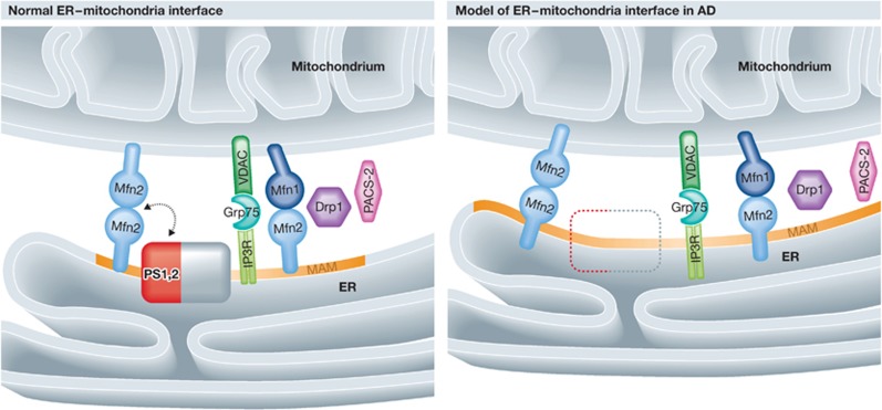
Alzheimer’s disease (AD) is characterized by the loss of hippocampal and cortical neurons as a consequence of the accumulation of amyloid-β (Aβ). Aβ is produced from the amyloid precursor protein (APP) by the γ-secretase complex components presenilin-1 (PS1) and -2 (PS2), which are mutated in genetic forms of AD. In this issue, Schon and coworkers show that PS1 and PS2 are located at the interface between mitochondria and endoplasmic reticulum (ER). In models of familial and sporadic AD, these two organelles are juxtaposed closely, affecting shared lipid metabolic pathways. The interface between mitochondria and ER emerges as a new potential determinant of AD pathogenesis.
Conflict of interest statement
The authors declare that they have no conflict of interest.
Figures
Comment on
-
Upregulated function of mitochondria-associated ER membranes in Alzheimer disease.EMBO J. 2012 Nov 5;31(21):4106-23. doi: 10.1038/emboj.2012.202. Epub 2012 Aug 14. EMBO J. 2012. PMID: 22892566 Free PMC article.
References
-
- D’Amelio M, Cavallucci V, Middei S, Marchetti C, Pacioni S, Ferri A, Diamantini A, De ZD, Carrara P, Battistini L, Moreno S, Bacci A, Ammassari-Teule M, Marie H, Cecconi F (2011) Caspase-3 triggers early synaptic dysfunction in a mouse model of Alzheimer’s disease. Nat Neurosci 14: 69–76 - PubMed
-
- de Brito OM, Scorrano L (2008) Mitofusin 2 tethers endoplasmic reticulum to mitochondria. Nature 456: 605–610 - PubMed
Publication types
MeSH terms
Substances
LinkOut - more resources
Full Text Sources
Medical
