. 2013 Jun;19(3):313-28.
doi: 10.1177/1073858412462607. Epub 2012 Oct 9.

A rat's whiskers point the way toward a novel stimulus-dependent, protective stroke therapy

Affiliations

A rat's whiskers point the way toward a novel stimulus-dependent, protective stroke therapy

Ron D Frostig et al. Neuroscientist. 2013 Jun.

Abstract

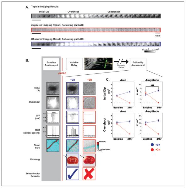
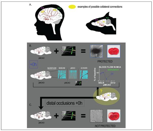
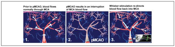
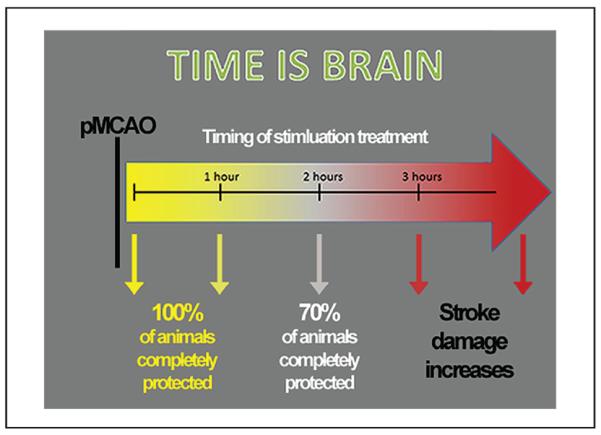
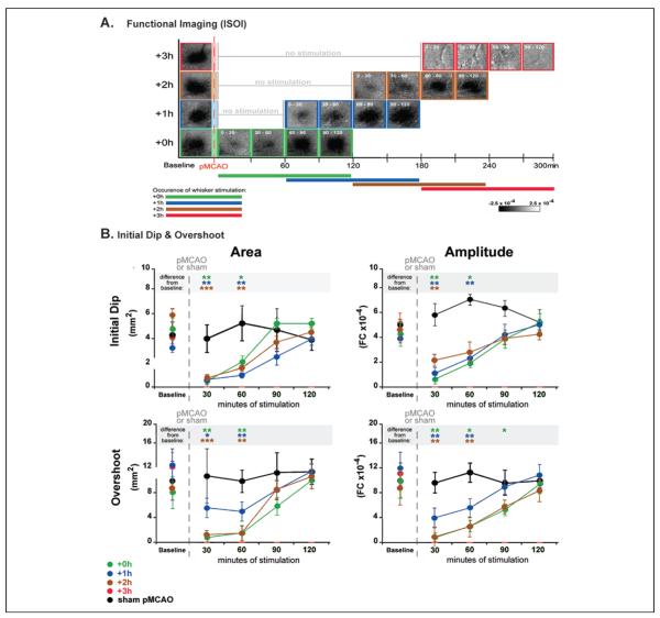
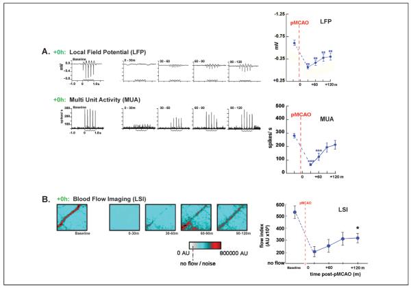
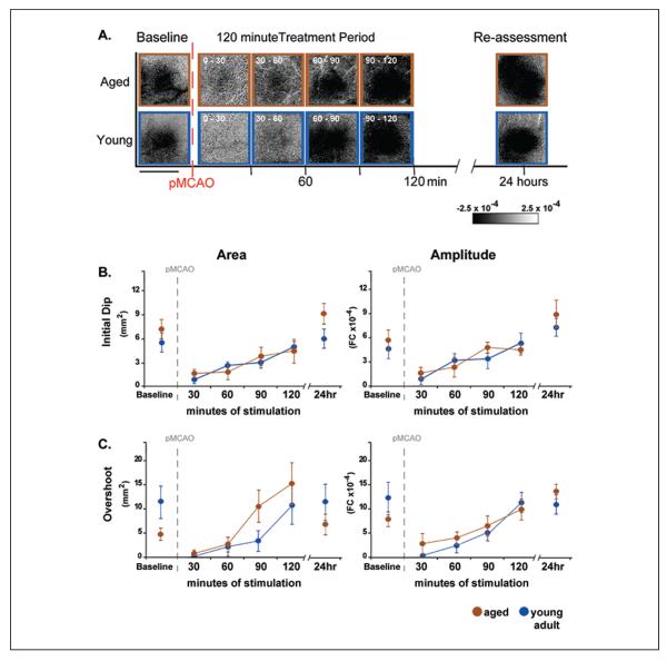
Stroke is the fourth leading cause of death in the United States and the leading cause of long-term disability. Ischemic stroke, due to an interruption in blood supply, is particularly prevalent; 87% of all strokes are ischemic. Unfortunately, current options for acute treatment are extremely limited and there is a great need for new treatment strategies. This review will discuss evidence that mild sensory stimulation can completely protect the jeopardized brain from an impending stroke in a rodent model. When delivered within the first 2 hours following ischemic onset, this stimulation results in complete protection, including a full reestablishment of cortical function, sensorimotor capabilities, and blood flow. Identical stimulation, however, initiated 3 hours following ischemic onset, results in an increase in damage compared with untreated animals. The protective effect is not specific to a single sensory modality, anesthesia, or age, and increasing evoked cortical activity by increasing stimulation accelerates recovery. Taken together, these findings demonstrate that cortical activity is a critical factor for protection and suggest a new, exciting potential avenue for the development of acute stroke treatment strategies that may produce a noninvasive, drug-free, equipment-free, and side effect-free means of protecting from ischemic stroke.

Keywords: ischemia; protection; reperfusion; rodent model; stimulation treatment; stroke.


Figures

Figure 1
Figure 1
Experimental design and representative cases. (A) Representative cases of a typical alternating triphasic evoked functional response to whisker stimulation (top, black), including the initial dip (dark patch), overshoot (white patch), and undershoot (dark patch). Next, the expected imaging result following permanent middle cerebral artery occlusion (pMCAO; middle, red), and the observed imaging result collected following pMCAO (bottom, blue). Each box represents 500 ms. Step function located to left of imaging examples indicates whisker stimulation. Scale bar, middle row right side, indicates 4 mm. Linear grayscale bar, located bottom right, indicates intrinsic signal strength ×10−4 fractional change (FC). (B) Top, schematic of the within animal experimental design. Each box denotes manipulations performed before and after pMCAO (denoted by red vertical line). Below, representative images from +0h and +3h subjects' initial dip and overshoot phase of the evoked functional response to whisker stimulation, local field potential (LFP), multi-unit activity (MUA), blood flow imaging (step function indicates stimulus delivery), 2,3,5-triphenyltetrazolium chloride (TTC; arrow indicates region of ischemia) histology collected before and 24 hours after pMCAO. Within the TTC images, note that the area devoid of staining (white) within the +3h subject's left cortex indicates ischemic infarct due to an occlusion of the left MCA. (C) The evoked functional response to whisker stimulation, quantified for immediate (+0h) and delayed (+3h) groups. In each plot, +0h (blue) and +3h (red) group data at pre-manipulation baseline is paired with +24-hour data. Means and standard errors are provided for the area (left column) and amplitude (right column) of the initial dip and overshoot of the evoked functional response to whisker stimulation before and 24 hours after pMCAO. A value of zero indicates no response to whisker stimulation. Asterisks indicate significant differences between baseline and 24-hour values for the +0h group (***P = .0086). Data in panels A, B, and C are from Lay and others (2010); used with permission of authors and publisher.
Figure 2
Figure 2
Major cerebral arteries of the human (left) and rat (right), and their potential collateral connection points. (A) Diagram representing the three major cerebral arteries of the human (left) and rat (right): anterior cerebral artery (ACA), middle cerebral artery (MCA), and posterior cerebral artery (PCA). Connection points between these three vessels, known as collateral vessels (examples marked in yellow) are potential locations for the redirection of blood flow from one major artery to another. (B) +0h animals (which received whisker stimulation immediately post-pMCAO) had intact structure and evoked functional response to whisker stimulation at 24 hours post-occlusion (representative functional imaging data and stained coronal brain slice showing normal function and lack of infarct shown at upper right). In addition, baseline level blood flow was observed within the branches of the occluded MCA 24 hours post–permanent MCA occlusion (post-pMCAO; after having dropped significantly in response to occlusion; see graph for quantification of blood flow drop following pMCAO and return following stimulation treatment). The question, based on our knowledge of collateral vessels and the return of blood flow and function in the protected +0h animals, was then, is collateral flow supporting this return and the protection observed? (C) +0h distal occlusion experiments, wherein stimulation was still delivered immediately post-occlusion, but the distal ends of MCA were occluded in addition to the standard base occlusion pMCAO. These animals no longer maintained functional cortical integrity and sustained infarct (arrow). These experiments demonstrate that patent distal branches of MCA are necessary for protective plasticity, suggesting collateral vessel blood flow as the mechanism for the protection observed in +0h animals. Data in panels B and C are from Lay and others (2010); used with permission of authors and publisher.
Figure 3
Figure 3
Mild sensory stimulation results in a redirection of blood flow into the ischemic region. Illustration of middle cerebral artery (MCA) blood flow before manipulation, during pMCAO, and following whisker stimulation treatment. MCA is pictured as the large, red, central vessel in each panel. The distal portions of MCA are indicated by the smaller branches extending from MCA. The direction of blood flow is indicated by the arrows. Neuron icons represent the underlying cortical tissue. (1) Prior to manipulation, blood flows up through MCA, into the anterior and posterior branches of the vessel, and into the most distal segments, supplying a large area of the cortex with blood. (2) Following permanent MCA occlusion (pMCAO), blood flow through MCA is interrupted, resulting in ischemia of the surrounding cortical tissue. (3) In response to whisker stimulation treatment, collateral vessels (pictured top left in each panel) form a new supply route, allowing blood to flow in reverse back into the ischemic territory.
Figure 4
Figure 4
Schematic of the effect of whisker stimulation on outcome depending on timing of administration. Complete protection is observed in all subjects when stimulation is delivered within the first hour following permanent middle cerebral artery occlusion (pMCAO), but only in about 70% of subjects when delivered at 2 hours post-pMCAO. By 3 hours post-pMCAO, whisker stimulation not only ceases to be beneficial, but actually exacerbates impending stroke damage.
Figure 5
Figure 5
Evoked functional response returns gradually on stimulation onset in all protected animals throughout the treatment period. (A) Following permanent middle cerebral artery occlusion (pMCAO), a return of cortical function is evident in +0h, +1h, and +2h groups during protective stimulation treatment, and a loss of all three phases of the evoked function response in animals that receive treatment 3 hours post-occlusion (+3h animals). Representative data from functional imaging of the initial dip for each group (+0h, +1h, +2h, and +3h) were arranged according to minutes of treatment delivered (treatment time is included as white font in the top left corner of each image). Groups +0h, +1h, and +2h all regained evoked functional response comparable to baseline after 90 minutes of whisker stimulation, whereas +3h animals never demonstrated any post-pMCAO cortical activity. (B) In each plot, group baseline is plotted with 120 minutes of post-occlusion stimulation period data. Means and standard errors are provided for the area (left) and amplitude (right) of the ipsi-ischemic C2 initial dip (top) and overshoot (bottom) phases of evoked functional response to whisker stimulation before and after pMCAO. +3h animals had no response to stimulation at any time point post-pMCAO. For all other groups (sham-pMCAO, +0h, +1h, and +2h), asterisks within the gray band above each plot indicate a significant difference from baseline, *P < .05, **P < .01, and ***P < .001. Data in panels A and B are from Lay and others (2011b); used with permission of authors and publisher.
Figure 6
Figure 6
Evoked neuronal activity returns gradually on stimulation onset following permanent middle cerebral artery occlusion (pMCAO). (A) Representative +0h animals' local field potential (LFP), and multi-unit activity (MUA) responses at baseline and during the stimulus period following pMCAO. Stepping function indicates stimulus delivery. Right, LFP and MUA mean and standard error is plotted prior to and post-occlusion. Asterisks indicated a significant difference from baseline, **P < .01 and ***P < .001. (B) Blood flow imaging (via laser speckle imaging; LSI) experiments demonstrate that post-pMCAO blood flow return in MCA is induced by whisker stimulation treatment. +0h group LSI color scaled images taken at baseline and following pMCAO during treatment at roughly 30-minute intervals. Scale bar indicates 1 mm. The red vessel diagonally traversing the image in each case is a cortical branch of MCA. Right, means and standard errors for +0h animals at baseline, following pMCAO, and during whisker stimulation. Dagger indicates a significant increase in +0h flow compared with value collected immediately following pMCAO, P < .01. Data in panels A and B are from Lay and others (2011b); used with permission of authors and publisher.
Figure 7
Figure 7
Return of cortical function in aged rats is equivalent to young adult rats. Return of cortical function in aged rats is equivalent to young adult rats. (A) Representative data from functional imaging of the initial dip in aged and young adult animals that underwent permanent middle cerebral artery occlusion (pMCAO) and received whisker stimulation treatment immediately post-occlusion. Functional imaging was conducted prior, during, and 24 hours following pMCAO and whisker stimulation treatment. Both young adult and aged rats regained evoked functional response comparable to baseline after 90 minutes of whisker stimulation treatment. Linear grayscale bar indicates intrinsic signal amplitude (fractional change FC ×10−4). Scale bar indicates 4 mm. (B, C) Group baseline is plotted with 120 minutes of post-occlusion stimulus period data and 24-hour re-assessment data for each graph. Means and standard errors are provided for the area (left) and amplitude (right) of the ipsiischemic C2 initial dip (B) and overshoot (C) phases of evoked functional response to whisker stimulation before and following pMCAO. Data in panels A, B, and C are from Lay and others (2012); used with permission of authors and publisher.

References

    1. Adibhatla RM, Hatcher JF. Lipid oxidation and peroxidation in CNS health and disease: from molecular mechanisms to therapeutic opportunities. Antioxid Redox Signal. 2010;12(1):125–69. - PubMed
    1. Baron JC. Mapping the ischaemic penumbra with PET: a new approach. Brain. 2001;124(Pt 1):2–4. - PubMed
    1. Bederson JB, Pitts LH, Tsuji M, Nishimura MC, Davis RL, Bartkowski H. Rat middle cerebral artery occlusion: evaluation of the model and development of a neurologic examination. Stroke. 1986;17(3):472–6. - PubMed
    1. Blumenfeld H. Neuroanatomy through clinical cases. Sinauer Associates; Sunderland, MA: 2002.
    1. Burnett MG, Shimazu T, Szabados T, Muramatsu H, Detre JA, Greenberg JH. Electrical forepaw stimulation during reversible forebrain ischemia decreases infarct volume. Stroke. 2006;37(5):1327–31. - PubMed

Publication types