Review
. 2012 Dec;32(12):2091-9.
doi: 10.1038/jcbfm.2012.139. Epub 2012 Oct 10.

Can restoring incomplete microcirculatory reperfusion improve stroke outcome after thrombolysis?

Affiliations
Review

Can restoring incomplete microcirculatory reperfusion improve stroke outcome after thrombolysis?

Turgay Dalkara et al. J Cereb Blood Flow Metab. 2012 Dec.

Abstract

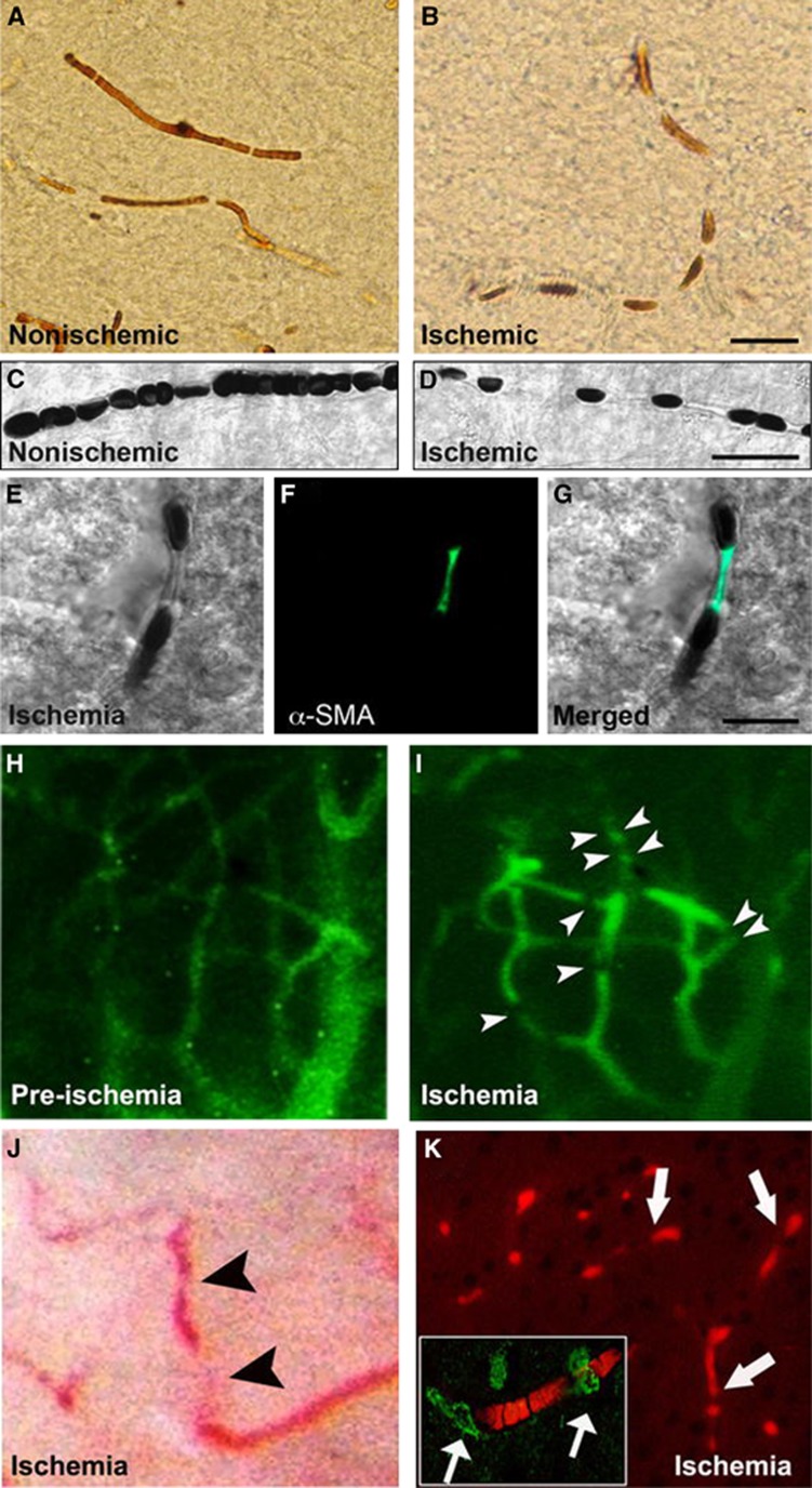
Substantial experimental data and recent clinical evidence suggesting that tissue reperfusion is a better predictor of outcome after thrombolysis than recanalization necessitate that patency of microcirculation after recanalization should be reevaluated. If indeed microcirculatory blood flow cannot be sufficiently reinstituted despite complete recanalization as commonly observed in coronary circulation, it may be one of the factors contributing to low efficacy of thrombolysis in stroke. Although microvascular no-reflow is considered an irreversible process that prevents tissue recovery from injury, emerging evidence suggests that it might be reversed with pharmacological agents administered early during recanalization. Therefore, therapeutic approaches aiming at reducing microvascular obstructions may improve success rate of recanalization therapies. Importantly, promoting oxygen delivery to the tissue, where entrapped erythrocytes cannot circulate in capillaries, with ongoing serum flow may improve survival of the underreperfused tissue. Altogether, these developments bring about the exciting possibility that benefit of reperfusion therapies can be further improved by restoring microcirculatory function because survival in the penumbra critically depends on adequate blood supply. Here, we review the available evidence suggesting presence of an 'incomplete microcirculatory reperfusion' (IMR) after focal cerebral ischemia and discuss potential means that may help investigate IMR in stroke patients after recanalization therapies despite technical limitations.


Figures

Figure 1
Figure 1
Cerebral ischemia induces intermittently spaced capillary constrictions, among which erythrocytes are entrapped. Brain sections in upper panels illustrate the cortical capillaries filled with horseradish peroxidase (HRP) injected before killing the mouse. In contrast to uninterruptedly filled nonischemic microvessels (A), the luminal HRP column is interrupted by nodal constrictions in the ischemic hemisphere (B). Differential interference contrast (DIC) images (CG) illustrate that erythrocytes are entrapped between the constricted segments and that the nodal capillary constrictions are colocalized with α-smooth muscle actin (α-SMA) immunopositive pericytes (green). (AG) Images were captured from the frontal cortex at 6 hours after 2 hours of middle cerebral artery occlusion (MCA) occlusion. Ischemia-induced capillary constrictions and erythrocyte entrapment can also be observed in vivo through a cranial window in mice under anesthesia (HJ). Microvessels made visible with systemically given fluorescein isothiocyanate (FITC)–dextran become better delineated after ischemia because of sluggish blood flow (allowing more fluorescence emitting), and numerous capillary constrictions appear (arrowheads) starting 1 hour after MCA occlusion (I). (H, I) Images were captured from the same cortical area before and during ischemia. Similarly, erythrocytes, which are hardly detectable under bright-field microscopy in vivo because of rapid blood flow, become visible and entrapped around the capillary constrictions during ischemia (J, arrowheads). Although it is difficult to show in vivo that all microvascular constrictions are colocalized with pericytes, processing of these brains ex vivo with NaBH4, which renders hemoglobin fluorescent (K, red), confirmed that constricted capillaries imaged during intravital microscopy were filled with trapped erythrocytes (arrows) and the capillary constrictions were colocalized with α-SMA immunopositive pericytes (green, arrows in inset). Reproduced from Dalkara et al with permission. The color reproduction of this figure is available at the Journal of Cerebral Blood Flow and Metabolism journal online.
Figure 2
Figure 2
Temporal evolution of cerebral blood flow changes on magnetic resonance image-derived lesion volumes after (A) 35 minutes, (B) 60 minutes, (C) 95 minutes, and (D) permanent middle cerebral artery occlusion. Tissue reperfusion is incomplete even after MCA occlusion as brief as 35 minutes, and further deteriorates with prolonged ischemia. *P<0.0001 compared with postreperfusion values. Adapted from Bardutzky et al with permission.
Figure 3
Figure 3
Absence of reperfusion, even in the setting of complete recanalization, may result in a large follow-up infarct volume. (A) Axial noncontrast computed tomography (CT) carried out on admission (2.5 hours after onset of symptoms) shows subtle hypoattenuation of the right putamen but no sulcal effacement in the right middle cerebral artery (MCA) territory. (B) Axial maximum-intensity projection image from computed tomography angiography (CTA) carried out admission shows occlusion of the M1 segment of the right MCA (arrow). Perfusion CT (PCT) maps show decreased cerebral blood volume (CBV) and cerebral blood flow (CBF) in the right frontal and temporal lobes and a larger region of prolonged mean transit time (MTT) that also involves the right anterior cerebral artery (ACA) territory. The region of decreased CBV corresponds to the infarct core, whereas the surrounding mismatch region of prolonged MTT represents the ischemic penumbra. The patient received endovascular thrombectomy with a MERCI device. (C) Axial maximum-intensity projection image from CTA and PCT carried out 6 hours after admission show that, despite complete recanalization of the right MCA, PCT maps show that the region of decreased CBV and CBF has expanded to include the right anterior ACA territory that was previously considered tissue at risk. MTT is still abnormally increased in the right superficial MCA territory and in a portion of the right ACA territory. (D) Axial noncontrast CT carried out 48 hours after admission shows marked hypoattenuation and edema in the territories matching the perfusion deficit on the reperfusion PCT. Reproduced from Soares et al with permission.

References

    1. Donnan GA, Davis SM, Parsons MW, Ma H, Dewey HM, Howells DW. How to make better use of thrombolytic therapy in acute ischemic stroke. Nat Rev Neurol. 2011;7:400–409. - PubMed
    1. Pfefferkorn T, Rosenberg GA. Closure of the blood-brain barrier by matrix metalloproteinase inhibition reduces rtPA-mediated mortality in cerebral ischemia with delayed reperfusion. Stroke. 2003;34:2025–2030. - PubMed
    1. Yang Y, Estrada EY, Thompson JF, Liu W, Rosenberg GA. Matrix metalloproteinase-mediated disruption of tight junction proteins in cerebral vessels is reversed by synthetic matrix metalloproteinase inhibitor in focal ischemia in rat. J Cereb Blood Flow Metab. 2007;27:697–709. - PubMed
    1. Al-Ali F, Jefferson A, Barrow T, Cree T, Louis S, Luke K, et al. The capillary index score: rethinking the acute ischemic stroke treatment algorithm. Results from the Borgess Medical Center Acute Ischemic Stroke Registry J Neurointerv Surgdoi:10.1136/neurintsurg-2011-010146in press). - DOI - PubMed
    1. Ito H. No-reflow phenomenon in patients with acute myocardial infarction: its pathophysiology and clinical implications. Acta Med Okayama. 2009;63:161–168. - PubMed

MeSH terms