. 2012 Oct 23;109(43):17531-6.
doi: 10.1073/pnas.1207238109. Epub 2012 Oct 9.

Functional characterization of pulmonary neuroendocrine cells in lung development, injury, and tumorigenesis

Affiliations

Functional characterization of pulmonary neuroendocrine cells in lung development, injury, and tumorigenesis

Hai Song et al. Proc Natl Acad Sci U S A. .

Abstract

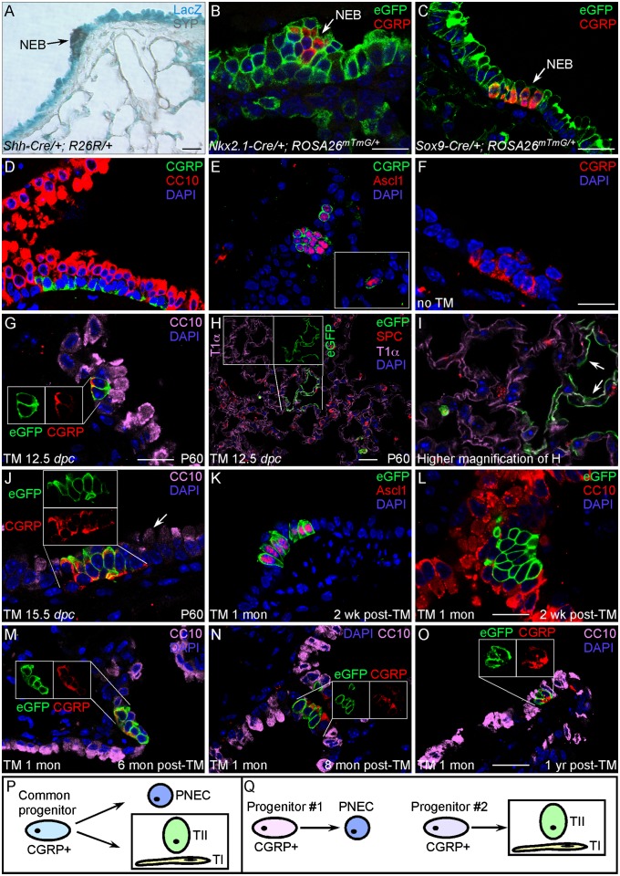
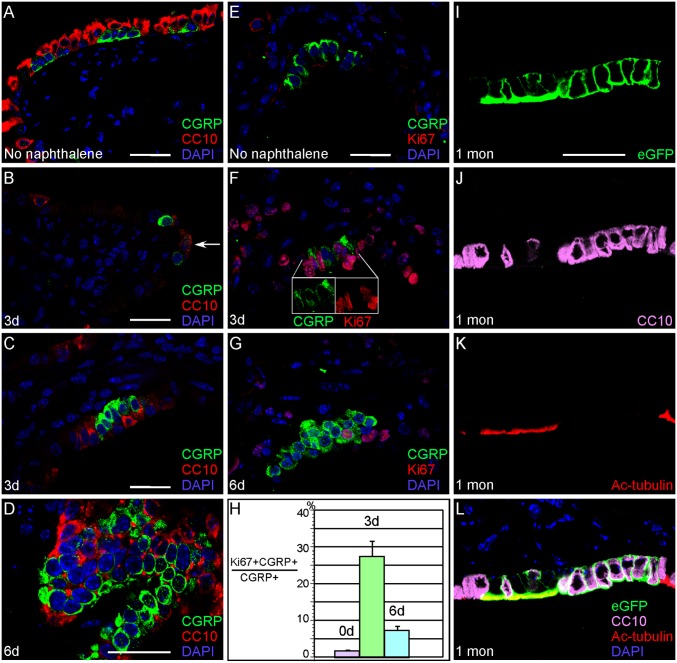
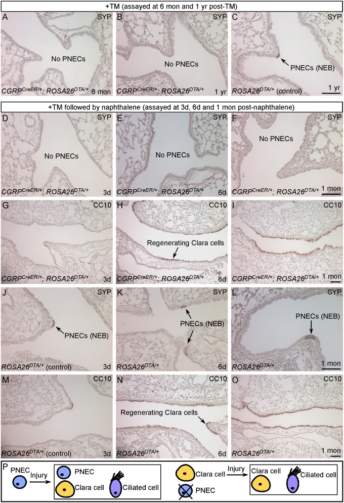
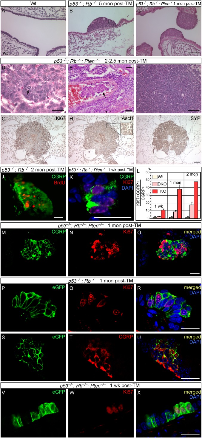
Pulmonary neuroendocrine cells (PNECs) are proposed to be the first specialized cell type to appear in the lung, but their ontogeny remains obscure. Although studies of PNECs have suggested their involvement in a number of lung functions, neither their in vivo significance nor the molecular mechanisms underlying them have been elucidated. Importantly, PNECs have long been speculated to constitute the cells of origin of human small-cell lung cancer (SCLC) and recent mouse models support this hypothesis. However, a genetic system that permits tracing the early events of PNEC transformation has not been available. To address these key issues, we developed a genetic tool in mice by introducing a fusion protein of Cre recombinase and estrogen receptor (CreER) into the calcitonin gene-related peptide (CGRP) locus that encodes a major peptide in PNECs. The CGRP(CreER) mouse line has enabled us to manipulate gene activity in PNECs. Lineage tracing using this tool revealed the plasticity of PNECs. PNECs can be colabeled with alveolar cells during lung development, and following lung injury, PNECs can contribute to Clara cells and ciliated cells. Contrary to the current model, we observed that elimination of PNECs has no apparent consequence on Clara cell recovery. We also created mouse models of SCLC in which CGRP(CreER) was used to ablate multiple tumor suppressors in PNECs that were simultaneously labeled for following their fate. Our findings suggest that SCLC can originate from differentiated PNECs. Together, these studies provide unique insight into PNEC lineage and function and establish the foundation of investigating how PNECs contribute to lung homeostasis, injury/repair, and tumorigenesis.


Conflict of interest statement

The authors declare no conflict of interest.

Figures

Fig. 1.
Fig. 1.
Lineage tracing of PNECs in embryonic and postnatal lungs. (A) β-gal (LacZ) staining of lung sections from Shh-Cre/+;R26R/+ adult mice. (B and C) Immunostaining of lung sections from Nkx2.1-Cre/+;ROSA26mTmG/+ or Sox9-Cre/+;ROSA26mTmG/+ adult mice. Extensive (nearly ubiquitous) labeling of epithelial cells by β-gal (blue) or eGFP (green) was observed. Labeled epithelial cells include clustered PNECs (NEBs; arrows), which were identified by anti-SYP (brown), anti-CGRP (red), or additional antibodies. (D) Immunostaining of lung sections from adult wild-type (WT) mice. PNECs (CGRP+) are found in clusters and in close association with Clara cells (CC10+). (E) Immunostaining of lung sections from adult WT mice. CGRP immunoreactivity coincides with that of Ascl1 (Mash1). (Inset) Solitary PNEC. (F) Immunostaining of lung sections from adult CGRPCreER/+;ROSA26mTmG/+ mice without TM injection. No eGFP-labeled cells were detected. (GI) Immunostaining of lung sections from adult CGRPCreER/+;ROSA26mTmG/+ mice [postnatal day 60 (P60)] exposed to tamoxifen at 12.5 dpc. Activation of CreER by tamoxifen during embryogenesis led to eGFP labeling of PNECs (G) as well as alveolar cells, including type I (T1α+) and type II (SP-C+) pneumocytes (H and I). (H Inset) Individual images of eGFP and CGRP immunostaining of the same cells. Arrows in I point to labeled type I cells. No Clara cells or other cell types were labeled by eGFP. (J) Immunostaining of lung sections from adult CGRPCreER/+;ROSA26mTmG/+ mice (P60) exposed to tamoxifen at 15.5 dpc. Only PNECs were labeled. Arrow points to unlabeled Clara cells. (Inset) Separate images of eGFP and CGRP immunostaining of the same cells. (KO) Immunostaining of lung sections from adult CGRPCreER/+;ROSA26mTmG/+ mice injected with tamoxifen at 1 mo of age and analyzed at different time points post-TM injection. (MO Insets) Individual images of eGFP and CGRP immunostaining of the same cells. (P and Q) A model of PNEC specification during lung homeostasis. (Scale bars: 25 μm for panels in each row.)
Fig. 2.
Fig. 2.
CGRP+ cells can give rise to Clara cells and ciliated cells during Clara cell regeneration induced by naphthalene administration. (AG) Immunostaining of lung sections from adult CGRPCreER/+;ROSA26mTmG/+ mice. CreER in PNECs (CGRP+) of adult lungs was activated by TM injection and followed by naphthalene injection to ablate Clara cells (CC10+) in these animals. Clara cells (A) were almost completely eliminated in this process (e.g., day 3 after naphthalene injection shown in B). Occasionally, cells that express both CGRP and CC10 can be found (arrow in B). Regeneration of surviving Clara cells was apparent by day 6 after naphthalene treatment (D). Naphthalene also induced PNEC proliferation as indicated by increased Ki67+ PNECs (F and G). A time course of changes in the proliferation rate of PNECs after naphthalene treatment is shown in H. (F Inset) Individual images of CGRP and Ki67 immunostaining of the same cells. (IL) Immunostaining of lung sections from adult CGRPCreER/+;ROSA26mTmG/+ mice 1 mo after naphthalene injection, which was performed following tamoxifen administration. The combined treatment of TM and naphthalene resulted in labeling of PNECs as well as Clara cells (CC10+) and ciliated cells [acetylated (Ac)-tubulin+] (IL). No other cell types were labeled by eGFP, which is in contrast to the stable lineage of PNECs during homeostasis. Note that L is a merged image of IK. (Scale bars: 25 μm for panels in each column.)
Fig. 3.
Fig. 3.
PNECs are dispensable for Clara cell regeneration during naphthalene-induced lung injury. (AC) Immunostaining of lung sections from adult CGRPCreER/+;ROSA26DTA/+ (experimental) and ROSA26DTA/+ (control) mice. Activation of CreER by TM induced DTA expression in CGRPCreER/+;ROSA26DTA/+ adult lungs, resulting in efficient killing of PNECs. PNECs (SYP+ or CGRP+) or NEBs could not be detected in these animals (A and B), whereas other cell types were unaffected (Fig. S5). Long-term tracing of PNECs in adult CGRPCreER/+;ROSA26DTA/+ mice showed no sign of PNEC replenishment. (DI) Immunostaining of lung sections from adult CGRPCreER/+;ROSA26DTA/+ mice. Activation of CreER by TM in adult lungs induced DTA expression, resulting in efficient PNEC ablation. These mice were subsequently treated with naphthalene to ablate Clara cells (CC10+). Despite the absence of PNECs (DF), Clara cell regeneration followed a time course (GI) similar to that in control mice (MO). Extensive Clara cell proliferation was observed by day 6 after naphthalene injection (H), and Clara cells had fully regenerated by 1 mo after the initial insult (I). (JO) Immunostaining of lung sections from adult ROSA26DTA/+ control mice that received an identical regimen of TM and naphthalene treatment. PNECs (arrows in JL) were unaffected, and the distribution of lung cell types and the kinetics of Clara cell regeneration (MO) are similar to those in WT mice. (P) A model of PNEC specification and function during lung injury. (Scale bars: 100 μm for panels in each row)
Fig. 4.
Fig. 4.
SCLC can originate from differentiated PNECs. (AI) Histology and immunostaining of lung sections from WT, CGRPCreER/+;p53f/f;Rbf/f [double knockout (DKO)], and CGRPCreER/+;p53f/f;Rbf/f;Ptenf/f [triple knockout (TKO)] adult mice. Cancer cells in triple-mutant mice displayed aberrant mitosis (arrow in D) and also showed signs of tumor invasiveness (arrow in E). Tumors are comprised of proliferating Ki67+ cells (G) and also expressed neuroendocrine markers such as Ascl1 (Mash1) (H) and SYP (I). (JO) Immunostaining of lung sections from WT, CGRPCreER/+;p53f/f;Rbf/f, and CGRPCreER/+;p53f/f;Rbf/f;Ptenf/f adult mice. PNECs lacking p53 and Rb displayed extensive PNEC proliferation as judged by Ki67 staining (J). Proliferating PNECs could be found 1 wk after TM injection (K) in triple-mutant mice, and the proliferation rate increased substantially with time (LO). (PX) Immunostaining of lung sections from WT, CGRPCreER/+;ROSA26mTmG/+;p53f/f;Rbf/f, and CGRPCreER/+;ROSA26mTmG/+;p53f/f;Rbf/f;Ptenf/f adult mice. TM injection selectively ablated p53/Rb or p53/Rb/Pten in PNECs, which were simultaneously labeled by eGFP. Inactivating p53 and Rb in PNECs (CGRP+) (T) led to extensive PNEC proliferation as observed by Ki67 staining (Q). Most, if not all PNECs, were also labeled by eGFP (P and S), suggesting that differentiated PNECs proliferated and subsequently led to tumor growth. Similarly, TM injection into CGRPCreER/+;ROSA26mTmG/+;p53f/f;Rbf/f;Ptenf/f mice labeled proliferating PNECs (V and W), which likely led to tumor development. (Scale bars: AC and FI, 100 μm; D, E, J, K, and MX, 20 μm.)

References

    1. Morrisey EE, Hogan BL. Preparing for the first breath: Genetic and cellular mechanisms in lung development. Dev Cell. 2010;18(1):8–23. - PMC - PubMed
    1. Domyan ET, Sun X. Patterning and plasticity in development of the respiratory lineage. Dev Dyn. 2011;240(3):477–485. - PMC - PubMed
    1. Warburton D, et al. Lung organogenesis. Curr Top Dev Biol. 2010;90:73–158. - PMC - PubMed
    1. Cardoso WV, Whitsett JA. Resident cellular components of the lung: Developmental aspects. Proc Am Thorac Soc. 2008;5(7):767–771. - PMC - PubMed
    1. Linnoila RI. Functional facets of the pulmonary neuroendocrine system. Lab Invest. 2006;86(5):425–444. - PubMed

Substances