

doi: 10.1002/adma.201203348.
Epub 2012 Oct 9.
Biopsy with thermally-responsive untethered microtools
Affiliations
- PMID: 23047708
- PMCID: PMC3832625
- DOI: 10.1002/adma.201203348
Item in Clipboard
Biopsy with thermally-responsive untethered microtools
Adv Mater.
.
Abstract
Thermally activated, untethered microgrippers can reach narrow conduits in the body and be used to excise tissue for diagnostic analyses. As depicted in the figure, the feasibility of an in vivo biopsy of the porcine bile duct using untethered microgrippers is demonstrated.
Copyright © 2013 WILEY-VCH Verlag GmbH & Co. KGaA, Weinheim.
Figures

(a–f) The fabrication and actuation schematic of μ-grippers. (a) A thin Cu layer was used as the sacrificial layer. (b) The pre-stressed Cr-Au bilayer was patterned. (c) Ferromagnetic Ni was electroplated as the rigid segments between the hinges and then covered with Au. (d) The thermo-sensitive polymeric trigger was patterned. (e) The μ-grippers were released from the substrate by dissolving the sacrificial layer. (f) The μ-grippers closed when exposed to the body temperature. (g–j) Bright field microscopy sequence showing thermal actuation of the μ-grippers at 37 °C within 10 minutes.

(a–b) Optical image of μ-grippers distributed on the bile duct opening of the porcine liver. Scale bar represents 200 μm. (c) Optical image of μ-grippers during retreival using a magnetic catheter. (d) Image of a retrieved μ-gripper with an excised tissue piece after staining with trypan blue. Scale bar represents 100 μm.
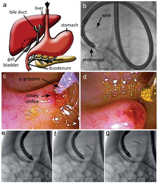
(a) A schematic diagram of the porcine upper gastrointestinal track; the endoscope entry is depicted with an arrow. (b) Fluoroscopic image showing the endoscope entering from the mouth (top) and reaching the duodenum. The guide wire that was advanced into the bile duct is also visible. Endoscopic images of (c) the delivery of the μ-grippers through a catheter into the porcine biliary orifice, and (d) the retrieval of the μ-grippers via a magnetic catheter. (e–g) Fluoroscopic image sequence of the retrieval magnet being maneuvered inside the bile duct.

(a) Optical image of the retrieved μ-grippers on the magnetic catheter tip. (b) The retrieved tissue after staining with trypan blue. Scale bars represent 200 μm. (c) H&E stained section of cells retrieved by the μ-grippers from the porcine bile duct. (d) Genomic DNA from the tissue obtained with the μ-grippers (G) compared to the negative control (N).
References
-
- Mack MJ. J Am Med Assoc. 2001;285:568. - PubMed
-
- Swanstrom LL, Whiteford M, Khajanchee Y. Surg Endosc. 2008;22:600. - PubMed
-
- Menciassi A, Quirini1 M, Dario P. Minim Invasiv Ther. 2007;16:91. - PubMed
-
- Kalloo AN, Singh VK, Jagannath SB, Niiyama H, Hill SL, Vaughn CA, Magee CA, Kantsevoy SV. Gastrointest Endosc. 2004;60:114. - PubMed
Publication types
MeSH terms
Substances
Grants and funding
LinkOut - more resources
Full Text Sources
Other Literature Sources
Research Materials
