. 2012 Nov 30;287(49):41288-96.
doi: 10.1074/jbc.M112.391417. Epub 2012 Oct 9.

Heparanase enhances the insulin receptor signaling pathway to activate extracellular signal-regulated kinase in multiple myeloma

Affiliations

Heparanase enhances the insulin receptor signaling pathway to activate extracellular signal-regulated kinase in multiple myeloma

Anurag Purushothaman et al. J Biol Chem. .

Abstract

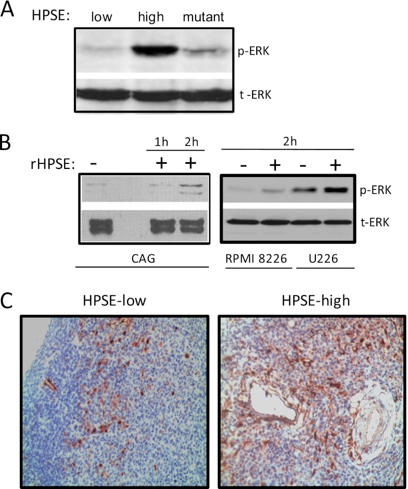
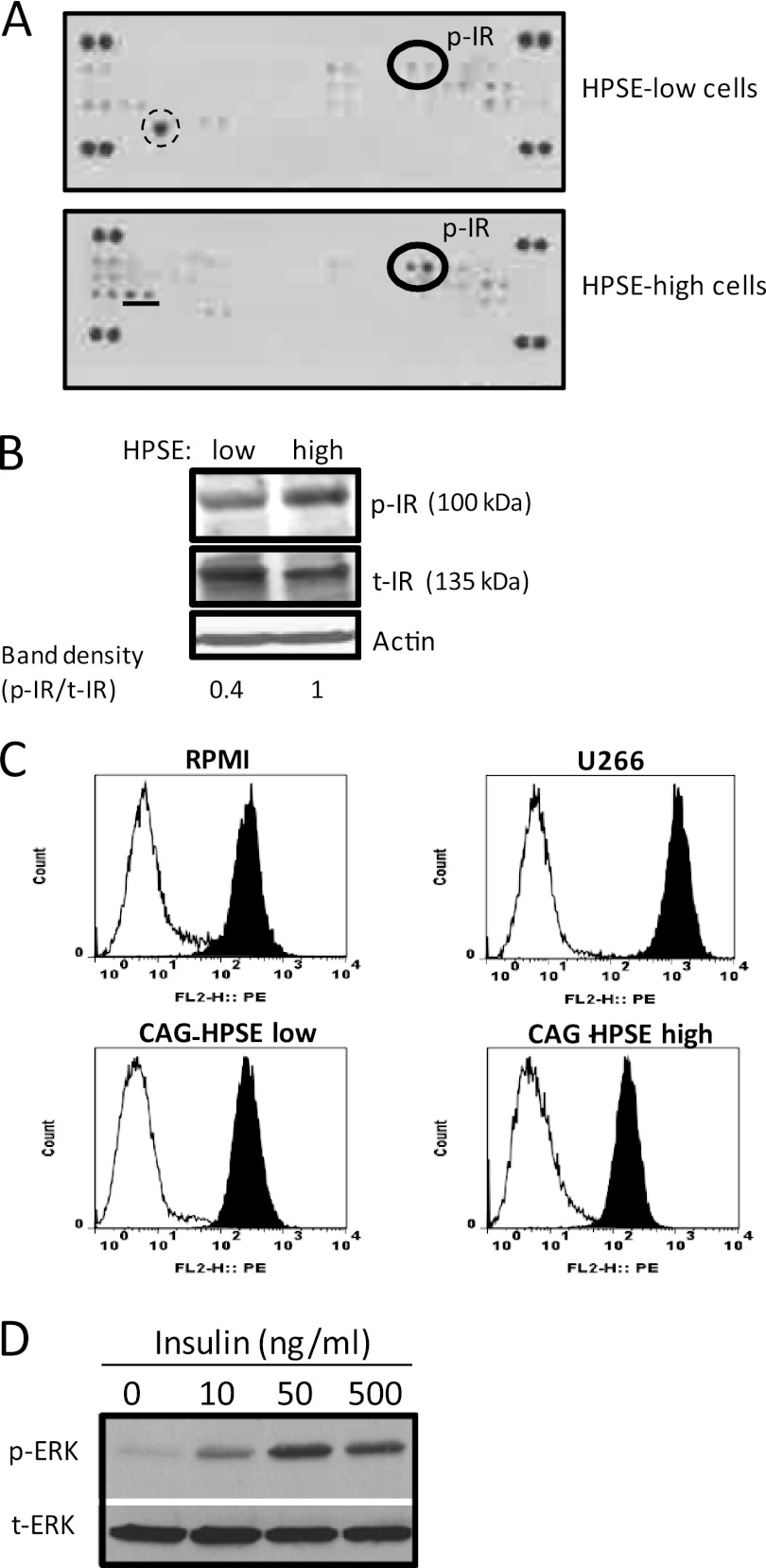
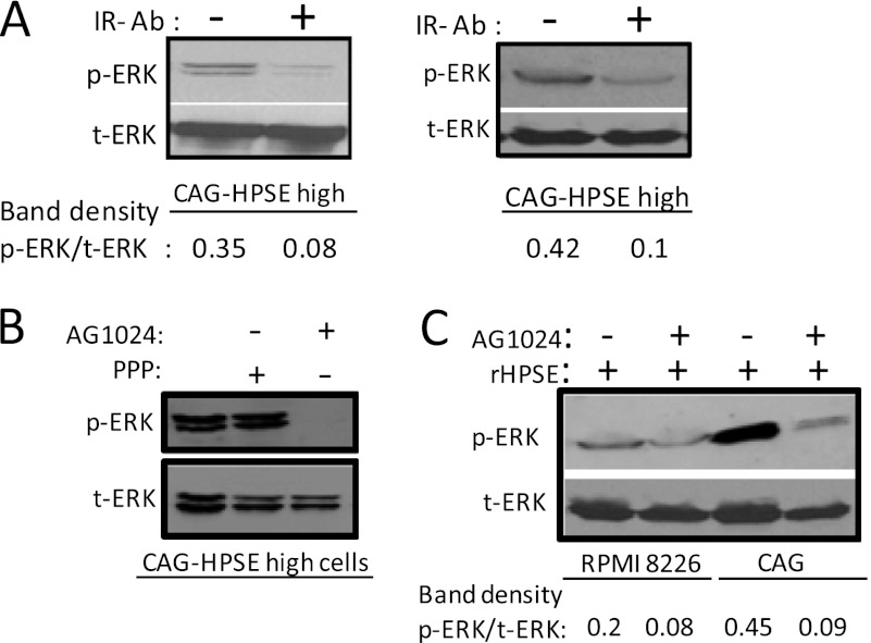
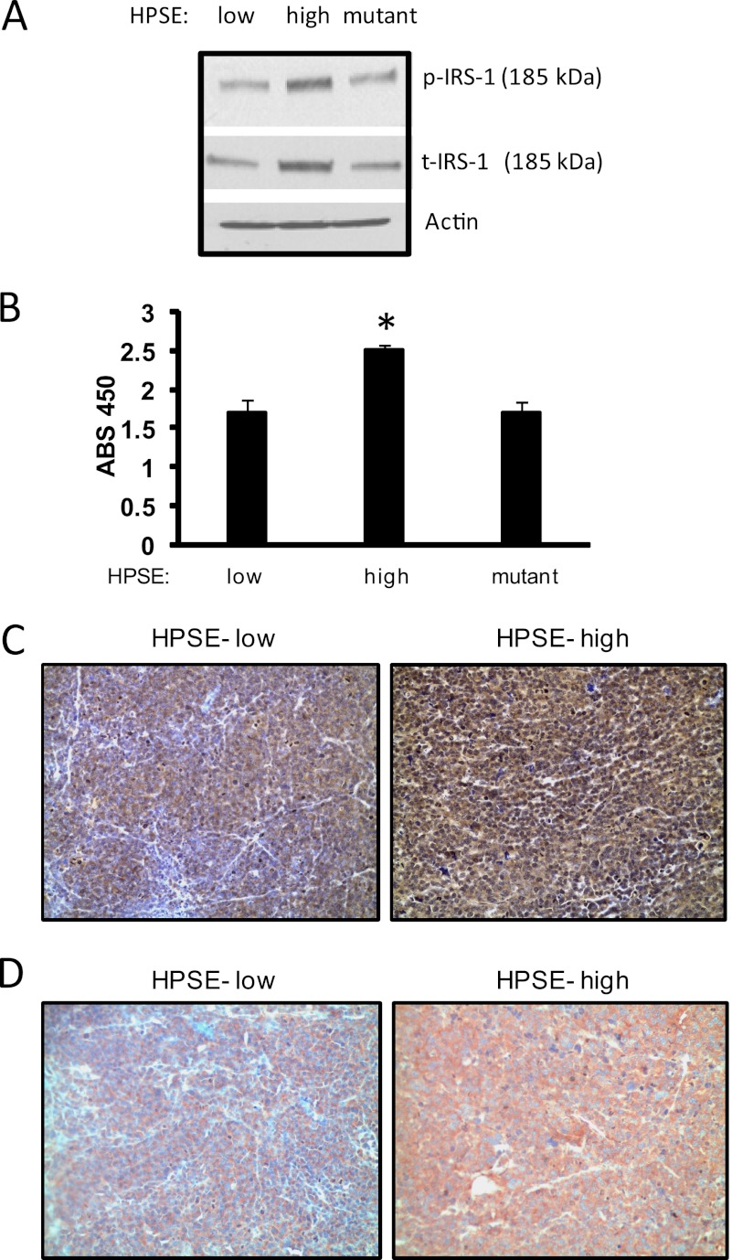
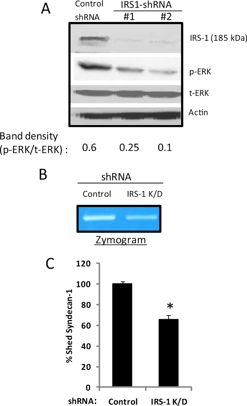
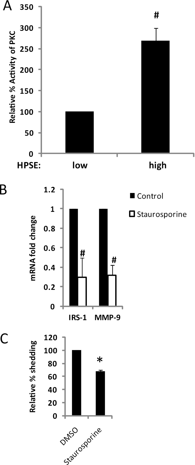
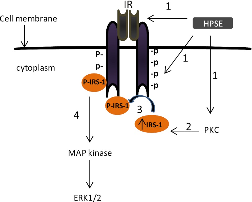
ERK signaling regulates proliferation, survival, drug resistance, and angiogenesis in cancer. Although the mechanisms regulating ERK activation are not fully understood, we previously demonstrated that ERK phosphorylation is elevated by heparanase, an enzyme associated with aggressive behavior of many cancers. In the present study, myeloma cell lines expressing either high or low levels of heparanase were utilized to determine how heparanase stimulates ERK signaling. We discovered that the insulin receptor was abundant on cells expressing either high or low levels of heparanase, but the receptor was highly phosphorylated in heparanase-high cells compared with heparanase-low cells. In addition, protein kinase C activity was elevated in heparanase-high cells, and this enhanced expression of insulin receptor substrate-1 (IRS-1), the principle intracellular substrate for phosphorylation by the insulin receptor. Blocking insulin receptor function with antibody or a small molecule inhibitor or knockdown of IRS-1 expression using shRNA diminished heparanase-mediated ERK activation in the tumor cells. In addition, up-regulation of the insulin signaling pathway by heparanase and the resulting ERK activation were dependent on heparanase retaining its enzyme activity. These results reveal a novel mechanism whereby heparanase enhances activation of the insulin receptor signaling pathway leading to ERK activation and modulation of myeloma behavior.


Figures

FIGURE 1.
FIGURE 1.
Heparanase promotes ERK phosphorylation in myeloma. A, protein lysates from CAG human myeloma cells expressing either low or high levels of heparanase (HPSE-low or HPSE-high) or mutant heparanase that lacks enzyme activity were subjected to Western blot analysis. Membranes were probed with anti-phospho-ERK (p-ERK) antibody or total ERK (t-ERK) antibody. B, serum-starved wild-type CAG, RPMI 8266, or U226 cells were treated with or without recombinant heparanase (rHPSE) for the indicated time points, and cell lysates were immunoblotted for p-ERK and total ERK. C, heparanase enhances ERK activation in myeloma tumors growing in vivo. Subcutaneous tumors in SCID mice formed by heparanase-low or heparanase-high cells were removed and immunostained with antibody to p-ERK. Original magnification, 1300×.
FIGURE 2.
FIGURE 2.
Insulin receptor (IR) is the prominent RTK activated by heparanase in myeloma cells. A, an antibody array system that simultaneously examines the relative tyrosine phosphorylation level of 42 different RTKs was utilized to analyze cell lysates from CAG cells expressing either low or high levels of heparanase. Membranes were probed using a phospho-tyrosine-specific antibody, dots containing phosphorylated insulin receptor (p-IR) are circled. The large single spot circled by dashed lines in heparanase (HSPE)-low cells is a nonspecific spot and does not correspond to an RTK present on the membrane. The spots represented by an underline in HPSE-high cells correspond to neurotrophic tyrosine kinase receptor type 1 (TrkA). This RTK, along with two other RTKs (c-Ret and FGFR2), is also activated in wild-type CAG myeloma cells, suggesting that these RTKs are not regulated by heparanase expression.3 The duplicate dots at each corner represent phospho-tyrosine positive controls. B, whole cell extracts were subjected to Western blot analysis and probed with antibodies against the total IR (t-IR) or phosphorylated IR (p-IR). The band density was determined by analysis of scanned images using ImageJ software. C, cell surface levels of IR were analyzed on myeloma cells by flow cytometry using an antibody specific for the α-subunit of IR. X-axis values represent arbitrary logarithmic fluorescent units of the phycoerythrin (PE)-tagged mAb. D, insulin stimulates ERK activation in myeloma cells. CAG cells were serum-starved overnight and then stimulated with insulin at the indicated concentration for 15 min. Aliquots of cell extracts that contained equivalent amounts of total protein were resolved by SDS gel electrophoresis and then immunoblotted using antibody specific for p-ERK or total ERK.
FIGURE 3.
FIGURE 3.
Blocking insulin receptor signaling inhibits ERK activation by heparanase. A, blocking IR decreases ERK activation in heparanase (HPSE)-high CAG cells. IR function blocking antibody (clone 47-9) or an isotype-matched control antibody was added to heparanase-high CAG cells. After overnight incubation at 37 °C, whole-cell lysates were prepared and subjected to immunoblotting for p-ERK and total (t) ERK. The two bands represent p44 and p42 MAPK (Erk1 and Erk2), respectively. The two blots represent replicates of the same experiment. B, HPSE-high CAG cells were treated overnight with an IGF-1R inhibitor picropodophyllin (PPP) or an insulin receptor tyrosine kinase activity inhibitor AG1024 (10 μm). After overnight incubation at 37 °C, whole-cell lysates were prepared and subjected to immunoblotting for p-ERK and total ERK. C, serum-starved wild-type CAG cells or RPMI 8226 cells were pretreated with or without AG1024 (30 μm) for 2 h followed by addition of recombinant HPSE (rHPSE) (250 ng/ml). After 2 h of recombinant HPSE treatment, cell lysates were prepared and were immunoblotted for p-ERK and total ERK.
FIGURE 4.
FIGURE 4.
Heparanase up-regulates IRS-1 levels in myeloma cells. An equal number of heparanase (HPSE)-low or heparanase-high or heparanase-mutant CAG cells were harvested, lysed, and subjected to Western blot analysis for phosphorylated IRS-1 (p-IRS-1) or total IRS-1 (t-IRS-1) (A) or quantified for IRS-1 protein level by ELISA (B). *, p ≤ 0.05 versus heparanase-low cells. Data are representative of three independent experiments. ABS, absorbance. C and D, heparanase enhances phosphorylation and total IRS-1 levels in myeloma tumors growing in vivo. Subcutaneous tumors formed by heparanase-low or heparanase-high cells were removed and immunostained for p-IRS-1 (C) and total IRS-1 (D) and counterstained with hematoxylin. Heparanase-high cells show intense staining for both p-IRS-1 and total IRS-1. Original magnification, 1300×.
FIGURE 5.
FIGURE 5.
Knockdown of IRS-1 decreases ERK activation, MMP-9 activity, and shedding of syndecan-1 in heparanase-high cells. A, heparanase (HPSE)-high CAG cells were infected with lentiviral vectors coding for control or IRS-1 knockdown shRNAs. Western blotting of cell extracts of stably infected cells demonstrates effective knockdown of IRS-1 expression and concomitant decrease in ERK activation. B, zymogram showing levels of MMP-9 in conditioned medium of control or IRS-1 knockdown (K/D) cells indicates that MMP-9 activity levels are reduced when IRS-1 is knocked down as compared with controls. C, knockdown of IRS-1 decreases shedding of syndecan-1 in heparanase-high cells. Control or IRS-1 knockdown cells were plated at equal density for 24 h, conditioned media were harvested, and the level of syndecan-1 was quantified by ELISA. Values represent means of triplicate determination ± S.D. *, p ≤ 0.05 versus control shRNA.
FIGURE 6.
FIGURE 6.
Heparanase-mediated up-regulation of PKC activity is responsible for enhanced IRS-1 expression in myeloma cells. A, whole-cell extracts isolated from CAG human myeloma cells expressing either low or high levels of heparanase (HPSE) were assayed for their level of PKC activity by an ELISA kit, which utilizes a synthetic PKC pseudosubstrate and a monoclonal antibody that recognizes the phosphorylated form of that peptide (#, p ≤ 0.001). Data are expressed as relative % activity ± S.D. B, inhibition of PKC activity suppresses IRS-1 and MMP-9 expression in heparanase-high cells. CAG heparanase-high cells were treated with 100 nm of PKC inhibitor staurosporine and the expression of IRS-1 and MMP-9 was assessed by real time PCR and normalized to 28 S rRNA levels. Data are from three separate experiments ± S.D. #, p ≤ 0.001 versus heparanase-high cells without staurosporine. C, suppression of PKC activity decreases syndecan-1 shedding. CAG cells expressing high levels of heparanase were plated at equal density, and treated with or without 100 nm of PKC inhibitor staurosporine. After 24 h, conditioned media were harvested, and the level of shed syndecan-1 was quantified by ELISA. Values represents means of triplicate determination ±S.D. *, p ≤ 0.05 versus HPSE-high cells without staurosporine. DMSO, dimethyl sulfoxide.
FIGURE 7.
FIGURE 7.
Proposed mechanism of heparanase-mediated activation of ERK. Step 1, elevation of heparanase (HPSE) expression in multiple myeloma cells triggers the phosphorylation/kinase activity of IR and PKC activity. Step 2, elevated PKC activity up-regulates IRS-1 expression. Step 3, high levels of IRS-1 and high IR tyrosine kinase activity up-regulates the phosphorylated levels of IRS-1. Step 4, high levels of phospho-IRS-1 up-regulate ERK activation.

References

    1. Barash U., Cohen-Kaplan V., Dowek I., Sanderson R. D., Ilan N., Vlodavsky I. (2010) Proteoglycans in health and disease: new concepts for heparanase function in tumor progression and metastasis. FEBS J. 277, 3890–3903 - PMC - PubMed
    1. Levy-Adam F., Ilan N., Vlodavsky I. (2010) Tumorigenic and adhesive properties of heparanase. Semin. Cancer Biol. 20, 153–160 - PMC - PubMed
    1. Ilan N., Elkin M., Vlodavsky I. (2006) Regulation, function, and clinical significance of heparanase in cancer metastasis and angiogenesis. Int. J. Biochem. Cell Biol. 38, 2018–2039 - PubMed
    1. Sanderson R. D., Yang Y., Suva L. J., Kelly T. (2004) Heparan sulfate proteoglycans and heparanase–partners in osteolytic tumor growth and metastasis. Matrix Biol. 23, 341–352 - PubMed
    1. Doweck I., Kaplan-Cohen V., Naroditsky I., Sabo E., Ilan N., Vlodavsky I. (2006) Heparanase localization and expression by head and neck cancer: correlation with tumor progression and patient survival. Neoplasia 8, 1055–1061 - PMC - PubMed

Publication types

MeSH terms