. 2012 Nov 23;287(48):40586-97.
doi: 10.1074/jbc.M112.411793. Epub 2012 Oct 9.

Spatiotemporal regulation of the ubiquitinated cargo-binding activity of Rabex-5 in the endocytic pathway

Affiliations

Spatiotemporal regulation of the ubiquitinated cargo-binding activity of Rabex-5 in the endocytic pathway

Yoshikatsu Aikawa et al. J Biol Chem. .

Abstract

Background: The regulatory mechanism underlying the interaction of the Rabex-5 MIU domain with ubiquitinated cargos remains unclear.

Results: Rabex-5 guanine nucleotide exchange factor (GEF) mutants affected interactions of ubiquitinated cargos.

Conclusion: GDP/GTP exchange in the GEF domain controls the MIU domain interactions with the ubiquitinated cargos.

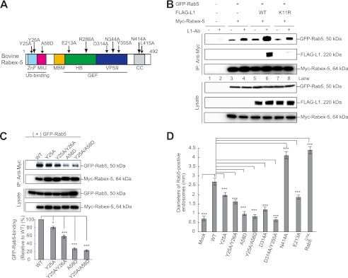
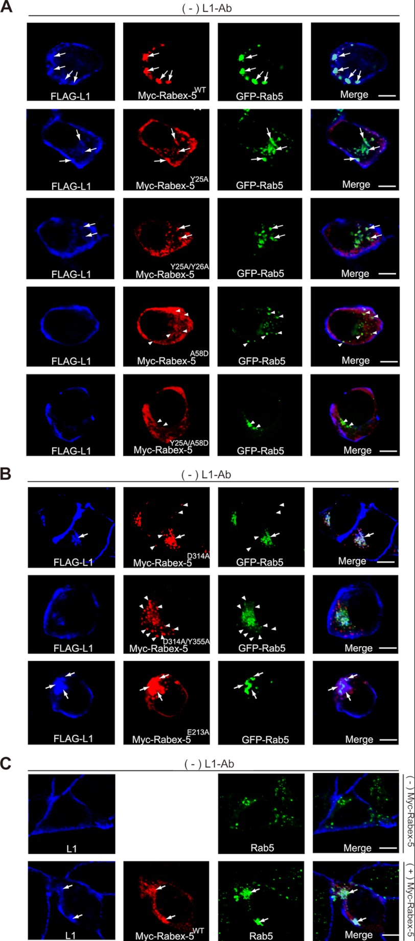
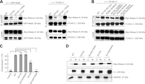
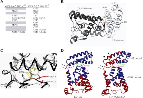
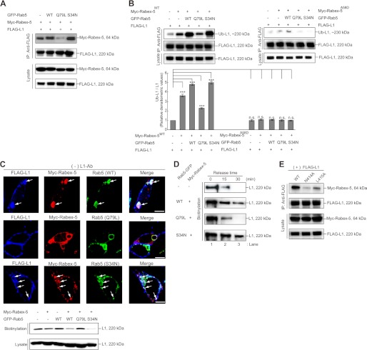
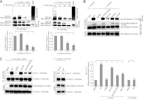
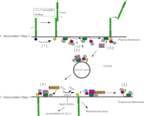
Significance: Rabex-5 GEF activity acts as an intramolecular switch for spatiotemporal trafficking of the ubiquitinated cargos. Ubiquitin (Ub)-dependent endocytosis of membrane proteins requires precise molecular recognition of ubiquitinated cargo by Ub-binding proteins (UBPs). Many UBPs are often themselves monoubiquitinated, a mechanism referred to as coupled monoubiquitination, which prevents them from binding in trans to the ubiquitinated cargo. However, the spatiotemporal regulatory mechanism underlying the interaction of UBPs with the ubiquitinated cargo, via their Ub-binding domains (UBDs) remains unclear. Previously, we reported the interaction of Rabex-5, a UBP and guanine nucleotide exchange factor (GEF) for Rab5, with ubiquitinated neural cell adhesion molecule L1, via its motif interacting with Ub (MIU) domain. This interaction is critical for the internalization and sorting of the ubiquitinated L1 into endosomal/lysosomal compartments. The present study demonstrated that the interaction of Rabex-5 with Rab5 depends specifically on interaction of the MIU domain with the ubiquitinated L1 to drive its internalization. Notably, impaired GEF mutants and the Rabex-5(E213A) mutant increased the flexibility of the hinge region in the HB-VPS9 tandem domain, which significantly affected their interactions with the ubiquitinated L1. In addition, GEF mutants increased the catalytic efficiency, which resulted in a reduced interaction with the ubiquitinated L1. Furthermore, the coupled monoubiquitination status of Rabex-5 was found to be significantly associated with interaction of Rabex-5 and the ubiquitinated L1. Collectively, our study reveals a novel mechanism, wherein the GEF activity of Rabex-5 acts as an intramolecular switch orchestrating ubiquitinated cargo-binding activity and coupled monoubiquitination to permit the spatiotemporal dynamic exchange of the ubiquitinated cargos.


Figures

FIGURE 1.
FIGURE 1.
UBDs regulate the Rab5-binding activity of Rabex-5. A, schematic diagram showing the domain structure of bovine Rabex-5. Light blue, ZnF; magenta, MIU; orange, membrane-binding motif (MBM); green, HB; blue, VPS9; and gray, CC domain. The arrows indicate the Rabex-5 mutants used in this study. B, N2a cells co-expressing the indicated plasmids were incubated in the presence or absence of L1-Ab. The cells were then lysed, and immunoblot analysis was performed. To control the functionality of the antibodies, cell lysates were applied (Lysate). C, cells co-expressing GFP-Rab5 with the indicated plasmids were lysed and analyzed by immunoblotting (upper). The bars represent the relative densitometric value of GFP-Rab5 to Rabex-5WT (lower). The data are mean ± S.E. in triplicate. *, p < 0.05; ***, p < 0.001. D, the diameters of the largest GFP-Rab5-labeled endosomes in 30 N2a cells expressing Rabex-5WT, its mutants, and Rab5Q79L were measured: the graph shows the mean and calculated S.E. ***, p < 0.001.
FIGURE 2.
FIGURE 2.
Impact of Rabex-5 mutants on the subcellular distributions of L1 and Rab5. A, N2a cells co-expressing the indicated plasmids in the absence of L1-Ab were stained with FLAG-tagged L1 (blue), Myc-tagged Rabex-5 (red), and GFP-Rab5 (green). The arrows indicate the accumulation of L1 on Rabex-5-positive endosomes. The arrowheads indicate that Rab5-positive endosomes do not co-localize with Rabex-5 UBDs mutants. Scale bar, 5 μm. B, N2a cells transfected with the indicated plasmids in the absence of L1-Ab were stained with FLAG-tagged L1 (blue), Myc-tagged Rabex-5 (red), and GFP-Rab5 (green). The arrows indicate the accumulation of L1 on Rabex-5-positive endosomes. The arrowheads indicate that Rabex-5 GEF mutants do not co-localize with Rab5-positive endosomes. Scale bar, 5 μm. C, N2a cells in the absence of L1-Ab were stained with endogenous L1 (blue) and endogenous Rab5 (green) (upper). N2a cells transfected with Myc-tagged Rabex-5 were stained with endogenous L1 (blue), Myc-tagged Rabex-5 (red), and endogenous Rab5 (green) (lower). Note that overexpression of Myc-tagged Rabex-5 induced the accumulation of endogenous L1 and enlarged the endogenous Rab5-positive endosomes. Scale bar, 5 μm.
FIGURE 3.
FIGURE 3.
Analysis of Rabex-5 GEF mutants. A, the cells were co-transfected with the indicated plasmids. Cell lysates were immunoprecipitated and analyzed by immunoblotting. B, cells co-expressing FLAG-tagged L1 with the indicated Myc-tagged Rabex-5 mutants were immunoprecipitated and analyzed by immunoblotting. The parentheses indicate human amino acid residues. C, fluorescence intensities were quantified, and the percentage of perinuclear to total fluorescence was plotted (right). ***, p < 0.001; n.s., not significant. D, immunoblot showing the membrane and cytosol distribution in N2a cells expressing the indicated plasmids and endogenous L1 and GAPDH, as described above. C, cytosol; M, membrane.
FIGURE 4.
FIGURE 4.
Structural modeling of the human Rabex-5 HB-VPS9 tandem domain. A, amino acid residues in human Rabex-5 located within 3 or 3.5 Å from the Oϵ2 atom of Glu-212 in the HB-VPS9 tandem domain. The parentheses indicate the bovine amino acid residues. B, ribbon diagram of the human HB (gray) and VPS9 domains (black) (PDB code 2OT3). Glu-212 (E213 in bovine) and Asn-343 (N344 in bovine) are highlighted as stick models in yellow and red, respectively. A dotted orange line indicates hydrogen bonds. C, structural comparison of human Rabex-5WT (black) and the Rabex-5E212A/N343A mutant (gray) in the HB-VPS9 tandem domains. The structure of the Rabex-5E212A/N343A mutant was calculated by MD simulation. A dotted orange line indicates hydrogen bonds. D, comparisons of the atomic fluctuations (B-factor) of the human Rabex-5 mutants with Rabex-5WT. Amino acid residues whose fluctuations are higher than that of the Rabex-5WT are shown in red. The arrows and arrowheads indicate the increment of fluctuations of the hinge region between the HB and VPS9 domains and the HB domain of Rabex-5E212A/N343A mutant, respectively.
FIGURE 5.
FIGURE 5.
Activation of Rabex-5 GEF activity decreased ubiquitinated L1 binding. A, N2a cells expressing the indicated plasmids were lysed and analyzed by immunoblotting. B, the accumulation of ubiquitinated L1 was examined by immunoprecipitation (IP) and analyzed by immunoblotting (upper). The bars represent the relative densitometric value of Ub-L1/L1 (lower). Note that a compatible amount of the ubiquitinated L1 in cells only expressing FLAG-tagged L1 was detected in cells co-expressing with Myc-tagged Rabex-5A58D. The data are mean ± S.E. in triplicate. ***, p < 0.001. C, N2a cells transfected with the indicated plasmids were stained with FLAG-tagged L1 (blue), Myc-tagged Rabex-5 (red), and GFP-Rab5 (green). The arrows indicate L1 accumulation on Rabex-5-positive endosomes (upper). Scale bar, 5 μm. N2a cells expressing the indicated plasmids were labeled with biotin and precipitated with streptavidin-agarose (lower). The amount of L1 on the surface was detected by anti-L1 antibodies. D, L1 recycling was measured. The internalized L1 (lane 1) after glutathione stripping of cells disappeared from the internal pools in mock cells and cells co-expressing GFP-Rab5Q79L within 15 min (lanes 2 and 3), whereas cells co-expressing GFP-Rab5WT and GFP-Rab5S34N exhibited the delayed disappearance of L1. The relative densitometric value of L1 divided by the incubation time was calculated as the release rate from endosomes (in the text). E, N2a cells expressing the indicated plasmids were lysed and analyzed by immunoblotting.
FIGURE 6.
FIGURE 6.
Determination of monoubiquitinated Rabex-5 levels. A, N2a cells co-expressing Myc-tagged Rabex-5 with HA-tagged Ub (left) or N2a cells co-expressing Myc-tagged Rabex-5, FLAG-tagged L1K11R, and HA-tagged Ub (right) were stimulated with L1-Ab. Immunoprecipitation (IP) and immunoblotting were performed as indicated (uppers). The mean amount of Ub-Rabex-5 normalized to Ub-Rabex-5 in the absence of L1-Ab is presented in the graphs (lower). The data are mean ± S.E. in triplicate. ***, p < 0.001. B, N2a cells were co-transfected with the indicated plasmids. Immunoprecipitation and immunoblotting were performed as indicated. C, the accumulation of ubiquitinated L1 was examined by immunoprecipitation and analyzed by immunoblotting (left). The bars represent the relative densitometric value of Ub-L1/L1 (right). The data are mean ± S.E. in triplicate. ***, p < 0.001.
FIGURE 7.
FIGURE 7.
A proposed mechanism for the spatiotemporal regulation of Rabex-5 in the endocytic trafficking of ubiquitinated L1. M, motif-interacting with Ub; Z, ZnF. Step 1, Src kinase is involved in the phosphorylation of a tyrosine-based motif, thereby regulating recruitment of the AP-2 protein. However, the dephosphorylation of this motif upon L1-L1 homophilic interactions results in the recruitment of AP-2, causing the clathrin-mediated endocytosis of L1 from the plasma membrane. The dephosphorylation of L1 also triggers the recruitment of an unidentified E3-ligase to ubiquitinate L1 on the plasma membrane. Step 2, the coupled monoubiquitination of Rabex-5, presumably in a switched off state, in which the MIU domain interacts with the mono-Ub moiety in cis, undergoes deubiquitination by an unidentified deubiquitinase upon L1 ligand stimulation, which might cause a Ca2+ influx. Then, monoubiquitinated Rabex-5 undergoes deubiquitination, converting Rabex-5 to a switched on state in which the MIU domain is positioned for trans interactions. Step 3, the interaction of ubiquitinated L1 with Rabex-5 recruits Rab5, which in turn triggers the endocytosis of ubiquitinated L1 from the plasma membrane. Next, the ubiquitinated L1·Rabex-5·GDP-Rab5 complex in an endocytic vesicle is transported to the endosomal membrane. Step 4, Rabex-5 turns on the GEF activity of the HB-VPS9 tandem domain by recruiting Rabaptin-5 to endosomes. Step 5, the conversion of GDP-Rab5 to GTP-Rab5 catalyzed by the GEF domain releases the Rabex-5·GTP-Rab5·Rabaptin-5 complex from the ubiquitinated L1 on the endosomes. Releasing the ubiquitinated L1 from the MIU domain allows this domain to interact with the mono-Ub moiety in cis and to return to the coupled monoubiquitinated Rabex-5 for another cycle.

References

    1. Mukhopadhyay D., Riezman H. (2007) Proteasome-independent functions of ubiquitin in endocytosis and signaling. Science 315, 201–205 - PubMed
    1. Raiborg C., Stenmark H. (2009) The ESCRT machinery in endosomal sorting of ubiquitylated membrane proteins. Nature 458, 445–452 - PubMed
    1. Hicke L., Schubert H. L., Hill C. P. (2005) Ubiquitin-binding domains. Nat. Rev. Mol. Cell Biol. 6, 610–621 - PubMed
    1. Hurley J. H., Lee S., Prag G. (2006) Ubiquitin-binding domains. Biochem. J. 399, 361–372 - PMC - PubMed
    1. Traub L. M., Lukacs G. L. (2007) Decoding ubiquitin sorting signals for clathrin-dependent endocytosis by CLASPs. J. Cell Sci. 120, 543–553 - PubMed

Publication types

MeSH terms

Substances

LinkOut - more resources