Comparative Study
. 2012 Nov 1;53(12):7476-83.
doi: 10.1167/iovs.12-10735.

Notch inhibition during corneal epithelial wound healing promotes migration

Affiliations
Comparative Study

Notch inhibition during corneal epithelial wound healing promotes migration

Asadolah Movahedan et al. Invest Ophthalmol Vis Sci. .

Abstract

Purpose: To determine the role of Notch signaling in corneal epithelial migration and wound healing.

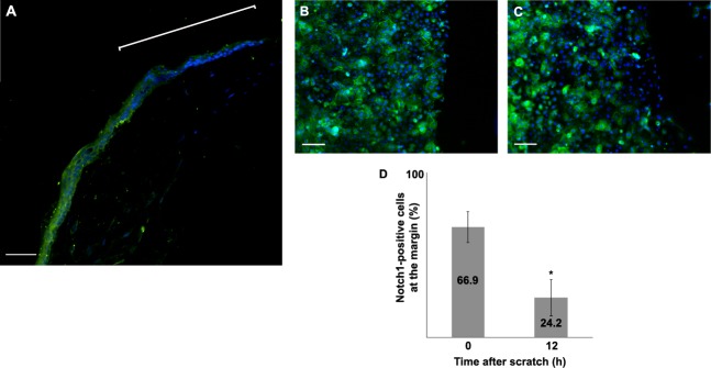
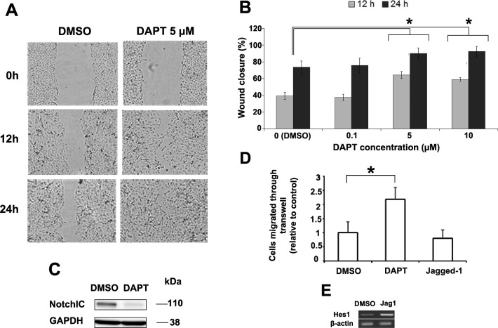
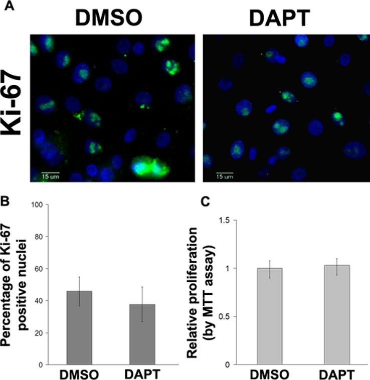
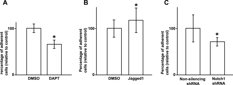
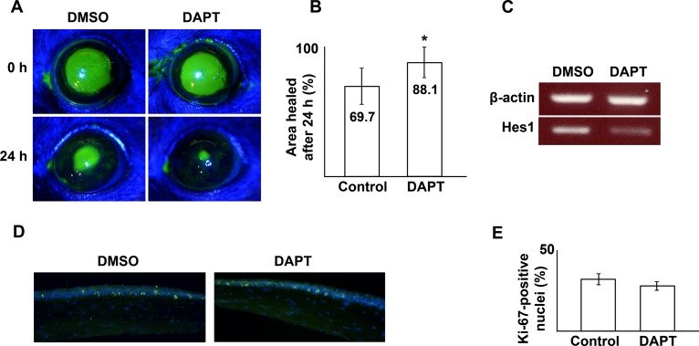
Methods: Immunolocalization of Notch1 was performed during epithelial wound healing in vivo in mouse corneal epithelial debridement wounds and in vitro in primary human corneal epithelial cells following a linear scratch wound. The effects of Notch inhibition, using the γ-secretase inhibitor N-(N-[3,5-difluorophenacetyl]-l-alanyl)-S-phenylglycine t-butyl ester (DAPT) or following stable transfection with Notch1-short hairpin RNA (shRNA), was evaluated in a scratch assay and transwell migration assay. Likewise, in vitro adhesion, proliferation and the actin cytoskeleton was examined. The DAPT effect was also evaluated in vivo in a mouse model of corneal epithelial wound healing.

Results: The expression of Notch1 was reduced at the leading edge of a healing corneal epithelium both in vivo and in vitro. Notch inhibition using DAPT and using Notch1-shRNA both enhanced in vitro migration in scratch and transwell migration assays. Consistent with this increased migratory behavior, Notch inhibited cells demonstrated decreased cell-matrix adhesion and enhanced lamellipodia formation. Notch inhibition by DAPT was also found to accelerate corneal epithelial wound closure in an in vivo murine model without affecting proliferation.

Conclusions: The results highlight the role of Notch in regulating corneal epithelial migration and wound healing. In particular, Notch signaling appears to decrease in the early stages of wound healing which contributes to cytoskeletal changes with subsequent augmentation of migratory behavior.


Conflict of interest statement

Disclosure: A. Movahedan, None; M. Majdi, None; N. Afsharkhamseh, None; H.M. Sagha, None; N.S. Saadat, None; K. Shalileh, None; B.Y. Milani, None; H. Ying, None; A.R. Djalilian, None

Figures

Figure 1.
Figure 1.
The expression of Notch1 was evaluated in the mouse corneal epithelium at 6 hours after a debridement wound. Notch1 staining was distinctly reduced in the cells near the leading edge (the bracket) (A). The expression of Notch1 was evaluated in primary human corneal epithelial cells subjected to a scratch assay at baseline (B) and after 12 hours (C). The percentage of cells that stained for Notch1 in the leading edge was 42% lower at 12 hours compared with immediately (0 hour) after the scratch (D) (P < 0.001). (Green: FITC, blue: DAPI); scale bar is 40 μm for (A) and 100 μm for (B) and (C). Asterisk represents statistically significant data.
Figure 2.
Figure 2.
Human corneal epithelial cells were subjected to a scratch assay and then treated with DAPT or DMSO (control) (A). The effect of DAPT concentration on scratch assay wound closure rate was measured (B). * Represents a statistically significant difference compared to control (P < 0.001). Western blot for Notch1IC confirmed that 10-μM DAPT can effectively inhibit Notch activation (C). HCE-T cells pretreated with DAPT migrated 2.2 times faster than control in transwell migration assay (P < 0.0001) while Jagged1 treated cells migrated 20% slower but did not reach statistical significance (P = 0.077) (D). Jagged1 treatment was found to activate Notch by inducing the expression of its downstream Hes1 (E). Asterisk represents statistically significant data.
Figure 3.
Figure 3.
HCE-T cells stably transfected with Notch1-shRNA demonstrating that Notch1 was knocked down by 66% by RT-PCR compared with NS shRNA (A) with a corresponding decrease in Notch1IC protein level by Western blot (B). Notch1-shRNA transfected HCE-T cells closed scratch wounds faster (P < 0.004) (C, D) and migrated 4 times faster in transwell migration assay (P < 0.0001) (E, F) compared with NS shRNA transduced control cells. Asterisk represents statistically significant data.
Figure 4.
Figure 4.
Primary human corneal epithelial cells were treated with DMSO or DAPT (5 μM) and subjected to Ki67 staining (A). The percentage of Ki67-positive cell nuclei was quantified. No difference was found between DAPT-treated and DMSO vehicle control cells (P = 0.254) (B). (Green: FITC; blue: DAPI), scale bar: 15 μm; Cell proliferation was also compared using MTT assay (P = 0.302) (C).
Figure 5.
Figure 5.
Notch inhibition with DAPT (5 μM) reduces corneal epithelial cell attachment to fibronectin-collagen coated plates by 34.5% compared with DMSO (control). (P < 0.001) (A). In contrast, Jagged1 treated cells were 18% more adherent to the matrix compared with control (P < 0.01) (B). HCE-T transduced with Notch1 (N1) shRNA likewise demonstrated 29% lower matrix attachment compared with NS shRNA transduced HCE-T cells (P < 0.001) (C). Asterisk represents statistically significant data.
Figure 6.
Figure 6.
F-Actin (rhodamine-phalloidin) staining of human corneal epithelial cells 6 hours after scratch demonstrating enhanced lamellipodia (arrows) formation in DAPT compared with control (AD). Cells treated with DAPT (10 μM) also demonstrate loss of marginal actin bundle (D, *) compared with control (C, *). HCE-T cells stably transfected with Notch1-shRNA similarly demonstrate enhanced lamellipodia at the wound age at 6 hours compared with NS shRNA transduced cell (E, F). Stable expression of the shRNA vector is evident based on GFP (green) expression, which often overlaps DAPI (blue) nuclear stain. Red: rhodamine-F-actin; scale bar: 100 μm for (A, B) and 50 μm for (E, F).
Figure 7.
Figure 7.
Treatment with DAPT (20 μM) accelerates wound healing after a 2.0-mm corneal epithelial debridement wound in mice (A, B) (P = 0.025). RT-PCR of the mouse corneal epithelium at 24 hours demonstrates reduced Hes1 in DAPT treated corneas, consistent with suppression of Notch signaling (C). Immunofluorescence examination of the proliferation marker, Ki67 (green: FITC), in the limbal area did not show a significant difference between DAPT-treated and control (DMSO) specimens at 24 hours post wounding (P = 0.102) (blue: DAPI nuclear stain) (D, E). Asterisk represents statistically significant data.

References

    1. Lu L, Reinach PS, Kao WW. Corneal epithelial wound healing. Exp Biol Med. 2001;226:653–664 - PubMed
    1. Yu FS, Yin J, Xu K, Huang J. Growth factors and corneal epithelial wound healing. Brain Res Bull. 2010;81:229–235 - PMC - PubMed
    1. Greenwald I. LIN-12/Notch signaling: lessons from worms and flies. Genes Dev. 1998;12:1751–1762 - PubMed
    1. Artavanis-Tsakonas S, Rand MD, Lake RJ. Notch signaling: cell fate control and signal integration in development. Science. 1999;284:770–776 - PubMed
    1. Struhl G, Greenwald I. Presenilin is required for activity and nuclear access of Notch in Drosophila. Nature. 1999;398:522–525 - PubMed

Publication types

MeSH terms