. 2012 Sep 14:3:290.
doi: 10.3389/fimmu.2012.00290. eCollection 2012.

The transcription of the alarmin cytokine interleukin-1 alpha is controlled by hypoxia inducible factors 1 and 2 alpha in hypoxic cells

Affiliations

The transcription of the alarmin cytokine interleukin-1 alpha is controlled by hypoxia inducible factors 1 and 2 alpha in hypoxic cells

Peleg Rider et al. Front Immunol. .

Abstract

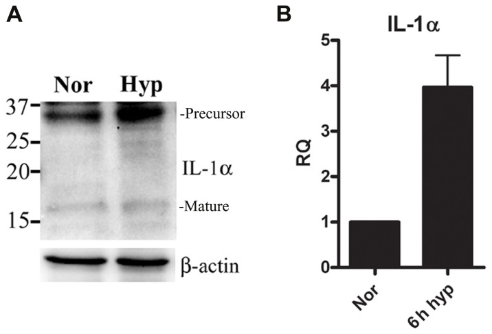
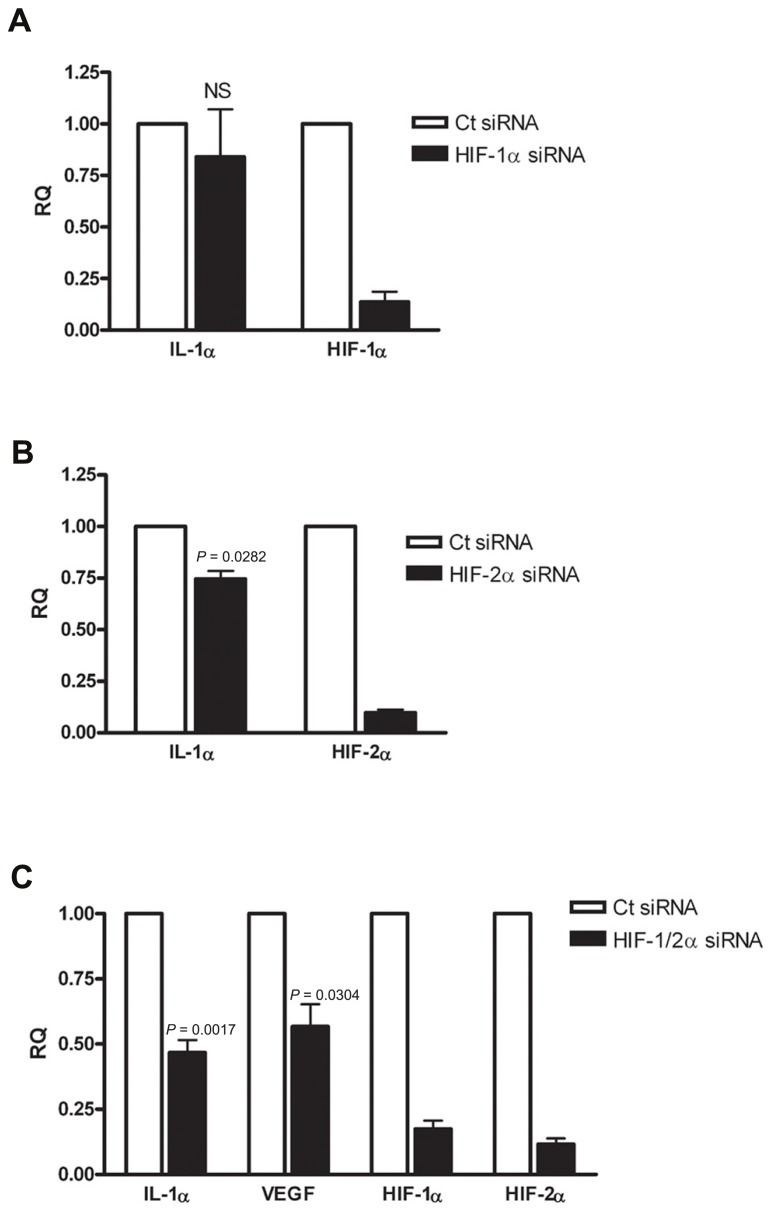
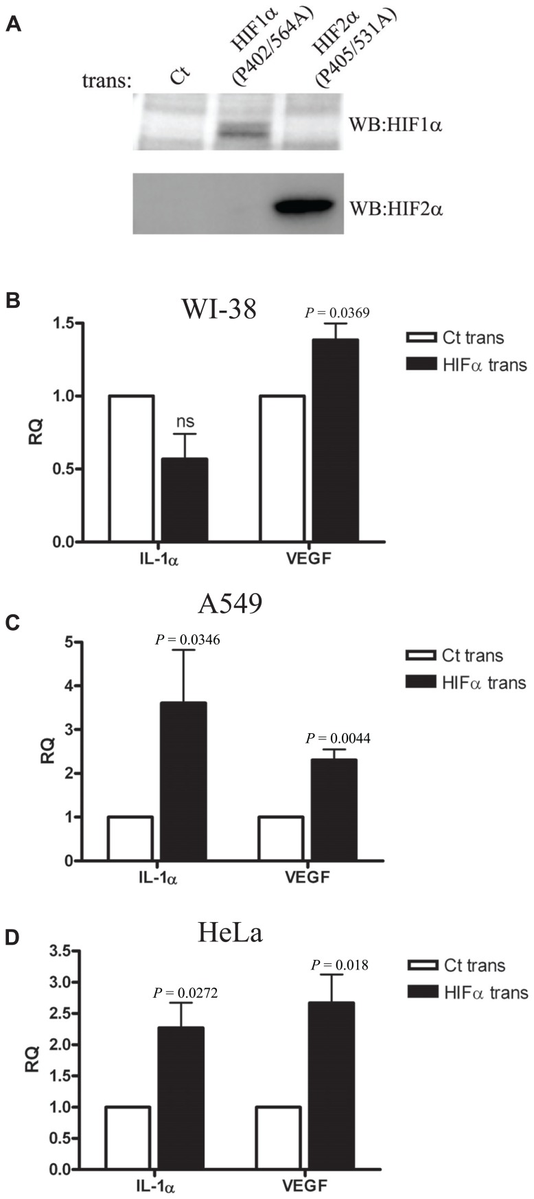
During hypoxia, cells undergo transcriptional changes to adjust to metabolic stress, to promote cell survival, and to induce pro-angiogenic factors. Hypoxia-induced factors (HIFs) regulate these transcriptional alterations. Failure to restore oxygen levels results in cell death by necrosis. IL-1α is one of the most important mediators of sterile inflammation following hypoxia-mediated necrosis. During hypoxia, IL-1α is up-regulated and released from necrotic cells, promoting the initiation of sterile inflammation. This study examined the role of IL-1α transcription in initiation of hypoxic stress and the correlation between IL-1α transcription and HIFα factors. In an epithelial cell line cultured under hypoxic conditions, IL-1α transcription was up-regulated in a process mediated and promoted by HIFα factors. IL-1α transcription was also up-regulated in hypoxia in a fibroblast cell line, however, in these cells, HIFα factors inhibited the elevation of transcription. These data suggest that HIFα factors play a significant role in initiating sterile inflammation by controlling IL-1α transcription during hypoxia in a differential manner, depending on the cell type.

Keywords: DAMPs; HIF-1α; HIF-2; IL-1; alarmin; cytokines and inflammation; sterile inflammation.


Figures

FIGURE 1
FIGURE 1
IL-1α transcription is up-regulated during hypoxia in A549 epithelial cells.(A) A549 cells were cultured in either normoxic or hypoxic conditions for 12 h. Cells lysates were analyzed by Western blot for IL-1α detection. Bands are marked as precursor or mature. (B) A549 cells were cultured in either normoxic or hypoxic conditions for 6 h, and were analyzed for relative quantification (RQ) of IL-1α transcription. Graph represents mean ± SD of three biological repeats.
FIGURE 2
FIGURE 2
Silencing HIFα proteins down-regulates IL-1α transcription in hypoxic A549 epithelial cells. A549 cells were transfected with HIF-1α siRNA (A) or HIF-2α siRNA (B) 24 h prior to hypoxia (6 h). Each graph represents mean ± SEM of three independent experiments. (C) Cells were transfected with both HIF-1α and HIF-2α mixed siRNA 24 h prior to hypoxia (6 h). Graph represents mean ± SEM of five independent experiments. P values of HIF-2α or HIF-1/2α siRNA-transfected cells vs. control (Ct) siRNA-transfected cells are annotated in the graphs.
FIGURE 3
FIGURE 3
Silencing HIFα proteins up-regulates IL-1α transcription in hypoxic WI-38 fibroblasts.(A) WI-38 fibroblasts were cultured in either normoxic or hypoxic conditions for 2, 4, or 6 h, and were analyzed by qRT-PCR for RQ of IL-1α transcription. Graph represents the average ± SEM of two different experiments. RQ of IL-1α transcription in WI-38 cells transfected with HIF-1α (B), HIF-2α (C) siRNA 24 h prior to 6 h culture in hypoxic conditions. Each graph represents mean ± SEM of three independent experiments. (D) Cells were transfected with both HIF-1α and HIF-2α mixed siRNA 24 h prior to hypoxia (6 h). Graph represents mean ± SEM of four independent experiments. P values of HIF-1/2α siRNA transfected cells vs. control (Ct) siRNA-transfected cells are annotated in the graphs.
FIGURE 4
FIGURE 4
Overexpression of HIFα proteins increases IL-1α transcription in A549 and HeLa cells but not in WI-38 fibroblasts.(A) Western blot using anti-HIF-1α or anti-HIF-2α antibodies of nuclear fractions obtained from HEK-T293 cells transfected with the annotated vectors. WI-38 (B), A549 (C), or HeLa (D) cells were transfected with either control vector or with HIF-1α (P402A/P564A) and HIF-2α (P405A/P531A), and 24 h later were analyzed for IL-1α and VEGF RQ of mRNA levels by real-time PCR. Graphs represent mean ± SEM of four independent experiments for each cell type. P values of HIFα-transfected cells vs. control (Ct) transfected are annotated in the graphs.

References

    1. Andersson U., Tracey K. J. (2011). HMGB1 is a therapeutic target for sterile inflammation and infection. Annu. Rev. Immunol. 29 139–162 - PMC - PubMed
    1. Basu S., Binder R. J., Suto R., Anderson K. M., Srivastava P. K. (2000). Necrotic but not apoptotic cell death releases heat shock proteins, which deliver a partial maturation signal to dendritic cells and activate the NF-kappa B pathway. Int. Immunol. 12 1539–1546 - PubMed
    1. Ben-Shoshan J., Afek A., Maysel-Auslender S., Barzelay A., Rubinstein A., Keren G., George J. (2009). HIF-1alpha overexpression and experimental murine atherosclerosis. Arterioscler. Thromb. Vasc. Biol. 29 665–670 - PubMed
    1. Berda-Haddad Y., Robert S., Salers P., Zekraoui L., Farnarier C., Dinarello C. A., Dignat-George F., Kaplanski G. (2011). Sterile inflammation of endothelial cell-derived apoptotic bodies is mediated by interleukin-1alpha. Proc. Natl. Acad. Sci. U.S.A. 108 20684–20689 - PMC - PubMed
    1. Bianchi M. E. (2007). DAMPs, PAMPs and alarmins: all we need to know about danger. J. Leukoc. Biol. 81 1–5 - PubMed