

Endoscopic Ultrasound-Guided Radiofrequency Ablation (EUS-RFA) of the Pancreas in a Porcine Model
- PMID: 23049547
- PMCID: PMC3459266
- DOI: 10.1155/2012/431451
Endoscopic Ultrasound-Guided Radiofrequency Ablation (EUS-RFA) of the Pancreas in a Porcine Model
Abstract
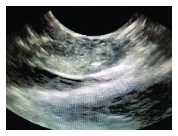
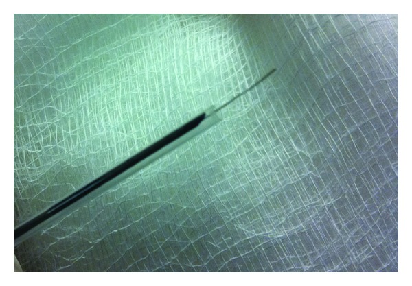
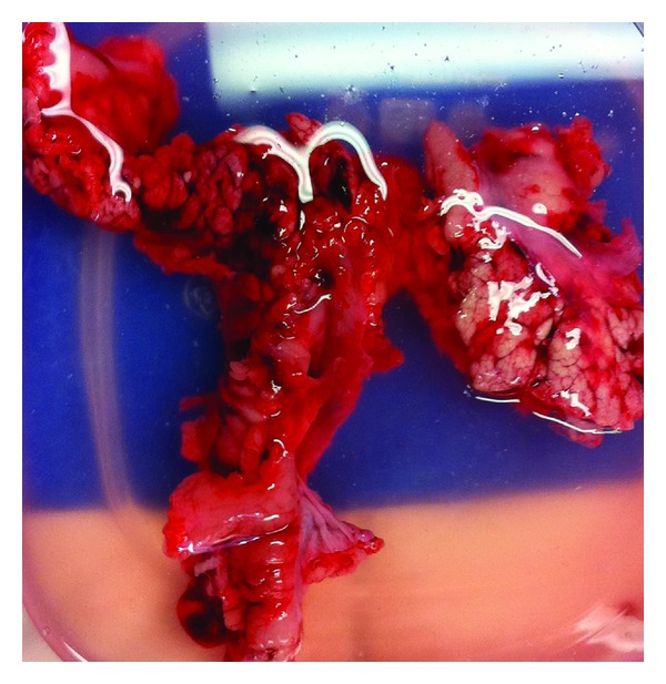
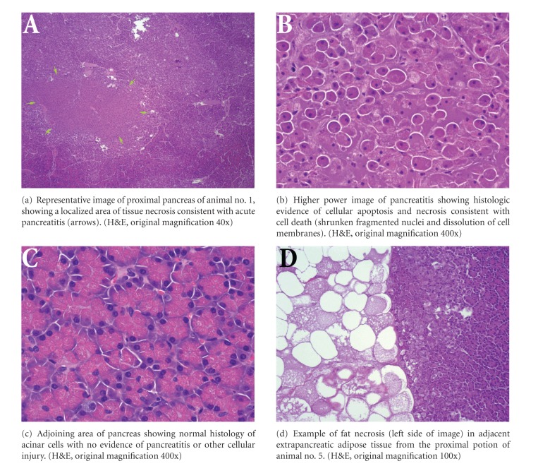
Backgrounds. Limited effective palliative treatments exist for pancreatic cancer which includes surgery or chemotherapy. Radiofrequency ablation (RFA) uses high frequency alternating current to ablate diseased tissue and has been used to treat various tumors. In this study, we evaluated a prototype probe adjusted to the EUS-needle to perform EUS-RFA to permit coagulative necrosis in the pancreas. Methods. Five Yucatan pigs underwent EUS-guided radiofrequency ablation of the head of their pancreas. Using an EUS-needle, RFA was applied with 6 mm and then 10 mm of the probe exposed at specific wattage for preset durations. Results. Only one pig showed moderate levels of pancreatitis (20% proximal pancreatitis). The other animals showed much lower areas of tissue damage. In 3 of the 5 pigs, the proximal pancreas showed greater levels of tissue injury than the distal pancreas, consistent with the proximity of the tissue to the procedure site. In 1 pig, both proximal and distal pancreas showed minimal pancreatitis (1%). There was minimal evidence of fat necrosis in intra-pancreatic and/or extra-pancreatic adipose tissue. Conclusion. EUS-guided RFA of the pancreatic head with the monopolar probe through a 19-gauge needle was well tolerated in 5 Yucatan pigs and with minimal amount of pancreatitis.
Figures
References
-
- Niederhuber JE, Brennan MF, Menck HR. The National Cancer Data Base report on pancreatic cancer. Cancer. 1995;76:1671–1677. - PubMed
-
- Warshaw AL, Fernandez-del Castillo C. Medical progress: pancreatic carcinoma. The New England Journal of Medicine. 1992;326(7):455–465. - PubMed
-
- Li D, Xie K, Wolff R, Abbruzzese JL. Pancreatic cancer. The Lancet. 2004;363(9414):1049–1057. - PubMed
-
- Carrara S, Arcidiacono PG, Albarello L, et al. Endoscopic ultrasound-guided application of a new hybrid cryotherm probe in porcine pancreas: a preliminary study. Endoscopy. 2008;40(4):321–326. - PubMed
-
- Wu Y, Tang Z, Fang H, et al. High operative risk of cool-tip radiofrequency ablation for unresectable pancreatic head cancer 1. Journal of Surgical Oncology. 2006;94(5):392–395. - PubMed
Grants and funding
LinkOut - more resources
Full Text Sources
Medical
