. 2012 Jun 1;6(6):391-397.
doi: 10.1038/nphoton.2012.104. Epub 2012 May 13.

Photovoltaic Retinal Prosthesis with High Pixel Density

Affiliations

Photovoltaic Retinal Prosthesis with High Pixel Density

Keith Mathieson et al. Nat Photonics. .

Abstract

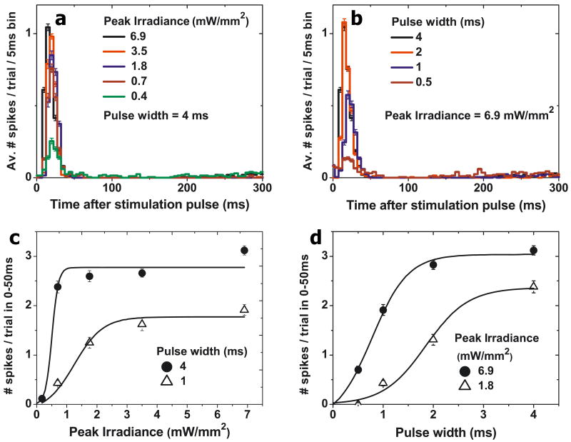
Retinal degenerative diseases lead to blindness due to loss of the "image capturing" photoreceptors, while neurons in the "image processing" inner retinal layers are relatively well preserved. Electronic retinal prostheses seek to restore sight by electrically stimulating surviving neurons. Most implants are powered through inductive coils, requiring complex surgical methods to implant the coil-decoder-cable-array systems, which deliver energy to stimulating electrodes via intraocular cables. We present a photovoltaic subretinal prosthesis, in which silicon photodiodes in each pixel receive power and data directly through pulsed near-infrared illumination and electrically stimulate neurons. Stimulation was produced in normal and degenerate rat retinas, with pulse durations from 0.5 to 4 ms, and threshold peak irradiances from 0.2 to 10 mW/mm(2), two orders of magnitude below the ocular safety limit. Neural responses were elicited by illuminating a single 70 μm bipolar pixel, demonstrating the possibility of a fully-integrated photovoltaic retinal prosthesis with high pixel density.


Figures

Figure 1
Figure 1
Simplified system diagram. A portable computer processes video images captured by a head-mounted camera. Video goggles then project these images onto the retina using pulsed infrared (880-915 nm) illumination. Finally, pixels in the subretinal photodiode array convert this light into local stimulation currents.
Figure 2
Figure 2
(a) A photodiode array consisting of 25μm pixels, each containing a ∼10 μm stimulating electrode (#1 in inset) surrounded by the photosensitive area of a single photodiode. Return electrode common to all pixels is situated on the back of the array. (b) An array with 3-diode pixels arranged in a hexagonal pattern. Pixels of 70 and 140 μm in size have been made. Central electrodes (#2 in inset) of 20 and 40 μm in diameter, respectively, are surrounded by 3 diodes connected in series, and by the common return electrode (#3 in inset). Pixels are separated by 5μm trenches to improve perfusion and for better isolation. 140 μm pixel is shown in the inset.
Figure 3
Figure 3
(a) Schematic of photovoltaic stimulation characterization system. Both healthy and degenerate rat retinas were placed between the stimulating and recording arrays, with the ganglion cell layer facing the recording electrodes. The photodiode array converts patterned 880-905 nm illumination into stimulation currents. (b) An IR pulse, with variable pulse width and intensity, creates a charge-balanced current waveform in each pixel of the photodiode array. The microelectrode array records the resultant stimulus artifact and retinal responses from each of the 512 electrodes. Stimulation is repeated at least 400 times for each setting. The artifact is then subtracted and the recorded action potentials undergo principal component analysis and automated clustering to attribute spiking waveforms to over 100 RGCs per experiment.
Figure 4
Figure 4
(a) Peristimulus time histograms showing the stimulated response of a wild type rat retina, using the triple-diode devices, to different peak irradiances at a fixed pulse width of 4ms. (b) Peristimulus time histograms for various pulse widths at a fixed irradiance of 6.9 mW/mm2. (c) Increase of retinal response with peak irradiance for two pulse widths. (d) Increase of retinal response with pulse width for two peak irradiances. All error bars denote the standard error of the mean.
Figure 5
Figure 5
(a) Peristimulus time histograms showing the elicited response of RCS rat retina, using the triple-diode devices, at a fixed NIR pulse width and (b) at a fixed light intensity. The number of elicited spikes increases with irradiance (c) and with pulse width (d). (e) Strength-duration plot for both wild type and RCS rat retinas, showing the average stimulation thresholds for 10 neurons in both the healthy and degenerate retinas (single-diode devices). The average values for the triple-diode devices are also plotted for 6 neurons over two preparations (WT) and 14 neurons in a single preparation (RCS).Hashed zones depict the range of standard deviation, while the whiskers show min and max for each data point. The optical safety limits for a single pulse and continuous retinal illumination (15 Hz repetition rate) with 905 nm light are also plotted. The error bars in parts a-d denote the standard error of the mean.
Figure 6
Figure 6
(a) Retinal histology of a flat polymer implant in the subretinal space of an RCS rat, with the numerically calculated current distribution from a 115 μm pixel (pixel schematics overlaid). (b) Retinal histology of a pillar array implant, overlaid with the numerically calculated current distribution from electrodes placed on the tops. Implants with pixel densities greater than 256 pixels/mm2 will likely require the use of such 3-D geometries to achieve sufficient proximity to target neurons.

References

    1. Smith W, et al. Risk factors for age-related macular degeneration: Pooled findings from three continents. Ophthalmology. 2001;108:697–704. - PubMed
    1. Haim M. Epidemiology of retinitis pigmentosa in Denmark. Acta Ophthalmol Scand Suppl. 2002:1–34. - PubMed
    1. Kim SY, et al. Morphometric analysis of the macula in eyes with disciform age-related macular degeneration. Retina. 2002;22:471–477. - PubMed
    1. Mazzoni F, Novelli E, Strettoi E. Retinal ganglion cells survive and maintain normal dendritic morphology in a mouse model of inherited photoreceptor degeneration. J Neurosci. 2008;28:14282–14292. 28/52/14282 [pii] 10.1523/JNEUROSCI.4968-08.2008. - PMC - PubMed
    1. Stone JL, Barlow WE, Humayun MS, de Juan E, Jr, Milam AH. Morphometric analysis of macular photoreceptors and ganglion cells in retinas with retinitis pigmentosa. Arch Ophthalmol. 1992;110:1634–1639. - PubMed