. 2012;7(9):e44885.
doi: 10.1371/journal.pone.0044885. Epub 2012 Sep 26.

Improving the extent of malignant glioma resection by dual intraoperative visualization approach

Affiliations

Improving the extent of malignant glioma resection by dual intraoperative visualization approach

Ilker Y Eyüpoglu et al. PLoS One. 2012.

Abstract

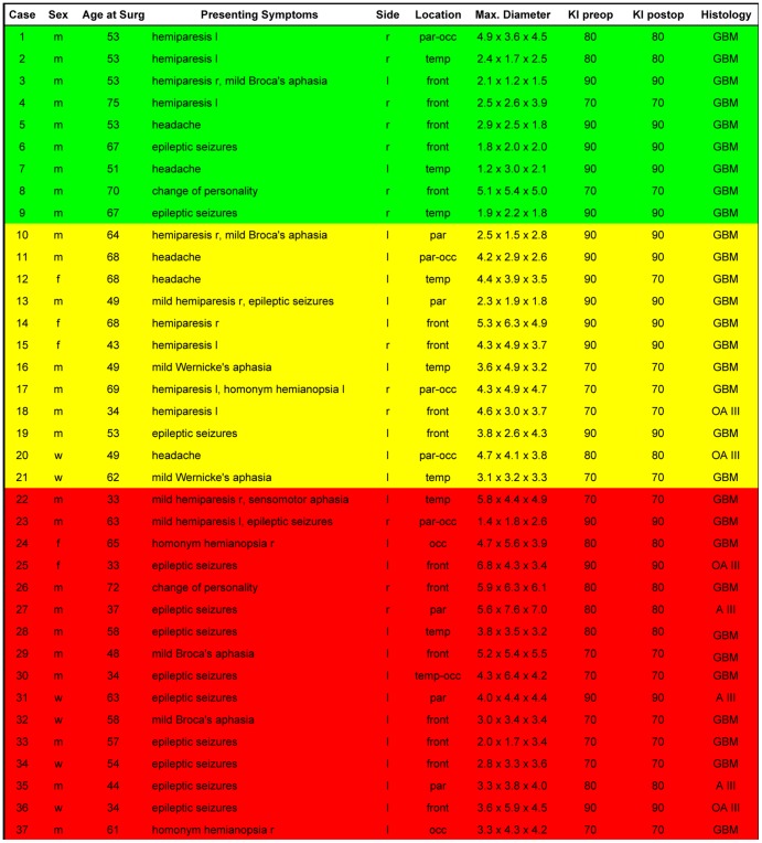
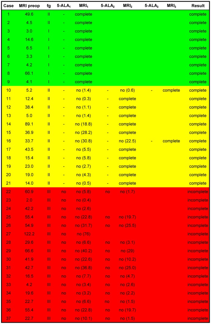
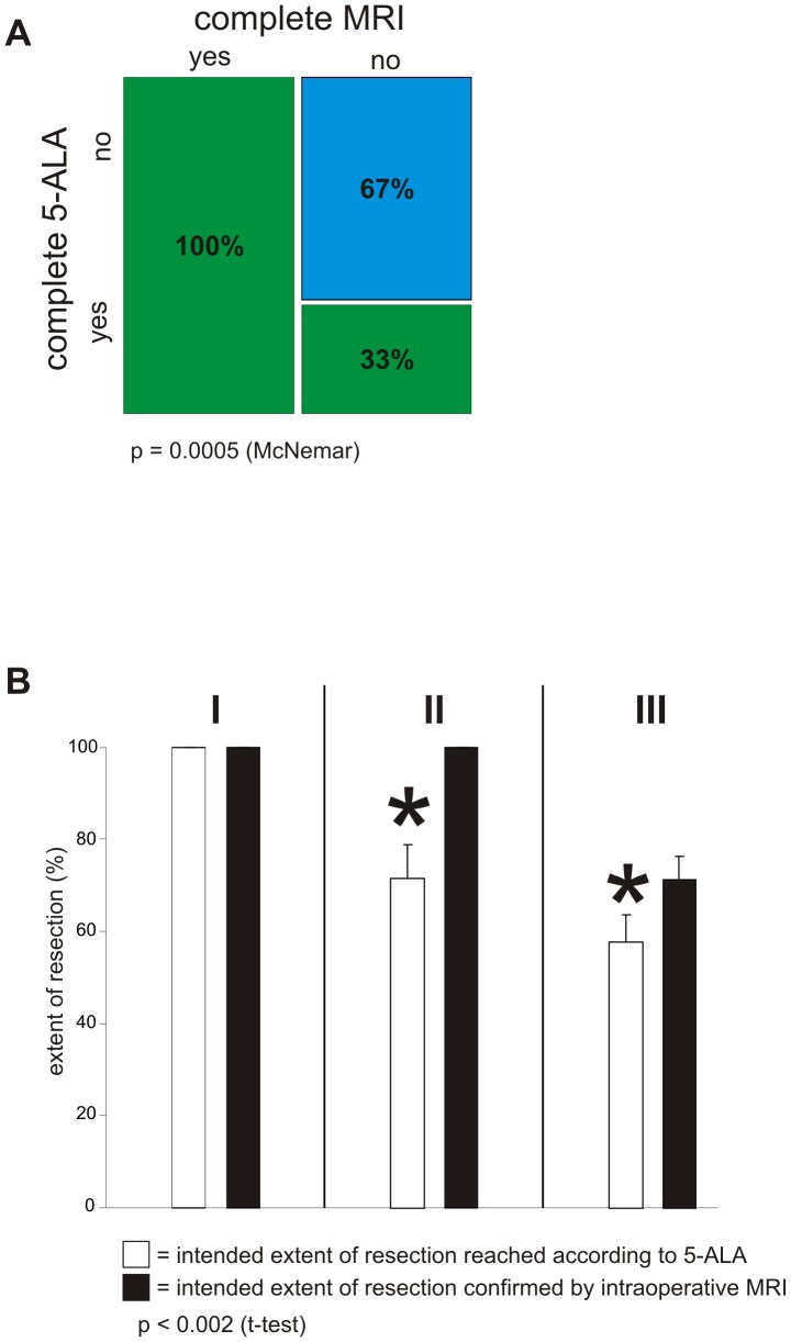
Despite continuing debates around cytoreductive surgery in malignant gliomas, there is broad consensus that increased extent of tumor reduction improves overall survival. However, maximization of the extent of tumor resection is hampered by difficulty in intraoperative discrimination between normal and pathological tissue. In this context, two established methods for tumor visualization, fluorescence guided surgery with 5-ALA and intraoperative MRI (iMRI) with integrated functional neuronavigation were investigated as a dual intraoperative visualization (DIV) approach. Thirty seven patients presumably suffering from malignant gliomas (WHO grade III or IV) according to radiological appearance were included. Twenty-one experimental sequences showing complete resection according to the 5-ALA technique were confirmed by iMRI. Fourteen sequences showing complete resection according to the 5-ALA technique could not be confirmed by iMRI, which detected residual tumor. Further analysis revealed that these sequences could be classified as functional grade II tumors (adjacent to eloquent brain areas). The combination of fluorescence guided resection and intraoperative evaluation by high field MRI significantly increased the extent of tumor resection in this subgroup of malignant gliomas located adjacent to eloquent areas from 61.7% to 100%; 5-ALA alone proved to be insufficient in attaining gross total resection without the danger of incurring postoperative neurological deterioration. Furthermore, in the case of functional grade III gliomas, iMRI in combination with functional neuronavigation was significantly superior to the 5-ALA resection technique. The extent of resection could be increased from 57.1% to 71.2% without incurring postoperative neurological deficits.


Conflict of interest statement

Competing Interests: The authors have declared that no competing interests exist.

Figures

Figure 1
Figure 1. Patient data depicted includes age, sex, presenting symptoms, tumor localization, the maximum dimensions of the tumor, the pre- and postoperative Karnofsky Performance Scale Index (KI) and the Neuropathological diagnosis according to the current WHO Classification System.
All included patients had suspected malignant gliomas (WHO grade III or IV), i.e. evidencing contrast enhancement.
Figure 2
Figure 2. The corresponding surgical data has been given with tumor volume and postoperative outcome.
Tumor volume was measured in cm3. Functional tumor localization (abbreviated as fg) was determined by preoperative MRI and classified according to Sawaya . Surgery was primarily carried out according to the 5-ALA signal, with corresponding iMRI resection controls carried out following disappearances of this signal. The residual tumor volume following each iMRI scan has been indicated in parentheses. The green color code has been used to depict complete resection according to both modalities during the first iMRI scan itself. Yellow has been used to depict complete resection requiring several iMRI scans. Red has been used to depict intentional, incomplete tumor resection.
Figure 3
Figure 3. Dual intraoperative visualization approach and anatomical view.
On the basis of a typical case of a tumor in the vicinity of an eloquent area, we demonstrate that the risk of missing tumor remnants covered by non-pathological tissue is eliminated through an iMRI control. A, The first iMRI scan carried out following the disappearance of the 5-ALA signal depicted a residual contrast enhancing area (marked by arrows). B, Tumor resection was resumed following re-segmentation and update of the neuronavigation. C, During resection of the intervening layer of non-pathological tissue, the 5-ALA signal reappeared (marked by arrows) and corresponded to the re-segmented contrast enhancing area.
Figure 4
Figure 4. Fluorescence guided tumor localization and eloquent area visualization.
On the basis of the same case, we demonstrate the potential dangers, above all of post-operative neurological deterioration, associated with resection carried out according to the 5-ALA signal alone without the safeguard of neuronavigation segmentation and iMRI scans. A, Following resection of the bulk of the tumor, a faint 5-ALA signal was detectable (marked by arrows). B, The corresponding neuronavigation segmentation however, demonstrated that the pyramidal tract was reached (marked by arrows), and that further resection would result in postoperative neurological deterioration. C, The corresponding iMRI control confirmed the close proximity of the resection margin to the pyramidal tract (depicted in pink).
Figure 5
Figure 5. Dual intraoperative visualization approach in different functional grade patients.
A, Extent of resection was determined by 5-ALA and the results verified through iMRI. This was defined as one sequence of the procedure. 21 sequences showing complete resection according to 5-ALA were confirmed by iMRI (complete 5-ALA: yes, complete MRI: yes – green bar, first column). 14 sequences showing complete resection according to 5-ALA could not be confirmed by iMRI, which detected residual tumor (complete 5-ALA: yes, complete MRI: no – green bar, second column). 29 sequences showed residual tumor both according to 5-ALA as well as iMRI (complete 5-ALA: no, complete MRI: no – blue bar, second column). The order of these sequences resulted in a p-value = 0.0005 (McNemar). B, Functional tumor localization was categorized according to Sawaya . Tumors in non-eloquent areas were defined as functional grade I [marked with I]. Functional grade II was defined as tumor localization close to an eloquent brain area [marked with II]. Functional grade III (given as III at the top) was defined as tumor localization in an eloquent brain area. Extent of resection was calculated as a percentage of prior tumor volume. Within the subgroup I the intended 100% resection was achieved by surgery with 5-ALA alone. In the subgroup II 5-ALA alone resulted in a tumor resection of 71.7% (±7.285 sem), whereas additional use of iMRI significantly increased tumor resection to 100% (p-value<0.002; Student's t-test). The subgroup III showed significant difference in extent of tumor resection. The results of tumor resection were 57.6% (±6.01 sem) achieved with 5-ALA alone, whereas a further tumor resection up to 71.2% (±5.257 sem) could be achieved through the additional use of iMRI.

References

    1. Wen PY, Kesari S (2008) Malignant gliomas in adults. N Engl J Med 359: 492–507. - PubMed
    1. Van Meir EG, Hadjipanayis CG, Norden AD, Shu HK, Wen PY, et al. (2010) Exciting new advances in neuro-oncology: the avenue to a cure for malignant glioma. CA Cancer J Clin 60: 166–193. - PMC - PubMed
    1. Kuhnt D, Becker A, Ganslandt O, Bauer M, Buchfelder M, et al. (2011) Correlation of the extent of tumor volume resection and patient survival in surgery of glioblastoma multiforme with high-field intraoperative MRI guidance. Neuro Oncol 13: 1339–1348. - PMC - PubMed
    1. Kubben PL, ter Meulen KJ, Schijns OE, ter Laak-Poort MP, van Overbeeke JJ, et al. (2011) Intraoperative MRI-guided resection of glioblastoma multiforme: a systematic review. Lancet Oncol 12: 1062–1070. - PubMed
    1. Stupp R, Mason WP, van den Bent MJ, Weller M, Fisher B, et al. (2005) Radiotherapy plus concomitant and adjuvant temozolomide for glioblastoma. N Engl J Med 352: 987–996. - PubMed

MeSH terms