. 2012;7(9):e45452.
doi: 10.1371/journal.pone.0045452. Epub 2012 Sep 25.

A novel group of Moraxella catarrhalis UspA proteins mediates cellular adhesion via CEACAMs and vitronectin

Affiliations

A novel group of Moraxella catarrhalis UspA proteins mediates cellular adhesion via CEACAMs and vitronectin

Darryl J Hill et al. PLoS One. 2012.

Abstract

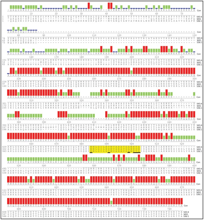
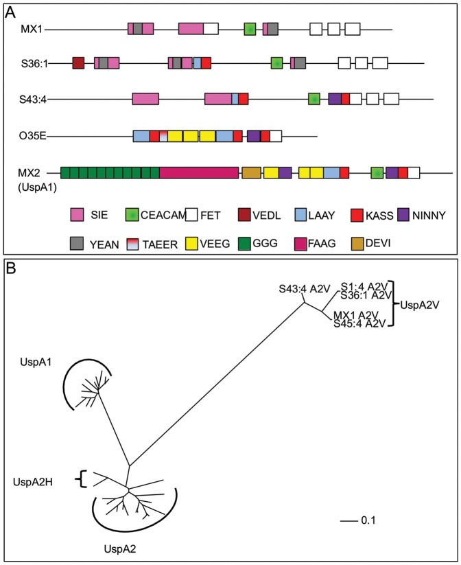
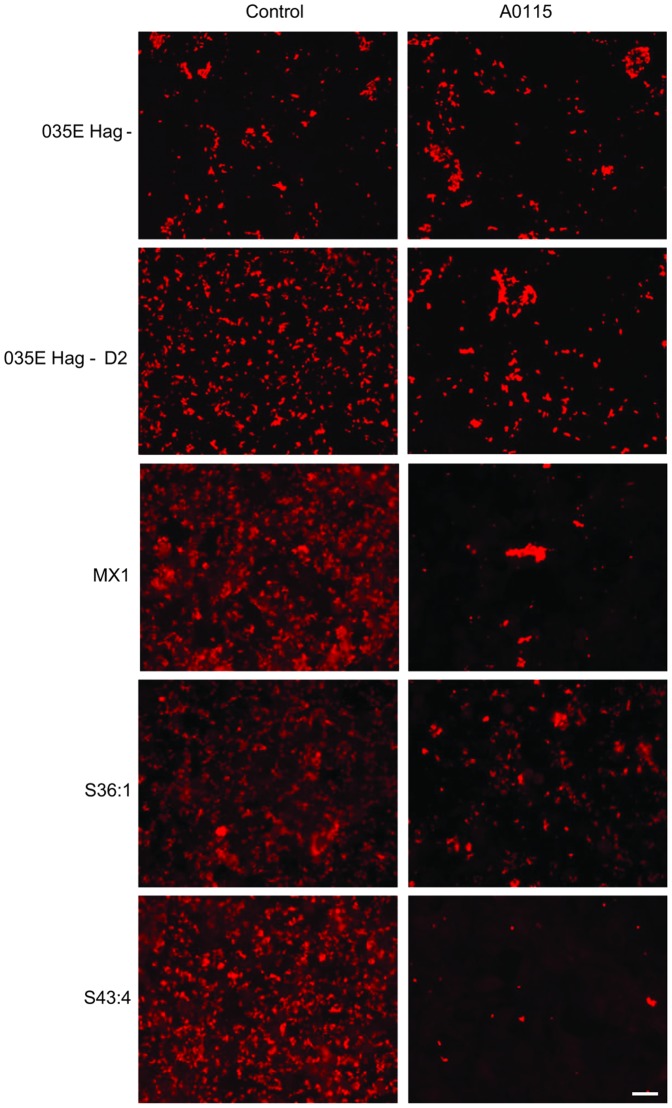
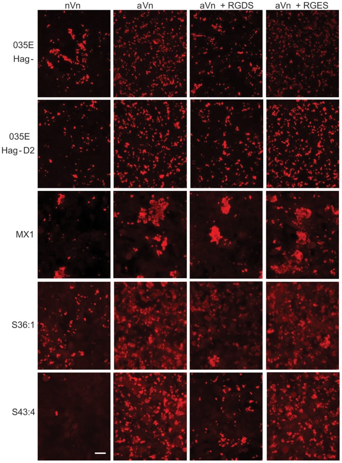
Moraxella catarrhalis (Mx) is a common cause of otitis media and exacerbation of chronic obstructive pulmonary disease, an increasing worldwide problem. Surface proteins UspA1 and UspA2 of Mx bind to a number of human receptors and may function in pathogenesis. Genetic recombination events in the pathogen can generate hybrid proteins termed UspA2H. However, whether certain key functions (e.g. UspA1-specific CEACAM binding) can be exchanged between these adhesin families remains unknown. In this study, we have shown that Mx can incorporate the UspA1 CEACAM1-binding region not only into rare UspA1 proteins devoid of CEACAM-binding ability, but also into UspA2 which normally lack this capacity. Further, a screen of Mx isolates revealed the presence of novel UspA2 Variant proteins (UspA2V) in ∼14% of the CEACAM-binding population. We demonstrate that the expression of UspA2/2V with the CEACAM-binding domain enable Mx to bind both to cell surface CEACAMs and to integrins, the latter via vitronectin. Such properties of UspA2/2V have not been reported to date. The studies demonstrate that the UspA family is much more heterogeneous than previously believed and illustrate the in vivo potential for exchange of functional regions between UspA proteins which could convey novel adhesive functions whilst enhancing immune evasion.


Conflict of interest statement

Competing Interests: MV is a named inventor on rD-7 related patent (WO2004/031236; WO2005/099337). This does not alter the authors' adherence to all the PLoS ONE policies on sharing data and materials.

Figures

Figure 1
Figure 1. CEACAM1 binding properties of M. catarrhalis strain 035E and its derivatives.
A) Western blot of Mx strains from right to left MX2 (used for comparison), 035E and 035E D2 and D1 overlaid with CEACAM1-Fc (CC1) or SIGLEC10-Fc (SIGLEC) as described in the methods. As expected, MX2 UspA1 bound to CEACAM1-Fc. No CEACAM binding protein was observed for 035E while its transformants D1 and D2 both bound to CEACAM1. Of these proteins, D2 migrated at a much higher molecular weight than expected for UspA1. B) SDS-PAGE gels stained with Coomassie (Gel) and corresponding Western blots (CC1 and CD33) overlaid with CEACAM1-Fc and CD33-Fc respectively. Bacterial lysates of 035E D1 and D2 were preincubated without (−FA) or with 70% formic acid (+FA) and then heated at 100°C for 5 min. In the case of D1, a high molecular weight band (*) is no longer seen in the gel after formic acid treatment and one prominent CEACAM-binding band was observed in the Western blot with or without prior formic acid treatment. Thus heat alone (−FA) appears to be sufficient to induce a level of dissociation of the protein and so affect the migration of the protein, whereas formic acid treatment results in its complete dissociation (+FA). In the case of D2, formic acid treatment was required for the dissociation of the D2 high molecular weight band in the gel and correlated with the appearance of a lower molecular weight CEACAM binding band (>). Note the laddering effect on the D2 CEACAM-binding blot in the absence of formic acid is characteristic of some oligomeric coiled coil adhesins. Whilst regions of interest are presented here, full gel and blot images are shown in Fig. S1.
Figure 2
Figure 2. Identification of uspA genes from M. catarrhalis CEACAM-binding variants.
PCR of Mx D1, D2, 035E and MX2 for uspa1 (upper panel) and uspA2 (lower panel). Compared to 035E, uspA1 showed an increase in size in D1, whilst uspA2 of D2 was larger than the parental 035E. Note uspA1 of D1was larger than both 035E and MX2 genes. Data are representative of PCR products obtained on several occasions.
Figure 3
Figure 3. Alignment of UspA variant proteins obtained in strain 035E on transformation with the MX2 uspA1 gene.
Sequences were aligned by pairwise alignment using the ClustalW method within MegAlign DNASTAR software. The overall consensus (Con) strength is shown by coloured histograms above each aligned amino acid; increasing height and colour (light blue–red) indicates increased consensus strength. Sections of alignments are indicated for 035E UspA1 amino acids 447–664 against D1 UspA1 (A). Alignment of 035E UspA2 amino acids 249–412 against D2 UspA2 (B). Note in both cases the insertion of amino acids equivalent to UspA1 of MX2 (defined in the results section) indicated by continuous runs of light blue histogram in each sequence. For reference the amino acid number corresponding to UspA1 (A) and UspA2 (B) from 035E are indicated on the left hand side of each respective alignment. The minimal CEACAM1 binding sequence of UspA1 from MX2 (TNLGILHSMVARAVGNNTQG) is highlighted in yellow.
Figure 4
Figure 4. Novel CEACAM-binding proteins of M. catarrhalis clinical isolates.
Representative Mx strains with novel CEACAM-binding variant proteins were subjected to SDS-PAGE and Western blotting. Gel was stained with Coomassie Blue (A) and corresponding Western blot overlaid with CEACAM1-Fc (B). As observed with 035E D2, the novel CEACAM-binding proteins migrate with a higher apparent molecular weight compared to UspA1 monomers (even after heating of bacterial lysates normally sufficient to dissociate UspA1 into its monomeric form). No CEACAM binding was observed to parental 035E. The laddering effect observed on the CEACAM-binding blot (B) is characteristic of some oligomeric coiled coil adhesins but is not detected by the less sensitive Coomassie Stain (A). (C–E) Several Mx strains were treated with formic acid prior to electrophoresis and the gel after electrophoresis was stained with Coomassie Blue (C) and the corresponding Western blots overlaid with CEACAM1-Fc (D) or anti-rD-7 polyclonal antiserum (E). Following treatment with formic acid, the CEACAM1-binding proteins migrate with higher Mr and react with the antiserum raised against the recombinant polypeptide rD-7 encompassing the UspA1 CEACAM-binding region of Mx strain MX2. No binding of either CEACAM1-Fc or anti-rD-7 was observed to MX13 lacking expression of both UspA1 and UspA2. (F) Western blot showing binding of anti-rD-7 antiserum to the protein co-precipitated using CEACAM1 (+CC1) compared to the control co-precipitation which used protein A- sepharose alone (−CC1). Bands were observed at ∼83 kDa and 90 kDa for MX1 and S43:4 respectively. UspA1 co-precipitated from MX2 migrated at ∼100 kDa and was detected by anti-rD-7 however, no protein detected by anti-rD-7 was co-precipitated from strain 035E. No CEACAM1 co-precipitated proteins were detected by the control antiserum (Control). Whilst regions of interest are presented here, full gel and blot images including Fc and antibody controls are shown in Fig. S2.
Figure 5
Figure 5. Identification of uspA genes from Mx CEACAM-binding variants.
PCR of Mx S11N:1, S1:4, S36:1, S43:4, S45:5 and MX1 as indicated for uspA1 (upper panel) and uspA2 (lower panel). uspA1 PCR gave no bands for uspA1 in strains S1:4, S36:1 and S43:4 whereas larger than expected bands were observed for S11N:1, S45:5 and MX1. PCR products were obtained for uspA2 for all strains tested. Data are representative of PCR products obtained on several occasions.
Figure 6
Figure 6. Alignment of UspA2V proteins of representative isolates of Mx.
Sequences were aligned by pairwise alignment using the ClustalW alignment method within MegAlign DNASTAR software. Strains are labelled at the right hand side of each sequence line. The top line below the histogram boxes indicates the consensus sequence (Con). The numbering above the alignment indicates positions within the consensus sequence and the numbers to the left hand side refer to the individual sequences. The overall consensus strength is shown by coloured histograms above each aligned amino acid; increasing height and colour indicates increased consensus strength (dark blue
Figure 7
Figure 7. Modular arrangement and phylogenetic relationship of UspA2V proteins.
The modular arrangements of UspA2V sequence type obtained are shown for MX1, S36:1 and S43:4 (A). Modules are largely based on those identified in previous work on UspA proteins , . Where possible, colour coding has been maintained with previous publications, with the exception that the minimal CEACAM binding domain identified is now included as a distinct refined module . Domains are scaled to indicate relative position and size within each protein sequence shown in Figure 6. Note that all protein sequences identified contain the CEACAM-binding domain as indicated. In addition, schematics for UspA2 of strain 035E and UspA1 of strain MX2 have been included for comparative purposes. B) Phylogenetic relationship of UspA2V proteins. A Phylip Tree file was generated by sequence alignments of UspA2V sequences against all complete UspA proteins in the NCBI protein database using MegAlign software. An unrooted phylogenetic tree was generated by viewing the Phylip Tree file in TreeView software. The UspA2V proteins appear to be distinct from UspA1 and UspA2/H proteins previously sequenced. Clusters of UspA1 and UspA2/H are labelled as indicated rather than individual sequences for clarity of the figure. Scale bar represents nucleotide substitutions per site.
Figure 8
Figure 8. Adherence of the Mx strains expressing CEACAM-binding variant UspA2 proteins to CEACAM1 expressed on A549 human lung epithelial cells.
A549 cells were pretreated with IFN-γ for 24 h prior to infection previously shown to upregulate CEACAM1 expression . Cells were infected with 035E Hag-, Hag- derivative of 035E D2 and clinical isolates MX1, S36:1 and S43:4 at an MOI of 100 for 1 hr at RT in medium 199 without serum. Infections were performed without (column 1), or with A0115 (anti-CEACAM binding polyclonal antibody; column 2). Following infection, monolayers were fixed, blocked, and incubated with antisera as described in experimental procedures and rhodamine conjugated secondary antibodies. As can be seen in the right columns, a dramatic reduction in Mx binding in the presence of A0115 occurred in all cases except the Hag- mutant of the 035E parental strain which lacks CEACAM-binding properties. Antibodies not directed against the N-domain of CEACAMs fail to inhibit the interaction of Mx with A549 cells (not shown here; [12]). Data are representative of duplicate infections performed on at least two separate occasions. Scale bar is 20 µm.
Figure 9
Figure 9. Evaluation of vitronectin-mediated adherence of Mx strains expressing CEACAM-binding UspA2 variant proteins to A549 human lung epithelial cells.
A549 cells were pretreated with IFN-γ for 24 h prior to infection to mimic epithelial inflammation. Cells were infected with 035E Hag-, Hag- derivative of 035E D2 and clinical isolates MX1, S36:1 and S43:4 at an MOI of 100 for 1 hr at RT. Infections were performed in medium 199 in the absence of serum but in the presence of A0115 to inhibit CEACAM-mediated interactions (see right column Figure 8 for binding in the absence of A0115). In addition, media contained from the left, native vitronectin (nVn; column 1), activated vitronectin (aVn; column 2), aVn and RGDS (column 3) and aVn and RGES (column 4). Following infection, fixed monolayers were treated as described in the legend to Figure 8. Higher levels of Mx binding in the presence of aVn compared to nVn could be seen in all cases. In addition, a reduction of aVn-mediated Mx binding to A549 cells was observed in the presence of RGDS but not the control tetrapeptide RGES. Data are representative of duplicate infections performed on at least two separate occasions. Scale bar is 20 µm.

Similar articles

Cited by

References

    1. Murphy TF, Parameswaran GI (2009) Moraxella catarrhalis, a human respiratory tract pathogen. Clin Infect Dis 49: 124–131. - PubMed
    1. Cripps AW, Otczyk DC, Kyd JM (2005) Bacterial otitis media: a vaccine preventable disease? Vaccine 23: 2304–2310. - PubMed
    1. Parameswaran GI, Wrona CT, Murphy TF, Sethi S (2009) Moraxella catarrhalis acquisition, airway inflammation and protease-antiprotease balance in chronic obstructive pulmonary disease. BMC Infect Dis 9: 178. - PMC - PubMed
    1. Mannino DM, Buist AS (2007) Global burden of COPD: risk factors, prevalence, and future trends. Lancet 370: 765–773. - PubMed
    1. de Vries SP, van Hijum SA, Schueler W, Riesbeck K, Hays JP, et al. (2010) Genome analysis of Moraxella catarrhalis strain RH4, a human respiratory tract pathogen. J Bacteriol 192: 3574–3583. - PMC - PubMed

Publication types

MeSH terms