

High prevalence of posterior polymorphous corneal dystrophy in the Czech Republic; linkage disequilibrium mapping and dating an ancestral mutation
- PMID: 23049806
- PMCID: PMC3458081
- DOI: 10.1371/journal.pone.0045495
High prevalence of posterior polymorphous corneal dystrophy in the Czech Republic; linkage disequilibrium mapping and dating an ancestral mutation
Abstract
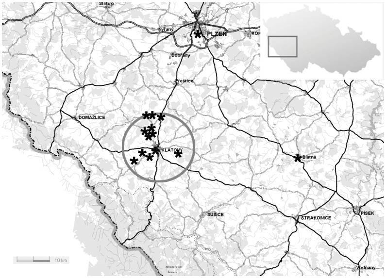
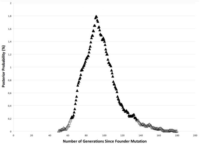
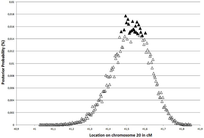
Posterior polymorphous corneal dystrophy (PPCD) is a rare autosomal dominant genetically heterogeneous disorder. Nineteen Czech PPCD pedigrees with 113 affected family members were identified, and 17 of these kindreds were genotyped for markers on chromosome 20p12.1- 20q12. Comparison of haplotypes in 81 affected members, 20 unaffected first degree relatives and 13 spouses, as well as 55 unrelated controls, supported the hypothesis of a shared ancestor in 12 families originating from one geographic location. In 38 affected individuals from nine of these pedigrees, a common haplotype was observed between D20S48 and D20S107 spanning approximately 23 Mb, demonstrating segregation of disease with the PPCD1 locus. This haplotype was not detected in 110 ethnically matched control chromosomes. Within the common founder haplotype, a core mini-haplotype was detected for D20S605, D20S182 and M189K2 in all 67 affected members from families 1-12, however alleles representing the core mini-haplotype were also detected in population matched controls. The most likely location of the responsible gene within the disease interval, and estimated mutational age, were inferred by linkage disequilibrium mapping (DMLE+2.3). The appearance of a disease-causing mutation was dated between 64-133 generations. The inferred ancestral locus carrying a PPCD1 disease-causing variant within the disease interval spans 60 Kb on 20p11.23, which contains a single known protein coding gene, ZNF133. However, direct sequence analysis of coding and untranslated exons did not reveal a potential pathogenic mutation. Microdeletion or duplication was also excluded by comparative genomic hybridization using a dense chromosome 20 specific array. Geographical origin, haplotype and statistical analysis suggest that in 14 unrelated families an as yet undiscovered mutation on 20p11.23 was inherited from a common ancestor. Prevalence of PPCD in the Czech Republic appears to be the highest worldwide and our data suggests that at least one other novel locus for PPCD also exists.
Conflict of interest statement
Figures
References
-
- Cibis GW, Krachmer JA, Phelps CD, Weingeist TA (1977) The clinical spectrum of posterior polymorphous dystrophy. Arch Ophthalmol 95: 1529–1537. - PubMed
-
- Laganowski HC, Sherrard ES, Muir MG (1991) The posterior corneal surface in posterior polymorphous dystrophy: a specular microscopical study. Cornea 10: 224–232. - PubMed
-
- de Felice GP, Braidotti P, Viale G, Bergamini F, Vinciguerra P (1985) Posterior polymorphous dystrophy of the cornea. An ultrastructural study. Graefes Arch Clin Exp Ophthalmol 223: 265–271. - PubMed
-
- Polack FM, Bourne WM, Forstot SL, Yamaguchi T (1980) Scanning electron microscopy of posterior polymorphous corneal dystrophy. Am J Ophthalmol 89: 575–584. - PubMed
Publication types
MeSH terms
Substances
Supplementary concepts
LinkOut - more resources
Full Text Sources
Miscellaneous
