. 2012;7(9):e45790.
doi: 10.1371/journal.pone.0045790. Epub 2012 Sep 25.

Neuroanatomical characterisation of the expression of the lipodystrophy and motor-neuropathy gene Bscl2 in adult mouse brain

Affiliations

Neuroanatomical characterisation of the expression of the lipodystrophy and motor-neuropathy gene Bscl2 in adult mouse brain

Alastair S Garfield et al. PLoS One. 2012.

Abstract

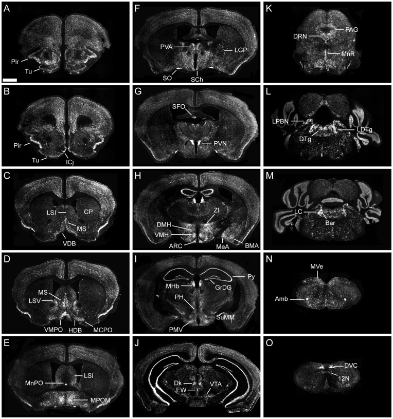
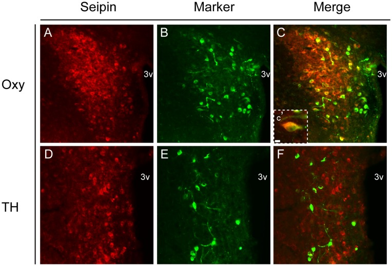
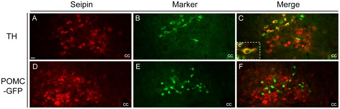
The endoplasmic reticulum localised protein seipin, encoded by the gene Berardinelli-Seip congenital lipodystrophy type 2 (BSCL2), serves a critical but poorly defined function in the physiology of both adipose and neural tissue. In humans, BSCL2 loss-of-function mutations cause a severe form of lipodystrophy, whilst a distinct set of gain-of-toxic-function mutations are associated with a heterogeneous group of neuropathies. However, despite the importance of seipin dysfunction to the pathophysiology of these conditions, little is known about its physiological role in adipocytes or neurons. BSCL2 mRNA has previously been identified in human and mouse brain, yet no definitive assessment of its expression has been undertaken. Here we comprehensively characterised the neuroanatomical distribution of mouse Bscl2 using complementary in situ hybridisation histochemistry and immunohistochemistry techniques. Whilst Bscl2 was broadly expressed throughout the rostral-caudal extent of the mouse brain, it exhibited a discrete neuroanatomical profile. Bscl2 was most abundantly expressed in the hypothalamus and in particular regions associated with the regulation of energy balance including, the paraventricular, ventromedial, arcuate and dorsomedial nuclei. Bscl2 expression was also identified within the brainstem dorsal vagal complex, which together with the paraventricular nucleus of the hypothalamus represented the site of highest expression. Further neurochemical profiling of these two nuclei revealed Bscl2/seipin expression within energy balance related neuronal populations. Specifically, seipin was detected in oxytocin neurons of the paraventricular nucleus of the hypothalamus and in catecholamine neurons of the dorsal vagal complex. These data raise the possibility that in addition to its role in adipose tissue development, seipin may also be involved in the central regulation of energy balance.


Conflict of interest statement

Competing Interests: The authors have declared that no competing interests exist.

Figures

Figure 1
Figure 1. Expression of Bscl2 mRNA in adult mouse brain.
Autoradiographical visualisation of Bscl2 mRNA in saggital section of the adult mouse brain using radioactive in situ hybridisation histochemistry. (A) Diagrammatic representation of the mouse Bscl2 locus detailing the location of the ISHH riboprobe (not to scale). (B) Endogenous Bscl2 expression as detected by a specific antisense riboprobe revealed strong expression within the basal forebrain (BF), hippocampus (Hippo), hypothalamus (Hypo), dorsal brainstem (dBS) and ventral brainstem (vBS). (C) Corresponding Bscl2 sense riboprobe control.
Figure 2
Figure 2. Neuroanatomical characterisation of Bscl2 mRNA in adult mouse brain.
Radioactive in situ hybridisation histochemistry analysis of Bscl2 mRNA distribution in coronal section across the rostral-caudal extent of adult mouse brain. Endogenous Bscl2 expression was detected throughout the brain, for full characterisation see Table 1. (A–O) 35S-labelled Bscl2 expression was detected in the piriform cortex (Pir), olfactory tubercle (Tu), islands of Calleja (ICj), caudate putamen (CP) lateral septal nucleus intermediate part (LSI), medial septal nucleus (MS), nucleus of the vertical limb of the diagonal band (VDB), lateral septal nucleus ventral part (LSV), nucleus of the horizontal limb of the diagonal band (HDB), magnocellular preoptic nucleus (MCPO), ventromedial preoptic nucleus (VMPO), median preoptic nucleus (MnPO), medial preoptic nucleus medial part (MPOM), paraventricular thalamic nucleus (PVA), lateral globus pallidus (LGP), supraoptic nucleus (SO), suprachiasmatic nucleus (SCh), subfornical organ (SFO), paraventricular nucleus of the hypothalamus (PVN), zona incerta (ZI), dorsomedial nucleus of the hypothalamus (DMH), ventromedial nucleus of the hypothalamus (VMH), arcuate nucleus of the hypothalamus (ARC), basomedial amygdaloid nucleus (BMA), medial amygdaloid nucleus (MeA), medial habenular (MHb), pyramidal cell layer of the hippocampus (py), granular layer of the dentate gyrus (GrDG), posterior hypothalamus (PH), supramammilliary nucleus medial part (SuMM), premammillary nucleus ventral part (PMV), nucleus of Darkschewitsch (Dk), Edinger-Westphal nucleus (EW), ventral tegmental area (VTA), dorsal raphe nucleus (DRN), periaqueductal grey (PAG), median raphe nucleus (MnR), lateral parabrachial nucleus (LPBN), dorsal tegmental nucleus (DTg), laterodorsal tegmental nucleus (LDTg), locus coeruleus (LC), Barrington’s nucleus (Bar), medial vestibular nucleus (MVe), ambiguous nucleus (Amb), dorsal vagal complex (DVC), hypoglossal nucleus (12N). Scale bar in (A) represents 1 mm and applies to all other images.
Figure 3
Figure 3. Bscl2 mRNA distribution within the PVN and DVC.
Radioactive in situ hybridisation histochemistry analysis of Bscl2 mRNA distribution in coronal section across three rostral-to-caudal levels of adult PVN and DVC. (A–C) 35S-labelled Bscl2 mRNA expression in the PVN demonstrating robust labelling in the ventral (A), medial magnocellular (B), lateral magnocellular (B, C) and posterior domains (C). Scattered Bscl2 labelled cells were expressed in the anterior and medial (B, C) parvicellular portion. (D–F) 35S-labelled Bslc2 mRNA expression in the DVC was highest within the 10N at the level of the area postrema. Within the NTS the preponderance of Bscl2 mRNA was localised to the medial and ventral domains (E, F). No expression was detected in the area postrema (E). 4v, fourth ventricle; 10N, dorsal motor nucleus of the vagus; 12N, hypoglossal nucleus; AP, area postrema; cc, central canal; PaAP, PVN anterior parvicellular; PaLM, PVN lateral magnocellular; PaMM, PVN medial magnocellular; PaMP, PVN medial parvicellular; PaPo, PVN posterior; PaV, PVN ventral; SolC, NTS commissural; SolDL, NTS dorsolateral; SolG, NTS gelatinous; SolIM, NTS intermediate; SolM, NTS medial; SolV, NTS ventral; SolVL, NTS ventrolateral.
Figure 4
Figure 4. Validation of seipin antibody in the PVN and DVC of adult mouse.
Dual Bscl2 fluorescent in situ hybridisation and seipin immunohistochemistry. (A–C) Co-localisation of Bscl2 mRNA and seipin in the magnocellular domain of the paraventricular nucleus of the hypothalamus (PVN) and (G–I) dorsal vagal complex (DVC). Co-localisation denoted by yellow colouring in panels C and I. Negative controls for fluorescent in situ hybridisation and immunohistochemical protocols in the PVN (D–F) and DVC (J–L) revealed no non-specific staining. Inserts (C′) and (I′) show high magnification images of cellular co-localisation. Scale bar in (A) represents 25 µm and applies to figures AL; scale bar in (C′) represents 10 µm and applies to I′.3v, third ventricle; cc, central canal.
Figure 5
Figure 5. Neurochemical profile of seipin positive neurons in the PVN of adult mouse.
Dual immunofluorescent colocalisation of seipin and (A–C) oxytocin (Oxy) and (D–E) tyrosine hydroxylase (TH) in the paraventricular nucleus of the hypothalamus (PVN). Colocalisation with oxytocin was evident within PVN (as denoted by yellow colouring in panel C and C′). No colocalisation was observed with TH. Scale bar in (A) represents 25 µm and applies to figures A–E; scale bar in (C′) represents 10 µm. 3v, third ventricle.
Figure 6
Figure 6. Neurochemical profile of seipin positive neurons in the DVC of adult mouse.
Dual immunofluorescent co-localisation of seipin and (A–C) tyrosine hydroxylase (TH) and (D–E) green fluorescent protein under the control of the pro-opiomelanocortin promoter (POMC-EGFP) in the nucleus of the solitary tract (NTS). Co-localisation with TH was evident within NTS (as denoted by yellow colouring in panel C and C′). No co-localisation was observed with GFP. Scale bar in (A) represents 25 µm and applies to figures A-E; scale bar in (C′) represents 10 µm. 3v, third ventricle. cc, central canal.

Similar articles

Cited by

References

    1. Cartwright BR, Goodman JM (2012) Seipin: from human disease to molecular mechanism. J Lipid Res 53: 1042–1055. - PMC - PubMed
    1. Fei W, Du X, Yang H (2011) Seipin, adipogenesis and lipid droplets. Trends Endocrinol Metab 22: 204–210. - PubMed
    1. Ito D, Suzuki N (2009) Seipinopathy: a novel endoplasmic reticulum stress-associated disease. Brain 132: 8–15. - PubMed
    1. Rochford JJ (2010) Molecular mechanisms controlling human adipose tissue development: insights from monogenic lipodystrophies. Expert Rev Mol Med 12: e24. - PubMed
    1. Magre J, Delepine M, Khallouf E, Gedde-Dahl T Jr, Van Maldergem L, et al. (2001) Identification of the gene altered in Berardinelli-Seip congenital lipodystrophy on chromosome 11q13. Nat Genet 28: 365–370. - PubMed

Publication types

MeSH terms

Substances

LinkOut - more resources