. 2012;7(9):e46126.
doi: 10.1371/journal.pone.0046126. Epub 2012 Sep 25.

Inhibition of doxorubicin-induced senescence by PPARδ activation agonists in cardiac muscle cells: cooperation between PPARδ and Bcl6

Affiliations

Inhibition of doxorubicin-induced senescence by PPARδ activation agonists in cardiac muscle cells: cooperation between PPARδ and Bcl6

Paola Altieri et al. PLoS One. 2012.

Abstract

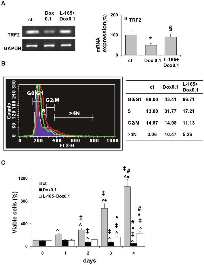
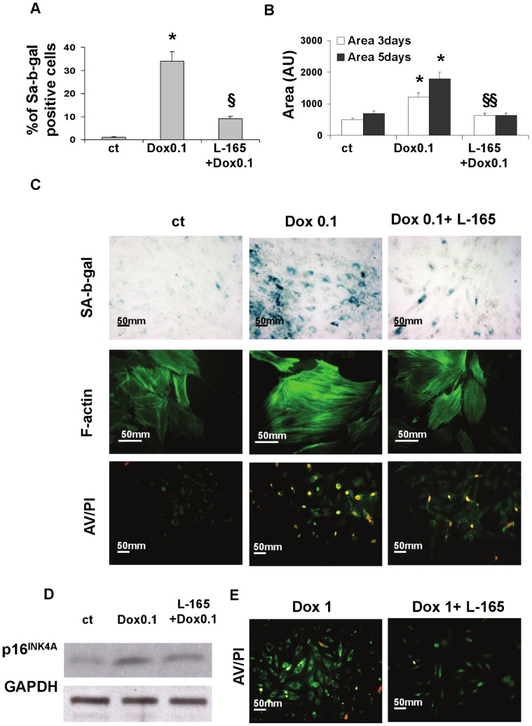
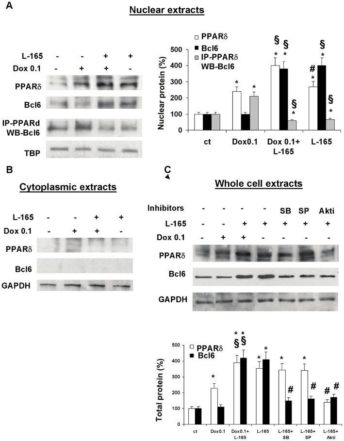
Senescence and apoptosis are two distinct cellular programs that are activated in response to a variety of stresses. Low or high doses of the same stressor, i.e., the anticancer drug doxorubicin, may either induce apoptosis or senescence, respectively, in cardiac muscle cells. We have demonstrated that PPARδ, a ligand-activated transcriptional factor that controls lipid metabolism, insulin sensitivity and inflammation, is also involved in the doxorubicin-induced senescence program. This occurs through its interference with the transcriptional repressor protein B cell lymphoma-6 (Bcl6). Low doses of doxorubicin increase the expression of PPARδ that sequesters Bcl6, thus preventing it from exerting its anti-senescent effects. We also found that L-165041, a specific PPARδ activator, is highly effective in protecting cardiomyocytes from doxorubicin-induced senescence through a Bcl6 related mechanism. In fact, L-165041 increases Bcl6 expression via p38, JNK and Akt activation, and at the same time it induces the release of Bcl6 from PPARδ, thereby enabling Bcl6 to bind to its target genes. L-165041 also prevented apoptosis induced by higher doses of doxorubicin. However, while experiments performed with siRNA analysis techniques very clearly showed the weight of Bcl6 in the cellular senescence program, no role was found for Bcl6 in the anti-apoptotic effects of L-165041, thus confirming that senescence and apoptosis are two very distinct stress response cellular programs. This study increases our understanding of the molecular mechanism of anthracycline cardiotoxicity and suggests a potential role for PPARδ agonists as cardioprotective agents.


Conflict of interest statement

Competing Interests: The authors have declared that no competing interests exist.

Figures

Figure 1
Figure 1. L-165041 prevents doxorubicin-induced changes in neonatal rat cardiomyocytes.
Incubation for 3 h with low dose doxorubicin 0.1 µM (Dox 0.1) modifies the expression levels of telomere binding factor 2 (TRF2), the cell cycle and cell viability. Pre-treatment for 2 h with L-165041 (L-165) prevents doxorubicin-induced modifications. (A) TRF2 mRNA evaluated 4 h after doxorubicin treatment. *p<0.05 versus control (ct), §p<0.05 versus Dox 0.1. (B) FACS analysis of cell cycle distribution 24 h after treatment with Dox 0.1. Filled line: untreated cells; red line: cells treated with Dox 0.1; green line: cells pre-treated with L-165041 and then treated with Dox 0.1. Percentages of cell subpopulations are summarized in the Table. (C) Cell viability determined by MTT Assay. ˆp<0.05 versus day 0. ‡p<0.05 versus day 1. • p<0.05 versus day 2. # p<0.05 versus day 3.
Figure 2
Figure 2. L-165041 prevents both senescence and apoptosis induced by doxorubicin.
Pre-treatment with the PPARδ agonist L-165041 (L-165) prevents both senescence induced by low (0.1 µM) doses of doxorubicin (Dox) (A,B,C), and apoptosis induced by high (1 µM) doses of Dox (E) in neonatal rat cardiomyocytes. (A) Percentage of SA-b-gal positive cells 3 days after treatment. *p<0.05 versus control (ct), §p<0.05 versus Dox 0.1. (B) Cell size, 3 and 5 days after treatment. *p<0.05 versus ct, §p<0.05 versus Dox 0.1. (C) Photographs showing cells (from top to bottom): SA-b-gal activity evaluated 3 days after treatment with Dox 0.1 (magnification, ×200), F-actin density (magnification, ×400), and AV/PI staining evaluated 24 h after treatment with Dox 0.1 (magnification, ×200). (D)Western blot analysis of p16 INK4a evaluated 24 h after treatment with Dox 0.1 µM. (E) AV/PI staining evaluated 24 h after treatment with Dox 1 (magnification, ×200).
Figure 3
Figure 3. Effects of L-165041 on the expression levels of PPAR isoforms.
Effects of L-165041 on PPARα, PPARγ and PPARδ evaluated in neonatal cardiomyocytes 4 h and 22 h after L-165041 treatment. *p<0.05 versus time 0, §p<0.05 versus 4 h.
Figure 4
Figure 4. Pre-treatment with p38, JNK and Akt inhibitors prevents the anti-senescent effects of L-165041.
The specific inhibitors of p38 (SB203580, SB), JNK (SP600125, SP), Akt (Akt1/2 kinase inhibitor, Akti) but not the inhibitor of ERK1/2 (PD98059, PD) reverse the effects of L-165041 (L-165) on doxorubicin-induced SA-b-gal activity. *p<0.05 versus untreated cells, §p<0.05 versus doxorubicin (Dox 0.1), # p<0.05 versus L-165+Dox 0.1.
Figure 5
Figure 5. Effects of L-165041 and/or doxorubicin 0.1 µM on MAPKs and Akt phosphorylation.
Both L-165041(L-165) and doxorubicin (Dox) 0.1 µM activate MAPKs and Akt. If cells are pre-treated with L-165, the doxorubicin-induced increase of pJNK and pAkt levels is inhibited, pERK levels are maintained sustained, while pp38 levels result higher than those induced by Dox 0.1 alone. (A) Time curve analysis of phosphorylated pp38, pJNK, pAKT, pERK1/2 and total p38, JNK, AKT, ERK1/2 evaluated by western blot. (B, C, D, E) Graphs showing values for pp38, pJNK, pAKT and pERK1/2 normalized to the amount of total enzyme.
Figure 6
Figure 6. L-165041 and doxorubicin regulate PPARδ and Bcl6 expression and interaction: the role of MAPKs and Akt.
H9c2 cells were pre-treated with or without L-165041 (L-165) for 2 h, then treated with or without doxorubicin (Dox) 0.1 µM for 3 h and analyzed after 24 h. Dox 0.1 increases PPARδ levels and enhances the interaction between Bcl6 and PPARδ but does not modify the amount of Bcl6. L-165041 increases the protein levels of both PPARδ and Bcl6 and induces the release of Bcl6 from PPARδ. Cytoplasmic extracts reveal nuclear localization of PPARδ and Bcl6. (A) Nuclear extracts were assayed by western blot analysis with PPARδ or Bcl6 antibodies; co-immunoprecipitation of PPARδ and Bcl6 (IP-PPARδ WB-Bcl6) was performed with anti- PPARδ followed by western blot using the anti-Bcl6 antibody. The bar graph shows the protein quantification expressed as a percentage. *p<0.05 versus control (ct), §p<0.05 versus Dox 0.1, # p<0.05 versus L-165141+Dox 0.1. (B) Cytoplasmic extracts were evaluated by western blot with PPARδ and Bcl6 antibodies. (C) The effects of SB203580 (SB), inhibitor of p38, SP600125 (SP), inhibitor of JNK and Akt1/2 kinase inhibitor (Akti), inhibitor of Akt, on the L-165041-induced increases of PPARδ (first line) and Bcl6 (second line). Whole cell extracts were assayed by western blot analysis. All inhibitors reverse the effects of L-165041 on Bcl6 levels, while only the Akt inhibitor reverses the up-regulation of PPARδ. The bar graph shows the protein quantification expressed as a percentage. *p<0.05 versus ct, §p<0.05 versus Dox 0.1, # p<0.05 versus L-165141.
Figure 7
Figure 7. Bcl6 plays a key role in cellular senescence.
Bcl6 siRNA(siBcl6) significantly increases the number of SA-b-gal positive cells in both unstressed and in 0.1 µM doxorubicin-treated cells, and it completely abolishes the anti-senescent effect of pre-treatment with L-165041 (L-165). siPPARδ completely abolishes the pro-senescent effect of 0.1 µM doxorubicin. H9c2 cells were transfected with Bcl6-, PPARδ specific or control (siCT) siRNA for 48 h before treatment with or without L-165041 for 2 h followed by treatment with or without 0.1 µM doxorubicin (Dox 0.1) for 3 h. Cells were assayed for protein amount (after 24 h), premature senescence (after 3 days) and apoptosis (after 24 h). (A) Western blot analysis with Bcl6 and PPARδ antibodies. (B) SA-b-gal activity (magnification ×200). (C) Bar graph illustrating the percentage of SA-b-gal positive cells. *p<0.05 versus corresponding siCT of the same treatment condition, (D) Bar graph illustrating the percentage of caspase-3 positive cells.
Figure 8
Figure 8. L-165041 protects H9c2 from doxorubicin-induced apoptosis through a Bcl6-independent mechanism.
(A) Pre-treatment with L-165141decreases the number of doxorubicin-induced apoptotic cells. Bcl6 siRNA (siBcl6) does not modify the number of activated caspase-3 positive cells in any of the treatment groups i.e., untreated cells, 1 µM doxorubicin-treated cells, and in cells pretreated with L-165041 and then incubated with doxorubicin. 24 h after treatment, cells were assayed by immunocytochemistry for cleaved caspase-3. Bar graph illustrates the percentage of cleaved-caspase-3 positive cells. *P<0.05 versus ct, §P<0.05 versus doxorubicin. (B) Effects of L-165041(L-165) and 1 µM doxorubicin (Dox1) on PPARδ and Bcl6 protein expression and on PPARδ and Bcl6 protein interaction. Doxorubicin increases PPARδ levels and down-regulates the amount of total and PPARδ bound Bcl6 (IP-PPARδ WB-Bcl6). Pre-treatment with L-165041 does not modify the effects induced by doxorubicin. H9c2 cells were pre-treated with or without L-165041 for 2 h, then treated with or without 1 µM doxorubicin for 3 h and analyzed after 24 h by western blot. The bar graph shows the protein quantification expressed as a percentage. *p<0.05 versus control (ct).

References

    1. Hershman DL, McBride RB, Eisenberger A, Tsai WY, Grann VR, et al. (2008) Doxorubicin, cardiac risk factors, and cardiac toxicity in elderly patients with diffuse B-cell non-Hodgkin’s lymphoma. J Clin Oncol 26: 3159–3165. - PubMed
    1. Auner HW, Tinchon C, Linkesch W, Halwachs-Baumann G, Sill H (2001) Correspondence re: O. J. Arola, et al., (2000) Acute doxorubicin cardiotoxicity involves cardiomyocyte apoptosis Cancer Res 60: 1789–1792. Cancer Res 61: 2335–2336. - PubMed
    1. Maejima Y, Adachi S, Ito H, Hirao K, Isobe M (2008) Induction of premature senescence in cardiomyocytes by doxorubicin as a novel mechanism of myocardial damage. Aging Cell 7: 125–136. - PubMed
    1. Spallarossa P, Altieri P, Aloi C, Garibaldi S, Barisione C, et al. (2009) Doxorubicin induces senescence or apoptosis in rat neonatal cardiomyocytes by regulating the expression levels of the telomere binding factors 1 and 2. Am J Physiol Heart Circ Physiol 297: H2169–H2181. - PubMed
    1. Hayflick L, Moorhead PS (1961) The serial cultivation of human diploid cell strains. Exp Cell Res 25: 585–621. - PubMed

Publication types