. 2012;7(9):e46171.
doi: 10.1371/journal.pone.0046171. Epub 2012 Sep 25.

R26R-GR: a Cre-activable dual fluorescent protein reporter mouse

Affiliations

R26R-GR: a Cre-activable dual fluorescent protein reporter mouse

You-Tzung Chen et al. PLoS One. 2012.

Abstract

Green fluorescent protein (GFP) and its derivatives are the most widely used molecular reporters for live cell imagining. The development of organelle-specific fusion fluorescent proteins improves the labeling resolution to a higher level. Here we generate a R26 dual fluorescent protein reporter mouse, activated by Cre-mediated DNA recombination, labeling target cells with a chromatin-specific enhanced green fluorescence protein (EGFP) and a plasma membrane-anchored monomeric cherry fluorescent protein (mCherry). This dual labeling allows the visualization of mitotic events, cell shapes and intracellular vesicle behaviors. We expect this reporter mouse to have a wide application in developmental biology studies, transplantation experiments as well as cancer/stem cell lineage tracing.


Conflict of interest statement

Competing Interests: The authors have declared that no competing interests exist.

Figures

Figure 1
Figure 1. The generation of R26R-GR mice.
(A) Gene targeting strategy for the Cre activable R26R-GR reporter allele. (B) The gene targeting vector contains a loxP flanked stop cassette, which can be excised in a Cre-expressing BNN132 E. coli, producing a predicted smaller PacI, AscI–restricted DNA fragment. (C) Southern blotting evidence for a R26 targeted mouse ES cell clone. (D) Transient transfection of a Cre-expressing plasmid, pOG231, activates the dual fluorescent protein reporter in the targeted ES cell clone. Scale bar: 15 µm. (E) Germline transmission demonstrated using a 3-primer PCR-based genotyping strategy. The presence of the wild-type allele results in a 622 bp PCR product amplified by P1 and P3 while the R26R-GR targeted allele is amplified by P2 and P3, leading to a 1,087 bp product. Lanes 1, 2, 5, 6, and 11 identify the R26R-GR heterozygous mice.
Figure 2
Figure 2. Ubiquitous dual fluorescent protein reporter expression from R26 resulting from a Sox2Cre-mediated activation event.
The R26R-GR male is crossed with a female Sox2Cre transgenic mouse to obtain compound heterozygous progeny with the dual fluorescent protein reporter activated in all tissues of an E10.5 embryo. (A, B, C) Whole mount images of E10.5 heterozygous R26R-GR (left) and Sox2Cre-activated R26R-GR (right) embryos under a fluorescent dissection microscope. The inactivated R26R-GR embryo shows neither EGFP nor mCherry signals whereas the compound heterozygous Sox2Cre/+; R26R-GR/+ embryo emits both green and red lights throughout the body. Scale bar: 1 mm. (D, E, F) Cross section of a E10.5 Sox2Cre/+; R26R-GR/+ embryo reveals ubiquitous expression of a nucleus localized EGFP and a cell membrane bound mCherry. Scale bar: 75 µm. (G, H, I) Higher magnifications from D–F pictures are shown. Scale bar: 25 µm.
Figure 3
Figure 3. Oocyte-specific dual fluorescent reporter labeling induced by a Zp3Cre transgene.
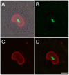
Frozen ovary section from an eight week old Zp3Cre/+; R26R-GR/+ female mouse illustrates a tissue-specific, Cre activable expression of the dual fluorescent reporter gene (GR). In the picture, the oocyte is the only cell in the follicle that shows both nuclear EGFP and cell membrane mCherry signals (D). Although the granulosa cells encompassing the oocyte and theta cells surrounding the follicle are observed in the DIC mode, EGFP channel, and mCherry channel overlaid image (A), they emit no fluorescent signals and they are absent in photos taken under EGFP channel (B) and mCherry channel (C). Scale bar: 20 µm.
Figure 4
Figure 4. Live images of homozygous R26-GR pre-implantation embryos at 2-cell, 4-cell, 8-cell, morula and blastocyst stages.
Scale bar: 25 µm. A 3D reconstructed movie taken on an E3.5 blastocyst is provided in the Movie S1.
Figure 5
Figure 5. Time–lapse imaging in an E14.5 heterozygous R26-GR mouse embryonic fibroblast primary culture.
Snapshots from a time-lapse imaging experiment (Movie S2). The elapsed times are given at the lower-left corner in each merged image. A cell division event is recorded in this experiment. Arrowheads indicate a dividing nucleus and her daughters. Scale bar: 50 µm.
Figure 6
Figure 6. Endoplasmic reticulum (ER) and endosomes labeled by mCherry-GPI in the intestinal epithelia of a P21 heterozygous R26-GR pup.
Immunohistochemistry using antibodies against a endosome-specific marker (EEA-1)(top row) and an ER-specific marker (calnexin) (bottom row) provides signals coincided with mCherry-GPI distributions (arrows). Scale bar: 5 µm.
Figure 7
Figure 7. Endocytic events colocalize with mCherry-GPI marked vesicles.
Cascade blue-labeled 10-kD dextran is found in mCherry-GPI marked vesicles in the intestinal epithelia of a P21 R26-GR heterozygote two hours after gavage feeding (arrows). Scale bar: 10 µm.

References

    1. Chalfie M, Tu Y, Euskirchen G, Ward WW, Prasher DC (1994) Green fluorescent protein as a marker for gene expression. Science 263: 802–805. - PubMed
    1. Shimomura O, Johnson FH, Saiga Y (1962) Extraction, purification and properties of aequorin, a bioluminescent protein from the luminous hydromedusan, Aequorea. J Cell Comp Physiol 59: 223–239. - PubMed
    1. Misgeld T, Kerschensteiner M, Bareyre FM, Burgess RW, Lichtman JW (2007) Imaging axonal transport of mitochondria in vivo. Nat Methods 4: 559–561. - PubMed
    1. Shaner NC, Campbell RE, Steinbach PA, Giepmans BN, Palmer AE, et al. (2004) Improved monomeric red, orange and yellow fluorescent proteins derived from Discosoma sp. red fluorescent protein. Nat Biotechnol 22: 1567–1572. - PubMed
    1. Shaner NC, Lin MZ, McKeown MR, Steinbach PA, Hazelwood KL, et al. (2008) Improving the photostability of bright monomeric orange and red fluorescent proteins. Nat Methods 5: 545–551. - PMC - PubMed

Publication types