. 2012;7(9):e46292.
doi: 10.1371/journal.pone.0046292. Epub 2012 Sep 26.

Intrahepatic infiltrating NK and CD8 T cells cause liver cell death in different phases of dengue virus infection

Affiliations

Intrahepatic infiltrating NK and CD8 T cells cause liver cell death in different phases of dengue virus infection

Jui-Min Sung et al. PLoS One. 2012.

Abstract

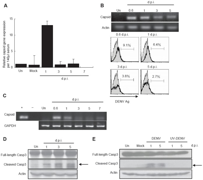
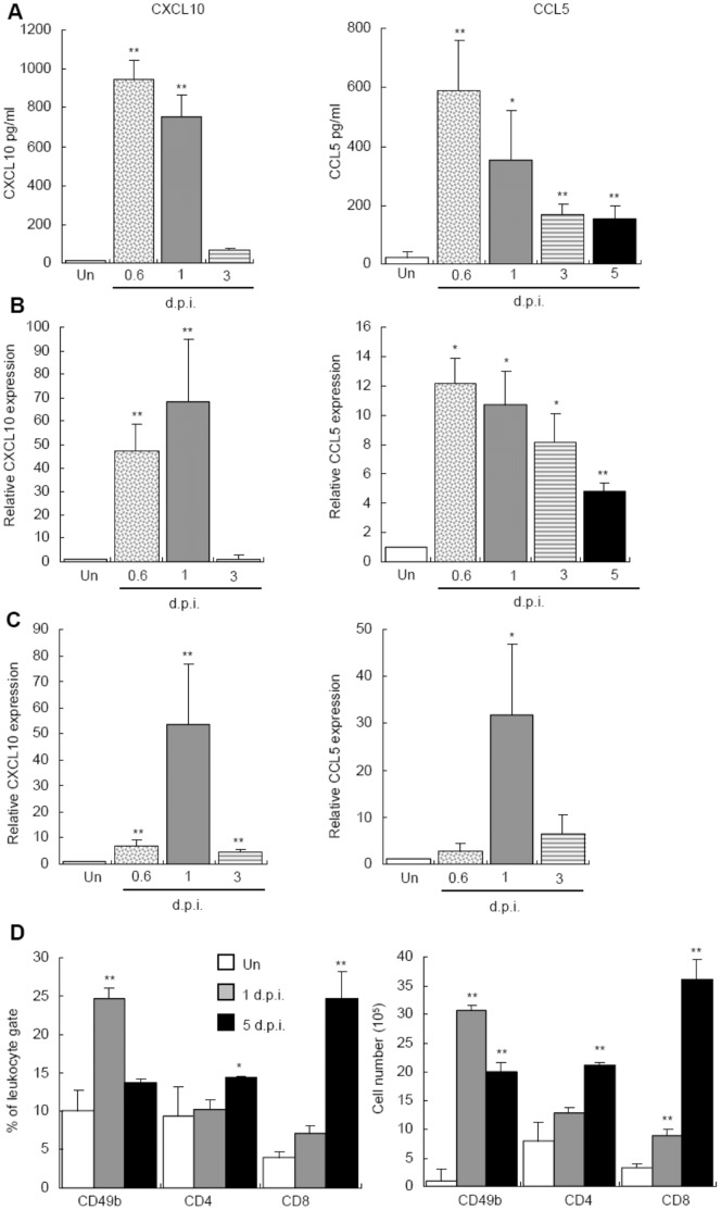
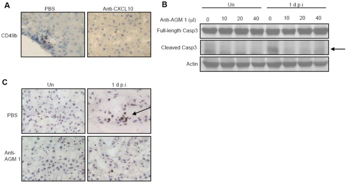
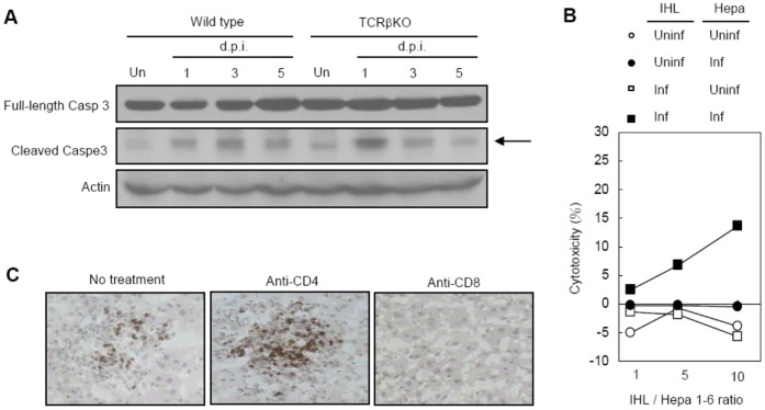
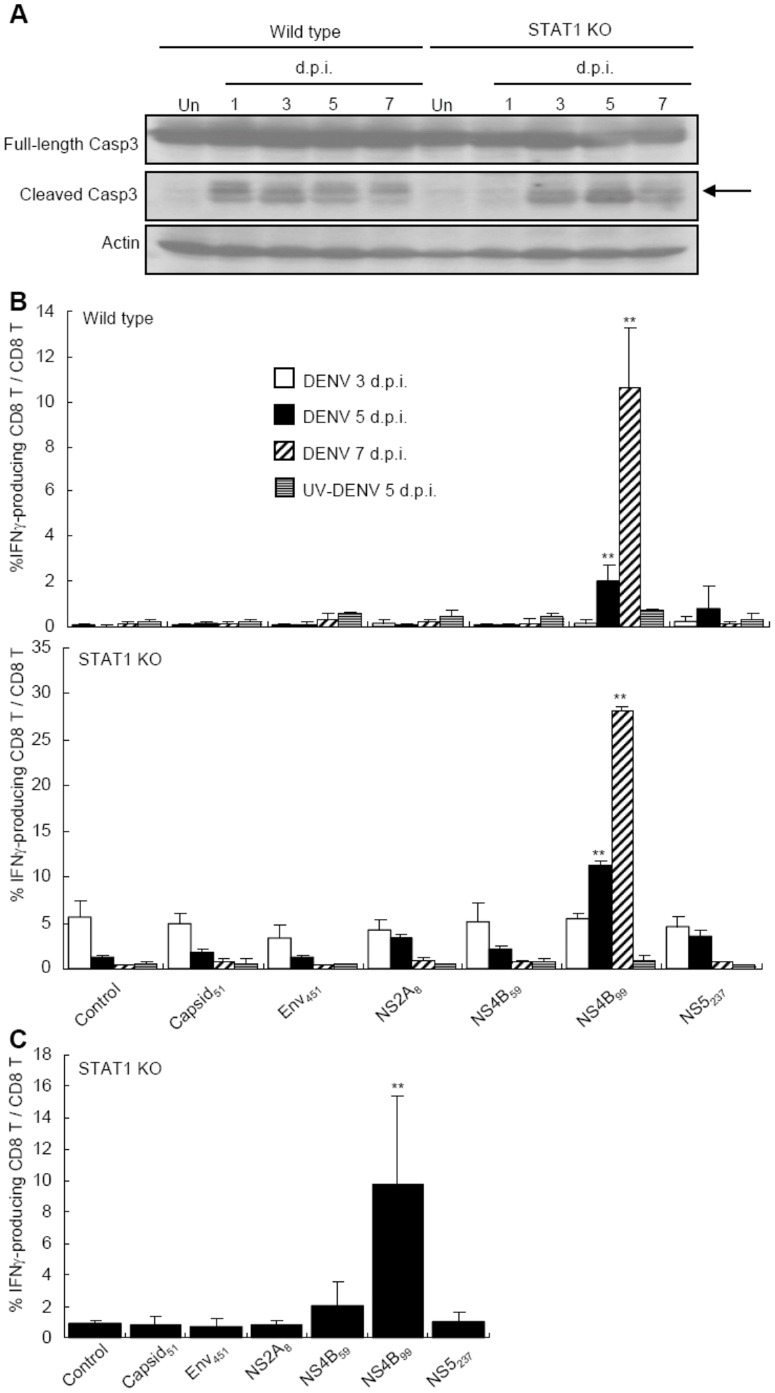
Elevated liver enzyme level is an outstanding feature in patients with dengue. However, the pathogenic mechanism of liver injury has not been clearly demonstrated. In this study, employing a mouse model we aimed to investigate the immunopathogenic mechanism of dengue liver injury. Immunocompetent C57BL/6 mice were infected intravenously with dengue virus strain 16681. Infected mice had transient viremia, detectable viral capsid gene and cleaved caspase 3 in the liver. In the mean time, NK cell and T cell infiltrations peaked at days 1 and 5, respectively. Neutralizing CXCL10 or depletion of Asialo GM1(+) cells reduced cleaved caspase 3 and TUNEL(+) cells in the liver at day 1 after infection. CD8(+) T cells infiltrated into the liver at later time point and at which time intrahepatic leukocytes (IHL) exhibited cytotoxicity against DENV-infected targets. Cleaved caspase 3 and TUNEL(+) cells were diminished in mice with TCRβ deficiency and in those depleted of CD8(+) T cells, respectively, at day 5 after infection. Moreover, intrahepatic CD8(+) T cells were like their splenic counterparts recognized DENV NS4B(99-107) peptide. Together, these results show that infiltrating NK and CD8(+) T cells cause liver cell death. While NK cells were responsible for cell death at early time point of infection, CD8(+) T cells were for later. CD8(+) T cells that recognize NS4B(99-107) constitute at least one of the major intrahepatic cytotoxic CD8(+) T cell populations.


Conflict of interest statement

Competing Interests: The authors have declared that no competing interests exist.

Figures

Figure 1
Figure 1. DENV administered intravenously causes transient viremia, liver cell infection and apoptosis.
Viral capsid gene expression in the serum of uninfected mice (Un) or mice at different time points after infection of DENV (d.p.i., days after infection) was determined by RT real-time PCR (n = 2 per time point in each experiment). (B) C6/36 cells were cultured in medium containing serum collected from uninfected or infected mice. Viral capsid gene expression was determined by RT-PCR. Beta-actin of C6/36 cells was used as internal control (top). Viral antigen expression was determined by rabbit anti-DENV antibody staining (bottom). Shaded histograms outlined by thin line and open histogram outlined by thick line represent cells cultured in serum from uninfected and infected mice respectively. Data presented are representative of two independent experiments (n = 3 per time point in each experiment). (C) Liver was collected from uninfected (Un) and infected mice. Viral capsid gene expressions were determined by RT-PCR. Data presented are representative of two independent experiments (n = 5 per time point in each experiment). (D and E) Liver was collected from uninfected (Un), mice infected with DENV and mice inoculated with otherwise equivalent UV-inactivated DENV (UV-DENV). Arrow points to cleaved caspase 3. Data presented are representative of five independent experiments (n = 3 per time point in each experiment).
Figure 2
Figure 2. Intrahepatic infiltration of NK and T cells coincides with CXCL10 and CCL5 productions in the liver.
(A) The levels of CXCL10 and CCL5 in sera collected from uninfected (Un) and infected mice were determined by ELISA. Data presented are from three independent experiments (n = 3 per time point in each experiment). (B) CXCL10 and CCL5 mRNAs in the liver were analyzed by RT-real-time PCR and normalized against GAPDH. Data presented are from three independent experiments (n = 3 per time point in each experiment). (C) Hepa 1–6 cells were infected with DENV at MOI of 1 and CXCL10 and CCL5 mRNA expressions were analyzed by RT-real-time PCR and normalized against GAPDH. Data presented are from three independent experiments (n = 2 per time point in each experiment). (D) IHLs were collected from uninfected (open) or infected mice at days 1 (shaded bar) and 5 (darkened) after infection. The percentage in total IHL population (left) and the absolute number (right) of CD3-CD49b+ NK cells, CD3+CD8+ and CD3+CD4+ T cells were analyzed by flow cytometry (n = 5 per time point in each experiment). * P value <0.05, ** P value <0.01 compared with uninfected groups.
Figure 3
Figure 3. Intrahepatic infiltrating NK cells cause liver cell death at day 1 after DENV infection.
(A) Liver cryosections collected from mice with or without anti-CXCL10 antibody at day 1 after DENV infection were stained with anti-CD49b antibody. Data are representative of three independent experiments (n = 3 per time point in each experiment). (B) Uninfected and infected mice at day 1 were treated with different volumes of anti-AGM1 antibody. Arrow points to cleaved capase 3. Data presented are representative of four independent experiments (n = 3 per time point in each experiment). (C) Liver cryosections collected from uninfected or infected mice with or without anti-AGM1 were stained with TUNEL reagents. Data are representative of three independent experiments (n = 3 per time point in each experiment).
Figure 4
Figure 4. Intrahepatic T cells are cytotoxic against DENV-infected hepatocytes.
(A) Liver lysates were collected from uninfected and infected wild type and TCRβ KO mice. Arrow points to cleaved capase 3. Data presented are representative of three independent experiments (n = 3 per time point in each experiment). (B) Uninfected (Uninf) or infected (Inf) Hepa 1–6 were co-cultured with IHLs isolated from uninfected (Uninf) or infected (Inf) mice at different ratio. Data presented are representative of three independent experiments (n = 3 per time point in each experiment). (C) Liver cryosections from infected mice at day 5 with or without anti-CD4 and anti-CD8 antibody were stained with TUNEL reagents. Data presented are representative of three independent experiments (n = 3 per time point in each experiment).
Figure 5
Figure 5. Intrahepatic as well as splenic CD8+ T cells recognize NS4B99–107 epitope.
(A) Liver lysates were collected from uninfected and infected wild type and STAT1 KO mice. Arrow points to cleaved caspase 3. Data presented are representative of three independent experiment (n = 3 per time point in each experiment). (B) Splenocytes were isolated from wild type and STAT1 KO mice at day 3 (empty), 5 (darkened), and 7 (hatched) after infection and at day 5 after UV-inactivated DENV injection (horizontal line). (C) IHLs were isolated from wild type and STAT1 KO mice at 5 days after DENV infection. Splenocytes and IHLs were stimulated with or without (control) indicated peptides. The percentages of IFNγ-producing CD8+ T cell within the total CD8+ T population were analyzed by flow cytometry. Data presented are representative of four independent experiments (n = 3 per time point in each experiment). ** P value <0.01 compared with the control group.

Similar articles

Cited by

References

    1. Beatty ME, Beutels P, Meltzer MI, Shepard DS, Hombach J, et al. (2011) Health economics of dengue: a systematic literature review and expert panel’s assessment. Am J Trop Med Hyg 84: 473–488. - PMC - PubMed
    1. Kuo CH, Tai DI, Chang-Chien CS, Lan CK, Chiou SS, et al. (1992) Liver biochemical tests and dengue fever. Am J Trop Med Hyg 47: 265–270. - PubMed
    1. Huerre MR, Lan NT, Marianneau P, Hue NB, Khun H, et al. (2001) Liver histopathology and biological correlates in five cases of fatal dengue fever in Vietnamese children. Virchows Arch 438: 107–115. - PubMed
    1. Kalayanarooj S, Vaughn DW, Nimmannitya S, Green S, Suntayakorn S, et al. (1997) Early clinical and laboratory indicators of acute dengue illness. J Infect Dis 176: 313–321. - PubMed
    1. Souza LJ, Alves JG, Nogueira RM, Gicovate Neto C, Bastos DA, et al. (2004) Aminotransferase changes and acute hepatitis in patients with dengue fever: analysis of 1,585 cases. Braz J Infect Dis 8: 156–163. - PubMed

Publication types

MeSH terms