. 2012 Feb;3(2):102-16.
doi: 10.1177/1947601912457024.

The EWS/FLI Oncogene Drives Changes in Cellular Morphology, Adhesion, and Migration in Ewing Sarcoma

Affiliations

The EWS/FLI Oncogene Drives Changes in Cellular Morphology, Adhesion, and Migration in Ewing Sarcoma

Aashi Chaturvedi et al. Genes Cancer. 2012 Feb.

Abstract

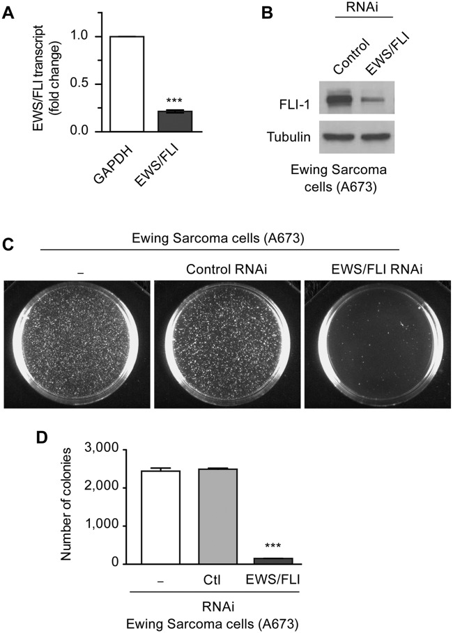
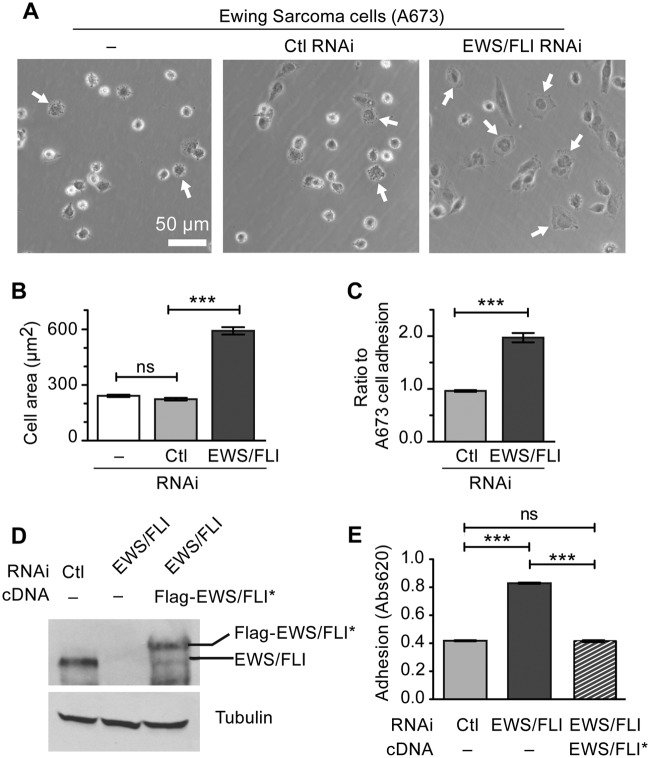
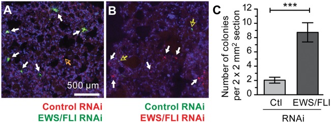
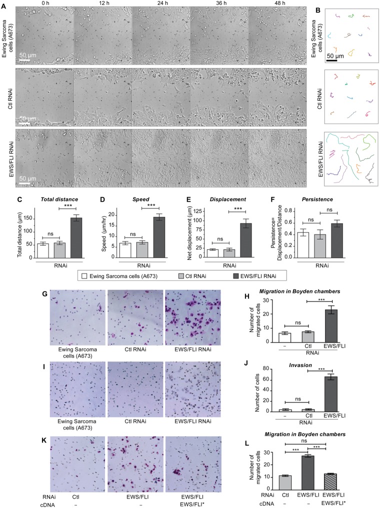
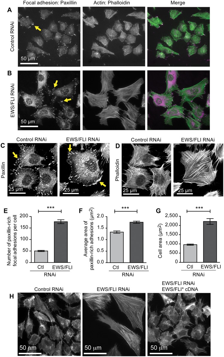
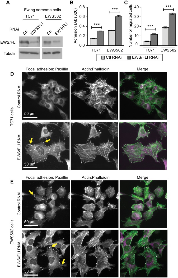
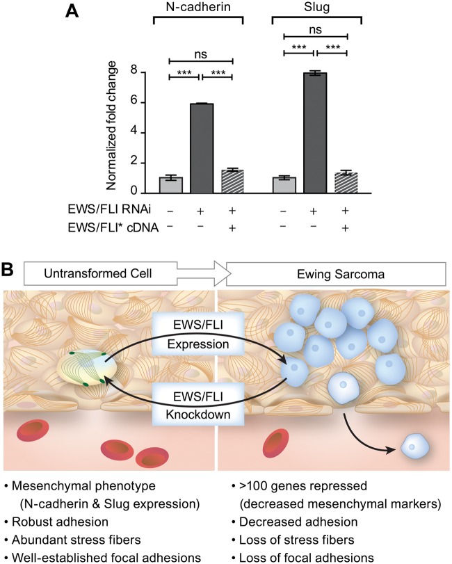
Ewing sarcoma is a tumor of the bone and soft tissue caused by the expression of a translocation-derived oncogenic transcription factor, EWS/FLI. Overt metastases are associated with a poor prognosis in Ewing sarcoma, but patients without overt metastases frequently harbor micrometastatic disease at presentation. This suggests that the metastatic potential of Ewing sarcoma exists at an early stage during tumor development. We have therefore explored whether the inciting oncogenic event in Ewing sarcoma, EWS/FLI, directly modulates tumor cell features that support metastasis, such as cell adhesion, cell migration, and cytoarchitecture. We used an RNAi-based approach in patient-derived Ewing sarcoma cell lines. Although we hypothesized that EWS/FLI might induce classic metastatic features, such as increased cell adhesion, migration, and invasion (similar to the phenotypes observed when epithelial malignancies undergo an epithelial-to-mesenchymal transition during the process of metastasis), surprisingly, we found the opposite. Thus, EWS/FLI expression inhibited the adhesion of isolated cells in culture and prevented adhesion in an in vivo mouse lung assay. Cell migration was similarly inhibited by EWS/FLI expression. Furthermore, EWS/FLI expression caused a striking loss of organized actin stress fibers and focal adhesions and a concomitant loss of cell spreading, suggesting that EWS/FLI disrupts the mesenchymal phenotype of a putative tumor cell-of-origin. These data suggest a new paradigm for the dissemination and metastasis of mesenchymally derived tumors: these tumors may disseminate via a "passive/stochastic" model rather than via an "active" epithelial-to-mesenchymal type transition. In the case of Ewing sarcoma, it appears that the loss of cell adhesion needed to promote tumor cell dissemination might be induced by the EWS/FLI oncogene itself rather than via an accumulation of stepwise mutations.

Keywords: EWS/FLI; Ewing sarcoma; cell adhesion; cytoskeleton; metastasis.


Conflict of interest statement

Declaration of Conflicting Interests: The author(s) declared no potential conflicts of interest with respect to the research, authorship, and/or publication of this article.

Figures

Figure 1.
Figure 1.
Retroviral RNAi mediated knockdown of EWS/FLI in Ewing sarcoma cells. (A) Retroviral mediated knockdown of endogenous EWS/FLI fusion transcripts in Ewing sarcoma A673 cells was measured by semiquantitative RT-PCR, with GAPDH transcripts as control. Transcripts were normalized as fold change compared with control A673 cells. (B) Western immunoblot with FLI-1 antibody detected EWS/FLI fusion protein in A673 cells with control RNAi and significant reduction of EWS/FLI protein with EWS/FLI RNAi. Tubulin antibody was used to show equivalent protein loading. (C) In soft agar transformation assays of A673 cells, the number of colonies formed in 4 weeks was unaffected by control RNAi but significantly reduced by EWS/FLI RNAi (D). ***P < 0.001.
Figure 2.
Figure 2.
EWS/FLI expression abrogates cell adhesion and spreading. (A) Ewing sarcoma A673 cells alone or with control or EWS/FLI RNAi were plated on a tissue culture dish for 2 hours and then evaluated for cell spreading by phase contrast microscopy. Arrows indicate cells adhering to the substratum, spreading, and losing their phase brightness. (B) Measurement of A673 cell area following 2 hours adhesion showed that EWS/FLI RNAi enhanced cell spreading compared with cells with control RNAi. (C) In a colorimetric adhesion assay of cells plated for 2 hours, the compromised adhesiveness of cells with control RNAi compared with the EWS/FLI RNAi was quantitated and shown to be approximately 2-fold. (D) Western immunoblot for EWS/FLI protein in A673 cells with control RNAi or EWS/FLI RNAi were compared with EWS/FLI RNAi cells reconstituted with Flag-tagged EWS/FLI* cDNA that is resistant to the RNAi. The reconstituted EWS/FLI levels approached those seen in control A673 cells, and the tubulin signal shows equivalent protein loading. (E) Cell adhesion over 2 hours was measured by a colorimetric assay for A673 cells with control RNAi (light gray) and EWS/FLI RNAi (dark gray) and showed that EWS/FLI knockdown increased cell adhesion 2-fold over control cells. Reconstituted cells programmed to express RNAi-resistant EWS/FLI* (striped) exhibited adhesion that was not statistically different (ns) than control cells. ***P < 0.001.
Figure 3.
Figure 3.
EWS/FLI expression compromises cell adhesion in vivo. A673 cells with control RNAi were colored red (DiI) and cells with EWS/FLI RNAi were colored green (DiO), mixed in 1:1 ratio, and then injected into mouse lateral tail veins. Twenty-four hours later the lungs were harvested and cryosectioned, and the red and green cells were counted. (A) Maximum intensity projection of confocal images of lung sections showing differentially labeled A673 cells with control RNAi (red, hollow arrows) and EWS/FLI RNAi (green, solid white arrows). Nuclei are stained with DAPI (blue). (B) Lung sections from additional experiment of mice injected with the cell colors switched to avoid bias of dyes (green-control RNAi, red-EWS/FLI RNAi). (C) Cell colonies were counted for 4 mm2 sections in 15 representative sections and showed more cells with EWS/FLI RNAi (solid white arrows in A and B) accumulated in lung parenchyma compared with cells with control RNAi (hollow arrows in A and B). ***P < 0.001.
Figure 4.
Figure 4.
Expression of EWS/FLI results in reduced cellular migration and invasion. Ewing sarcoma A673 cells with control RNAi or EWS/FLI RNAi were evaluated for directed cell migration, random motility, chemotaxis, and invasion. (A) Cell migration into wounds scraped across cell monolayers was monitored by time lapse microscopy for 48 hours. A673 cells with EWS/FLI RNAi reached the midline of the wound within 24 hours, whereas the other A673 cells did not migrate effectively. (B) Random cell motility was tracked by time lapse microscopy (15-minute intervals for 8 hours), and representative cells tracks are shown. Analysis for total distance (C), speed (D), displacement (E), and persistence (F) was performed with MetaMorph software, and showed enhanced motility but no EWS/FLI-dependent difference in persistence in A673 cells with EWS/FLI RNAi. (G, H) Cells in serum-free media were plated onto Boyden transwell migration filters and then given 24 hours to migrate through pores (small circles on images) toward a high serum environment, fixed, and stained (blue). Compared with control cells, significantly more cells with EWS/FLI RNAi migrated. (I, J) Transwell invasion through Matrigel matrix (24 hours) indicated significant increase in cellular invasiveness upon knockdown of EWS/FLI. (K,L) A673 cells with control (light gray bar) or EWS/FLI RNAi (dark gray bar) were compared with EWS/FLI RNAi cells reconstituted with Flag-tagged EWS/FLI* cDNA (striped bar) that is resistant to the RNAi in transwell migration assays. Quantitation of the number of migrated cells indicated the EWS/FLI-dependence of this cell migration difference. ***P < 0.001, ns is not significantly different.
Figure 5.
Figure 5.
EWS/FLI expression affects cellular morphology and cytoarchitecture. Widefield immunofluorescent images of A673 cells with (A) control RNAi or (B) EWS/FLI RNAi stained for focal adhesions (paxillin antibody) and for actin filaments (phalloidin) showed a striking EWS/FLI-dependent difference in cellular morphology, the actin cytoskeleton network, and focal adhesions. (C) Paxillin-rich focal adhesions and actin cytoskeletons (D) in cells with EWS/FLI or with EWS/FLI knockdown were analyzed. Both the number (E) and size (F) of focal adhesions were lower in A673 cells with control RNAi compared with cells with EWS/FLI RNAi, consistent with the compromised adhesion phenotype of Ewing sarcoma cells. (G) Cell area was measured in cells with control RNAi or EWS/FLI RNAi plated for 24 hours and then fixed and stained with phalloidin. Cell area of Ewing sarcoma A673 cells was significantly smaller than for cells with EWS/FLI knockdown. (H) Phalloidin-stained actin cytoskeleton in A673 cells with control RNAi, EWS/FLI RNAi, or EWS/FLI RNAi plus reconstituted Flag-EWS/FLI* showed that the loss of actin stress fibers is EWS/FLI-dependent. ***P < 0.001.
Figure 6.
Figure 6.
Ewing sarcoma cells TC71 and EWS502 exhibit EWS/FLI-dependent loss of adhesion, migration, and cytoarchitecture. (A) Cell lysates from Ewing sarcoma cell lines TC71 and EWS502 with retroviral mediated RNAi knockdown for control or EWS/FLI were evaluated by western immunoblot with FLI-1 antibody, and significant decreases in EWS/FLI expression were confirmed. (B) Ewing sarcoma cells with control RNAi (light gray bar) or EWS/FLI RNAi (dark gray bar) were seeded onto tissue culture plastic (uncoated (EWS502) or precoated with 10 µg/ml fibronectin (TC71)) and allowed to adhere for 2 hours, and then adherent cells were quantitated by a colorimetric assay. (C) Boyden chamber transwell inserts were precoated with fibronectin (5 µg/ml TC71; 1 µg/ml EWS502) and seeded with cells that were allowed 24 hours to migrate through the insert pores. EWS/FLI knockdown enhanced the number of migratory cells in both cell lines. (D) TC71 and (E) EWS502 cells with EWS/FLI or with EWS/FLI knockdown were stained for paxillin-rich focal adhesions and actin stress fibers (phalloidin). Cells with EWS/FLI RNAi displayed more pronounced focal adhesions and actin stress fibers. ***P<0.001
Figure 7.
Figure 7.
Model for EWS/FLI-dependent coupling of transformation to acquisition of potential for tumor cell dissemination. (A) RT-PCR analysis of mesenchymal markers, N-cadherin and Slug, in Ewing sarcoma cells programmed to express control RNAi, EWS/FLI RNAi, or EWS/FLI RNAi and an RNAi-insensitive EWS/FLI* construct. ***P<0.0001, ns is not statistically different. (B) In this model, EWS/FLI-induced oncogenic transformation simultaneously causes downregulation of cellular adhesion. Consistent with this model, EWS/FLI knockdown caused cells to return to a more normalized mesenchymal phenotype, including expression of mesenchymal markers, robust adhesion, pronounced stress fibers, and well-established focal adhesions. We postulate that these events prime the tumor cells for dissemination and may account for the early acquisition of metastatic potential in Ewing sarcoma.

References

    1. Valastyan S, Weinberg RA. Tumor metastasis: molecular insights and evolving paradigms. Cell. 2011;147:275-92 - PMC - PubMed
    1. Chaffer CL, Weinberg RA. A perspective on cancer cell metastasis. Science. 2011;331:1559-64 - PubMed
    1. Nguyen DX, Bos PD, Massague J. Metastasis: from dissemination to organ-specific colonization. Nat Rev Cancer. 2009;9:274-84 - PubMed
    1. Yang J, Weinberg RA. Epithelial-mesenchymal transition: at the crossroads of development and tumor metastasis. Dev Cell. 2008;14: 818-29 - PubMed
    1. Guilford P, Hopkins J, Harraway J, et al. E-cadherin germline mutations in familial gastric cancer. Nature. 1998;392:402-5 - PubMed

LinkOut - more resources