. 2012:2:700.
doi: 10.1038/srep00700. Epub 2012 Oct 3.

Alzheimer brain-derived tau oligomers propagate pathology from endogenous tau

Affiliations

Alzheimer brain-derived tau oligomers propagate pathology from endogenous tau

Cristian A Lasagna-Reeves et al. Sci Rep. 2012.

Abstract

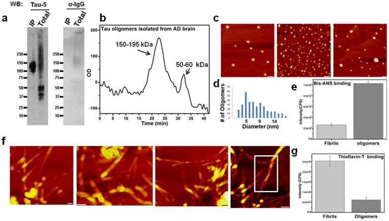
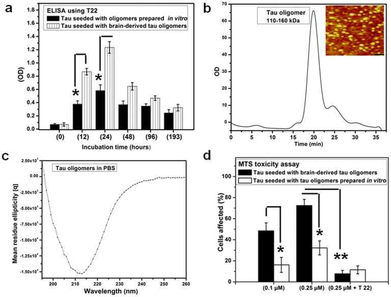
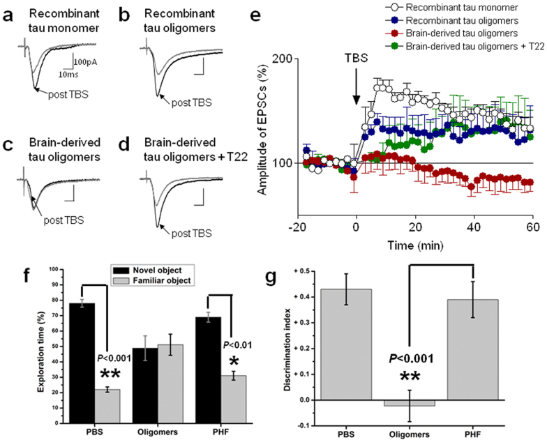
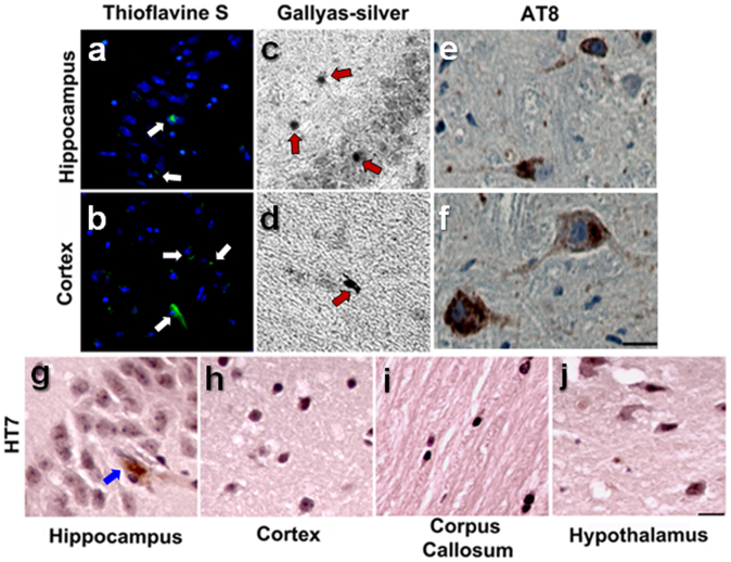
Intracerebral injection of brain extracts containing amyloid or tau aggregates in transgenic animals can induce cerebral amyloidosis and tau pathology. We extracted pure populations of tau oligomers directly from the cerebral cortex of Alzheimer disease (AD) brain. These oligomers are potent inhibitors of long term potentiation (LTP) in hippocampal brain slices and disrupt memory in wild type mice. We observed for the first time that these authentic brain-derived tau oligomers propagate abnormal tau conformation of endogenous murine tau after prolonged incubation. The conformation and hydrophobicity of tau oligomers play a critical role in the initiation and spread of tau pathology in the naïve host in a manner reminiscent of sporadic AD.


Conflict of interest statement

R. K. has patent applications on the compositions and methods related to tau oligomers and antibodies.

Figures

Figure 1
Figure 1. Isolation and characterization of tau oligomers from AD brains.
Tau oligomers were isolated by immunoprecipitation (IP) using anti-tau oligomer antibody T22 from the PBS soluble fractions of AD brain homogenates. (a) Western blot using Tau 5 showed that the isolated material (IP) was indeed tau oligomers, without monomer contamination which was abundant in the (Total) PBS soluble fractions from AD brains. The same blot was performed using a control anti-IgG antibody, which failed to detect any non-specific bands. (b) FPLC chromatogram of brain-derived tau oligomers; the main peak is ~150–195 kDa (tau dimer/trimer). The same protocol was used for further purification of oligomers isolated by IP. (c) AFM analysis showing brain-derived tau oligomers isolated from 3 different AD brains. (d) Size distribution histogram of AD brain-derived tau oligomers shows that the majority of these oligomers have a diameter of 4–8 nm. (e) Brain-derived oligomers have higher affinity for Bis-ANS than brain-derived fibrils, which implies that oligomers display more hydrophobic patches. (f) AFM images of brain-derived PHF isolated from the sarcosyl insoluble fractions of AD brain homogenates, the representative images show that they are classical PHF with many twists (white rectangle). (g) Brain-derived PHF show strong ThT binding affinity as compared to brain-derived oligomers, which confirms fibrillar nature of PHF.
Figure 2
Figure 2. Brain-derived tau oligomers propagate in vitro.
(a) Monomeric recombinant full-length tau was seeded (70/1 wt/wt) by either brain-derived oligomers (striped bars) or tau oligomers prepared in vitro (black). AD brain-derived tau oligomers induced aggregation of recombinant tau monomer as quantified by ELISA using T22; brain derived tau oligomers are more potent seeds than the oligomers prepared in vitro, after 24 hrs incubation monomeric tau fully assembled into oligomers. (b) Tau oligomers prepared using brain-derived oligomer seeds (24 hrs) were characterized by SEC; it is clear that at this time point the solution contains homogeneous population of tau oligomers (dimer/trimer). This was confirmed by AFM (inset). Scale bar, 10 nm. (c) Circular dichroism (CD) spectroscopy of tau oligomers formed after 24 hrs incubation (0.2 mg/ml in PBS) demonstrates that tau oligomers are β-sheet rich with minimum ellipticity (~213 nm). (d) Tau oligomers prepared using brain-derived oligomer seeds were more toxic to cells (at a much lower concentration) than oligomers prepared using seeds prepared from recombinant tau in vitro. Preincubation of tau oligomers with T22 (1:4 (mol/mol) for 1 hr) eliminated their toxicity as assessed by dye reduction assay using neuroblastoma cells. (** P <0.001; * P <0.01, two-way ANOVA).
Figure 3
Figure 3. Brain-derived tau oligomers are potent inhibitors of long term potentiation (LTP) in hippocampal brain slices, and repress the recognition memory in vivo.
Original recordings of excitatory postsynaptic currents (EPSCs) evoked in individual CA1 pyramidal cells before and after theta burst stimulation (TBS). The individual neurons were recorded in (a) a control hippocampal slice pretreated with recombinant tau monomer, (b) in a hippocampal slice pretreated with recombinant tau oligomers, (c) in a hippocampal slice pretreated with brain-derived tau oligomers, and (d) in a hippocampal slice pretreated with brain-derived tau oligomers preincubated with tau oligomer-specific antibody (T22). Each trace is the average of 8–10 EPSCs. Scale bars, 100 pA, 10 ms. (e) Time course of LTP averaged across the sample of neurons recorded in (a, n = 6 neurons), (b, n = 5 neurons), (c, n = 7 neurons), and (d, n = 6 neurons). Each symbol shows the mean ± SEM of 10 EPSCs at every 30 s. Brain-derived tau oligomers, but not brain-derived PHF, impair object recognition memory in vivo. (f) Brain-derived tau oligomers and PHF isolated and characterized as shown in figure 1 were injected into the hippocampus bilaterally (0.6 µg each). Mice were tested 3 days post injection. Mice injected with PBS, and PHF spent significantly more time investigating the novel object, whereas mice injected with brain-derived tau oligomers showed memory impairment, as evidenced by their inability to recognize the novel object and hence spending equal time investigating both objects. (g) Histograms show the discrimination index corresponding to the data in (f).
Figure 4
Figure 4. Brain-derived tau oligomers are potent seeds inducing the aggregation and propagation of endogenous tau in vivo.
Only mice injected with brain-derived tau oligomers displayed significant widespread tau pathology 11 months post injection. Tau pathology was detected using well established methods, including ThS and Gallyas silver, as well as immunohistochemistry using AT8 and HT7 antibodies. (a–f) NFT in the hippocampal CA1 region and frontal cortex. Positive staining with Gallyas silver, ThS, and AT8 (specific for Ser202/Thr205 phosphorylated tau) established the presence of hyperphosphorylated NFT deposits and the spreading of the pathology to areas far from the injection site (hippocampus). (g–j) HT7 staining (specific for human tau) was confined to the injection site (g), demonstrating that tau deposits in neighboring areas are comprised of endogenous murine tau and not derived from the original inocula. Scale bars 12 µm.

References

    1. Brundin P., Melki R. & Kopito R. Prion-like transmission of protein aggregates in neurodegenerative diseases. Nat Rev Mol Cell Biol 11, 301–307 (2010). - PMC - PubMed
    1. Frost B. & Diamond M. I. Prion-like mechanisms in neurodegenerative diseases. Nat Rev Neurosci 11, 155–159 (2010). - PMC - PubMed
    1. Meyer-Luehmann M. et al. Exogenous induction of cerebral beta-amyloidogenesis is governed by agent and host. Science 313, 1781–1784 (2006). - PubMed
    1. Eisele Y. S. et al. Peripherally applied Abeta-containing inoculates induce cerebral beta-amyloidosis. Science 330, 980–982 (2010). - PMC - PubMed
    1. Clavaguera F. et al. Transmission and spreading of tauopathy in transgenic mouse brain. Nat Cell Biol 11, 909–913 (2009). - PMC - PubMed

Publication types

MeSH terms