

doi: 10.3978/j.issn.2072-1439.2012.09.04.
Thoracoscopic anatomic pulmonary resection
Affiliations
- PMID: 23050119
- PMCID: PMC3461081
- DOI: 10.3978/j.issn.2072-1439.2012.09.04
Item in Clipboard
Thoracoscopic anatomic pulmonary resection
J Thorac Dis.
2012 Oct.
No abstract available
Figures
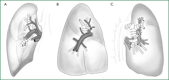
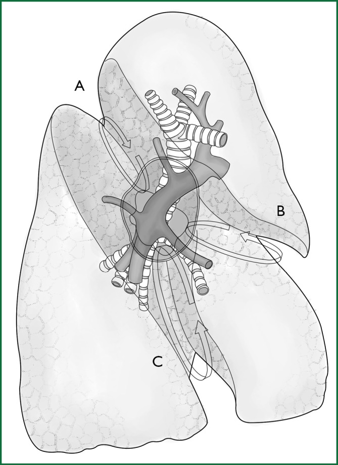
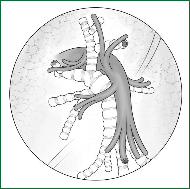
Anatomy of the right lung. (A) Frontal view of the right pulmonary lobe; (B) lateral view of the right pulmonary lobe; and (C) lateral-rear view of the right pulmonary lobe.
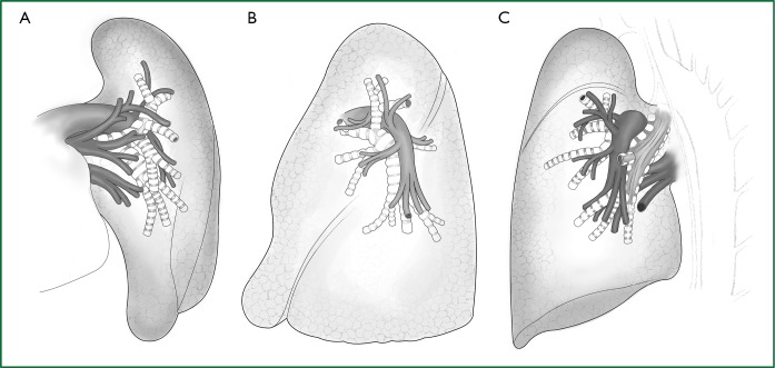
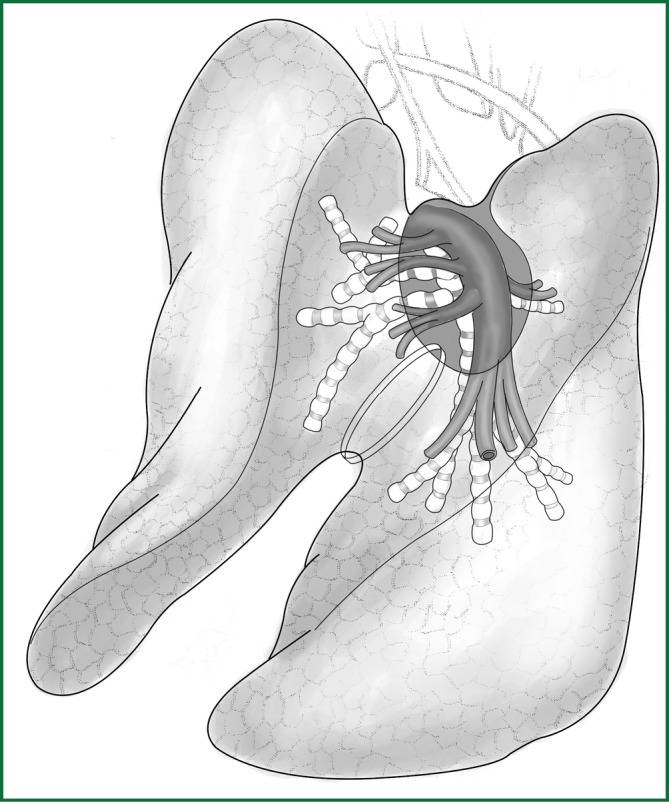
Anatomy of the left lung. (A) Frontal view of the left pulmonary lobe; (B) lateral view of the left pulmonary lobe; and (C) lateral-rear view of the left pulmonary lobe.
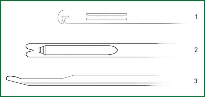
The different designs of knot pushers. 1. The knot pusher with a “C”-shaped head; 2. the knot pusher with a “Y”-shaped head; and 3. the lateral tube of a knot pusher.
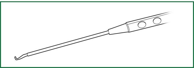
The appearance of the electric hook designed by Jianxing He.
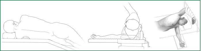
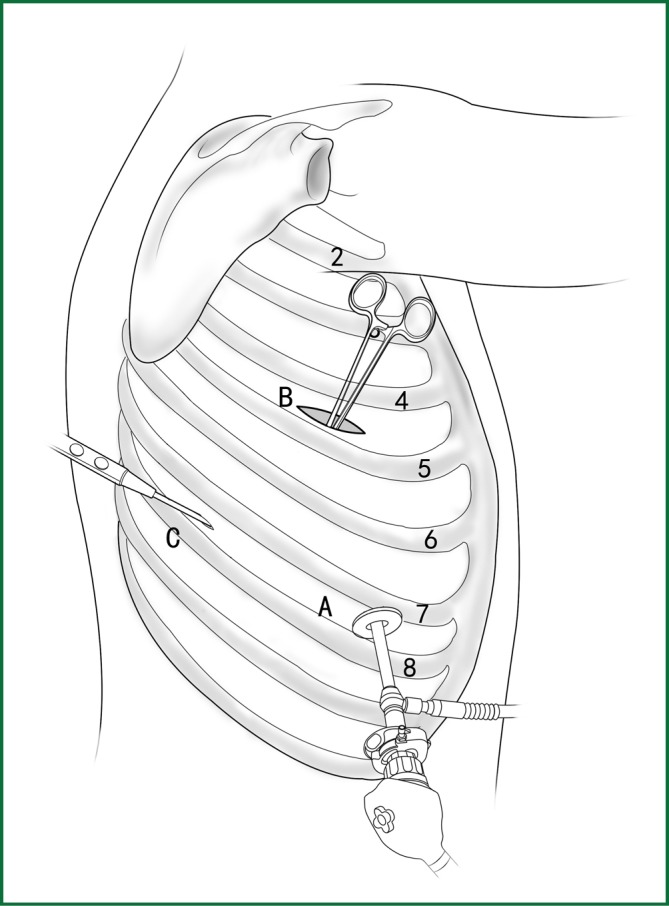
Surgical position.
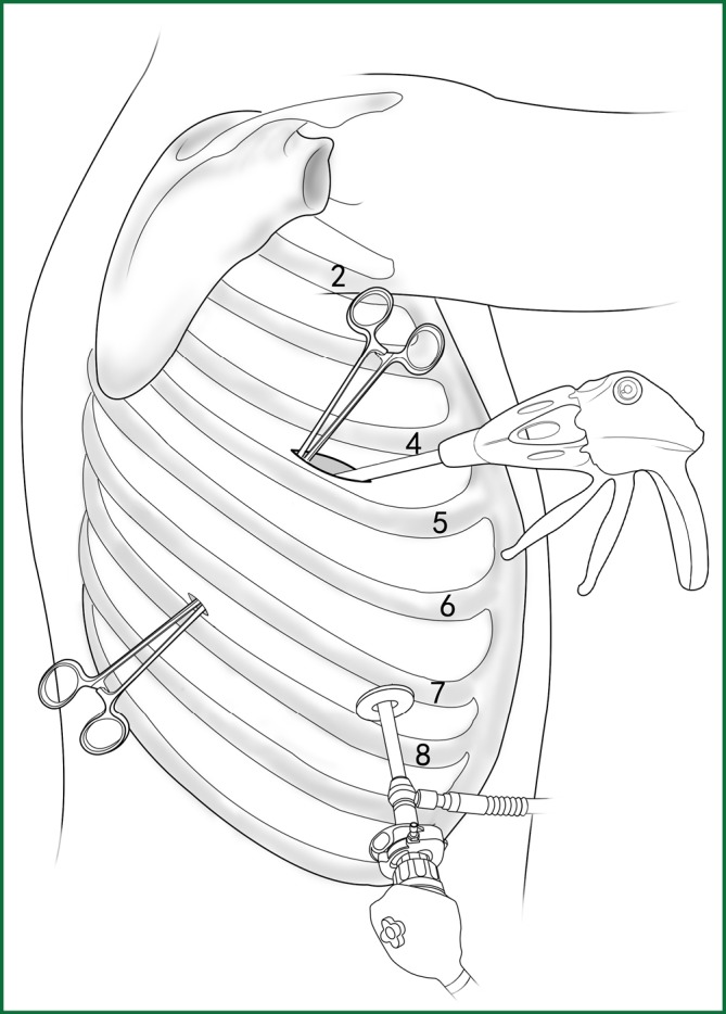
The distribution of incisions in a VATS lobectomy.
The fissures of the right lobe. (A) The upper part of the right oblique fissure. The arrow crosses the gap between the recurrent branch of the right upper pulmonary artery and the dorsal segment of the right lower pulmonary artery; (B) The horizontal fissure. The arrow crosses the gap above the right middle pulmonary artery; and (C) The lower part of the right oblique fissure. The arrow crosses the gap between the right middle pulmonary artery and the basilar segment of the right lower pulmonary artery.
The fissures of the left lobe. The lower part of the left oblique fissure. The arrow crosses the gap between the lingual segment of the left upper pulmonary artery and the basilar segment of the left lower pulmonary artery.
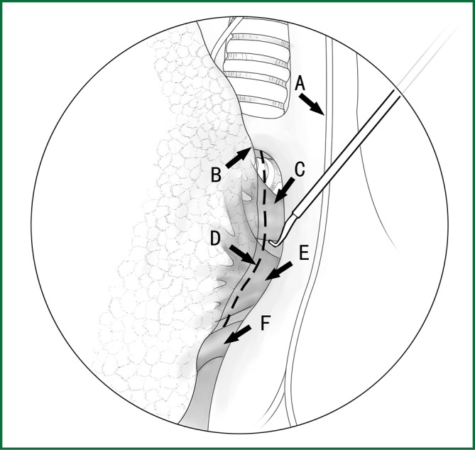
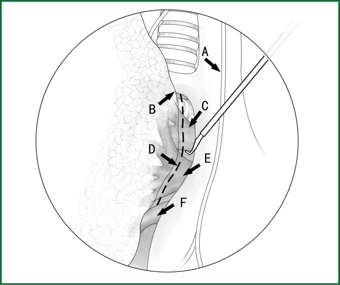
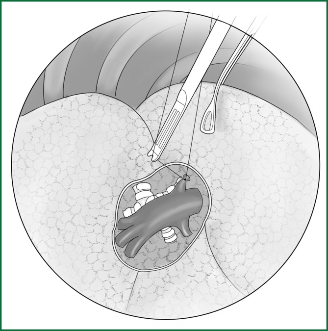
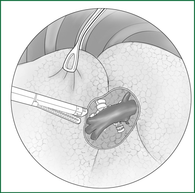
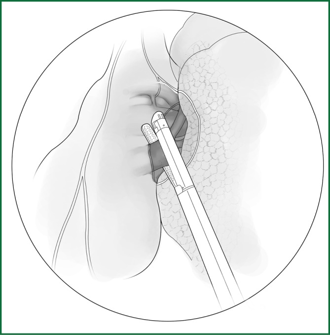
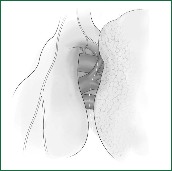
The incision of the mediastinal pleura. (A) Phrenic nerve; (B) lung edge; (C) pulmonary artery; (D) cutting line; (E) the upper pulmonary vein; and (F) the lower pulmonary vein.
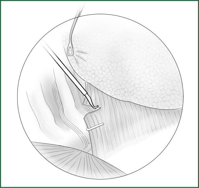
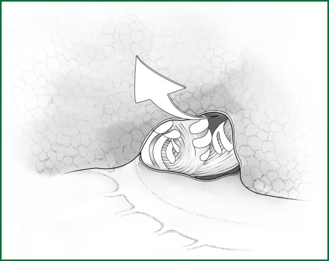
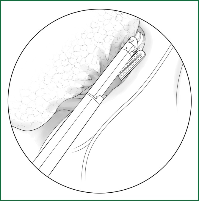
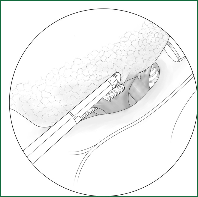
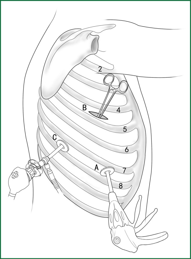
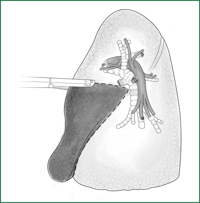
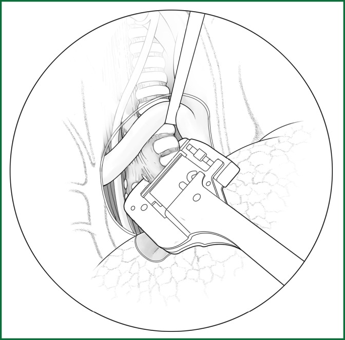
Incision of the lower pulmonary ligament.
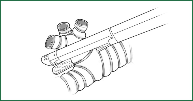
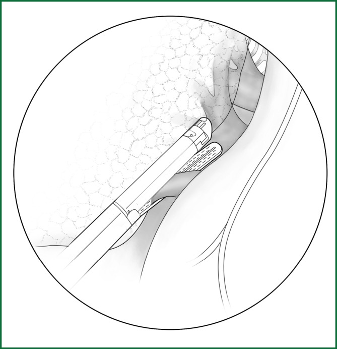
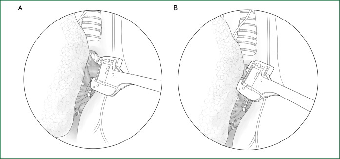
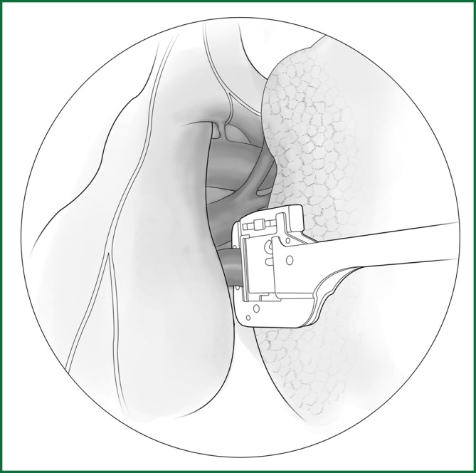
The location of the various devices when loosening the lower pulmonary ligament.
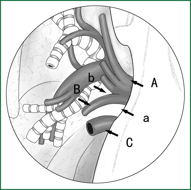
Locating the fissure using the location of the lobar veins. A. The lower pulmonary vein; B. the middle-lobe branch of the upper pulmonary vein; and C. the upper pulmonary vein. a. the gap for the cutting of the oblique fissure and b. the gap for the cutting of the horizontal fissure.
Locating the fissure using the location of the lobar bronchus. The arrow shows the gap between the right upper lobar bronchus and the middle bronchus.
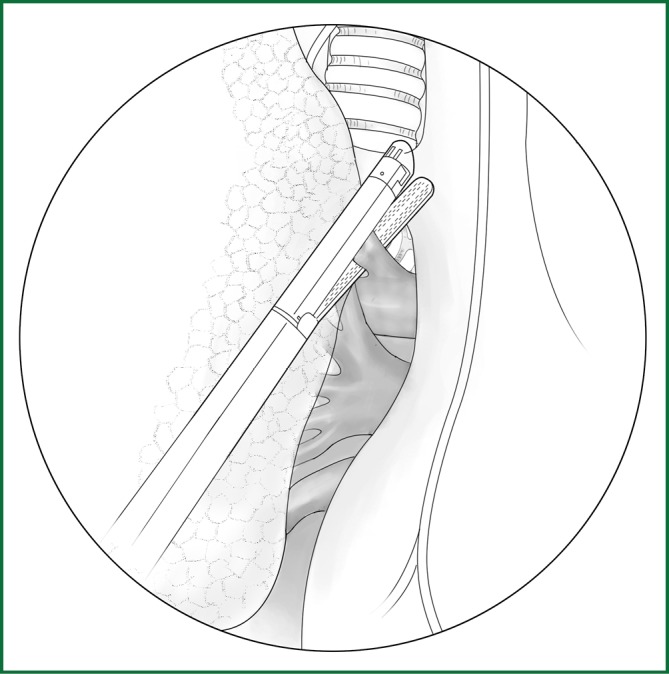
The fissure is cut with the guiding tube.
The locations of the various devices when cutting the fissure.
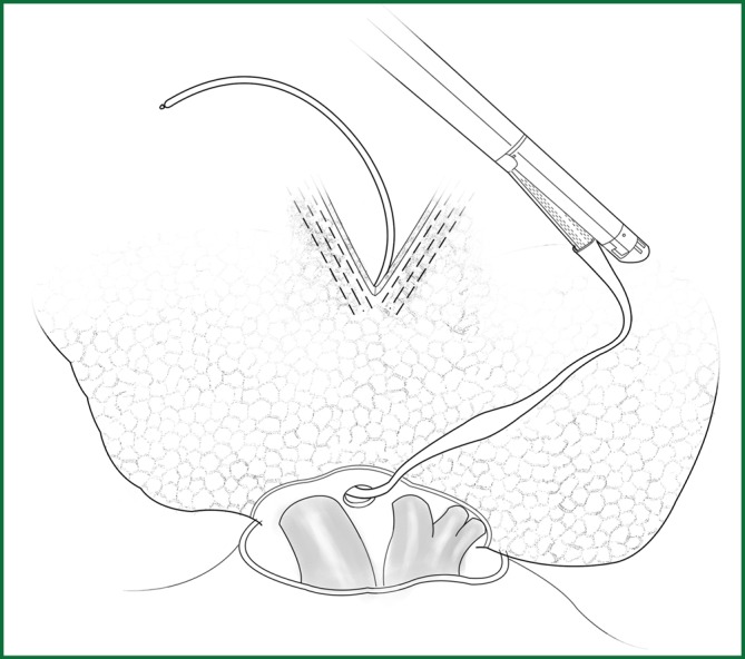
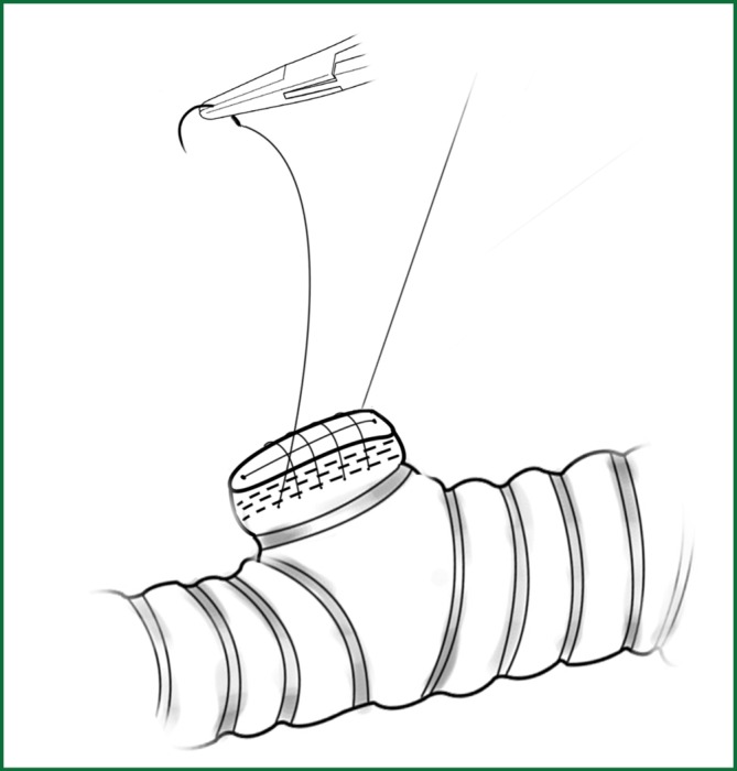
The edges of lung tissue are sutured with a continuous back and forth stitching using prolene thread.
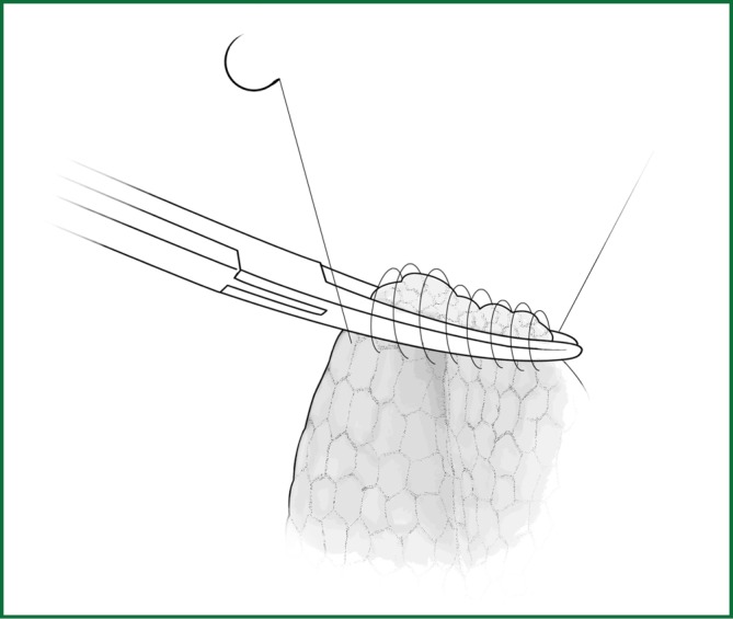
The silk ligation of blood vessels using the knot pusher.
Suture of blood vessels.
Clamping blood vessels with a titanium clip.
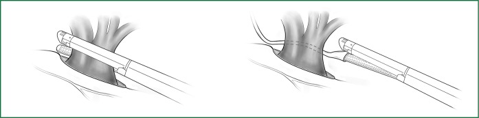
Ligating the blood vessels using the cutting stapler. Up: direct ligation of the blood vessel and Down: ligation of the blood vessel under the guidance of the pediatric urethral catheter.
Severing of the bronchus using the cutting stapler.
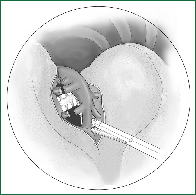
Cut open the mediastinal pleura of the hilum. (A) Phrenic nerve; (B) lung edge; (C) pulmonary artery; (D) incision line; (E) upper pulmonary vein; and (F) lower pulmonary vein.
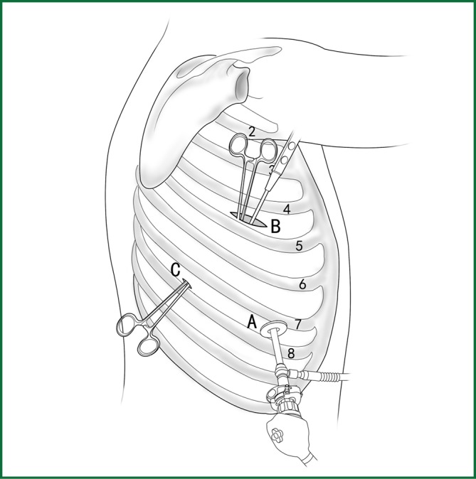
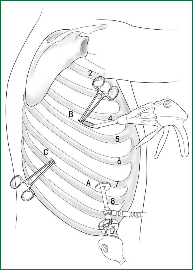
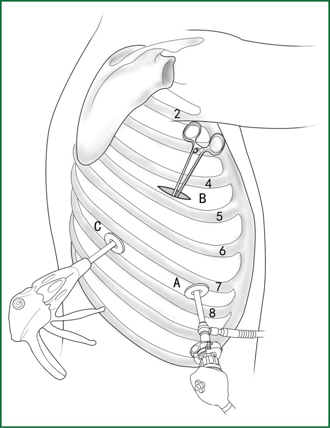
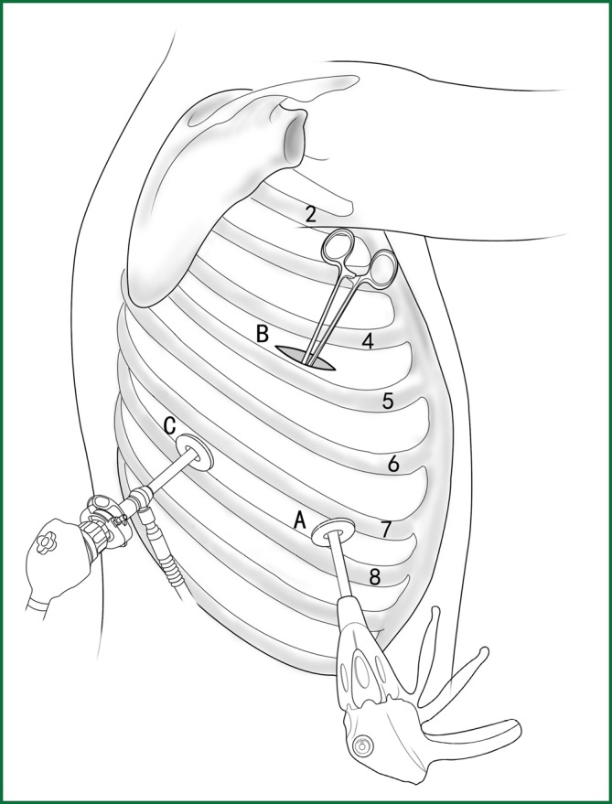
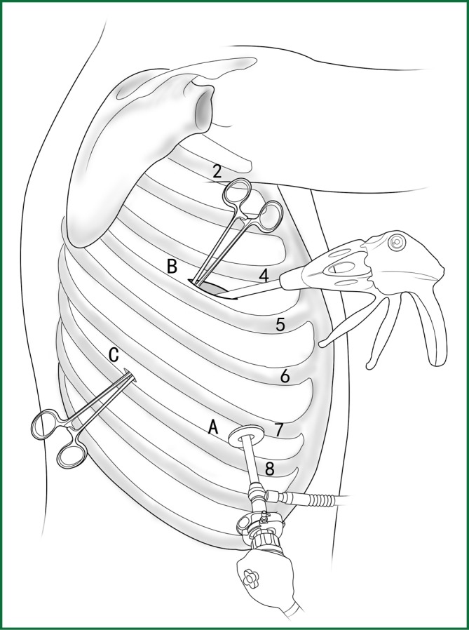
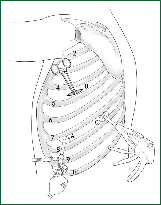
The location of the various devices. Incision A is used for thoracoscopic viewing; the ring forceps is used to lift and pull the lobe via incisions B and C; the mediastinal pleura is cut by the electric hook via incision B.
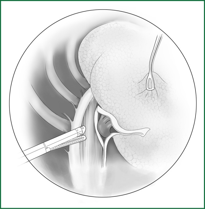
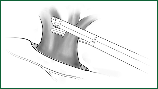
Ligating the right upper pulmonary vein using an endoscopic cutting stapler.
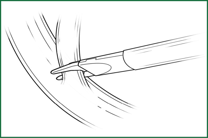
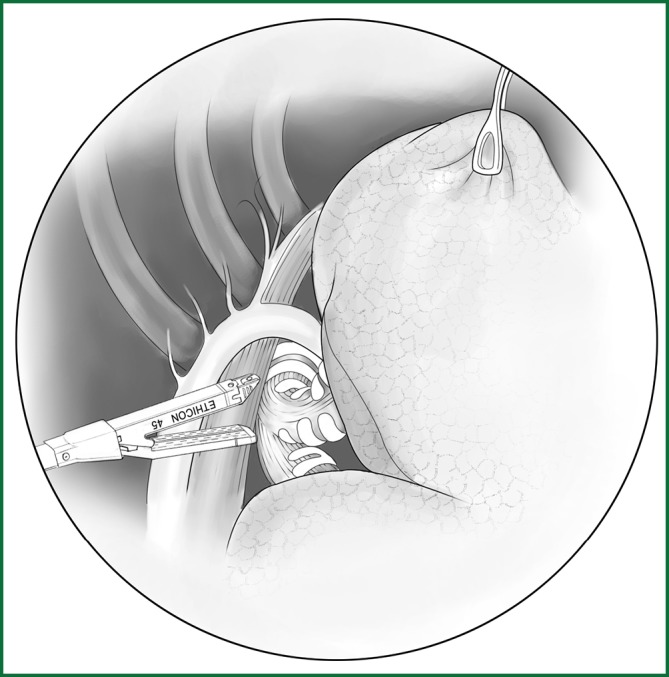
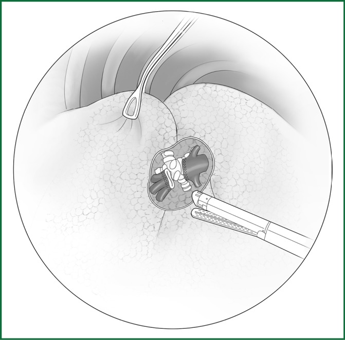
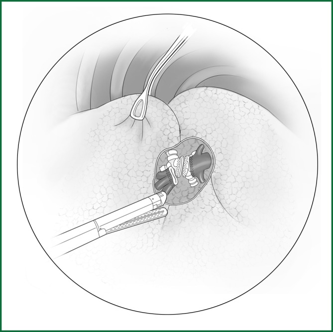
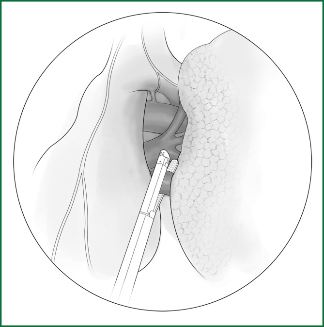
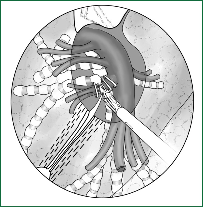
The apicoanterior branch of the right upper pulmonary artery is severed with an endoscopic cutting stapler.
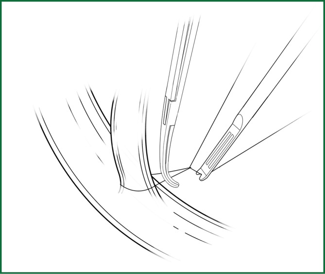
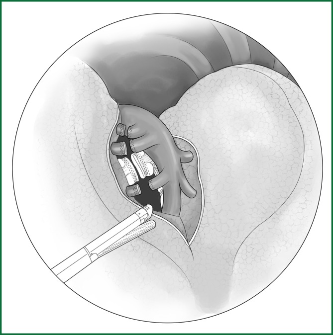
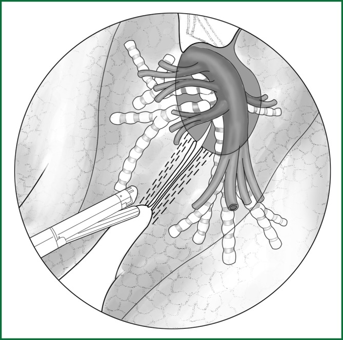
The ascending branch of the right upper pulmonary artery is ligated with a knot pusher.
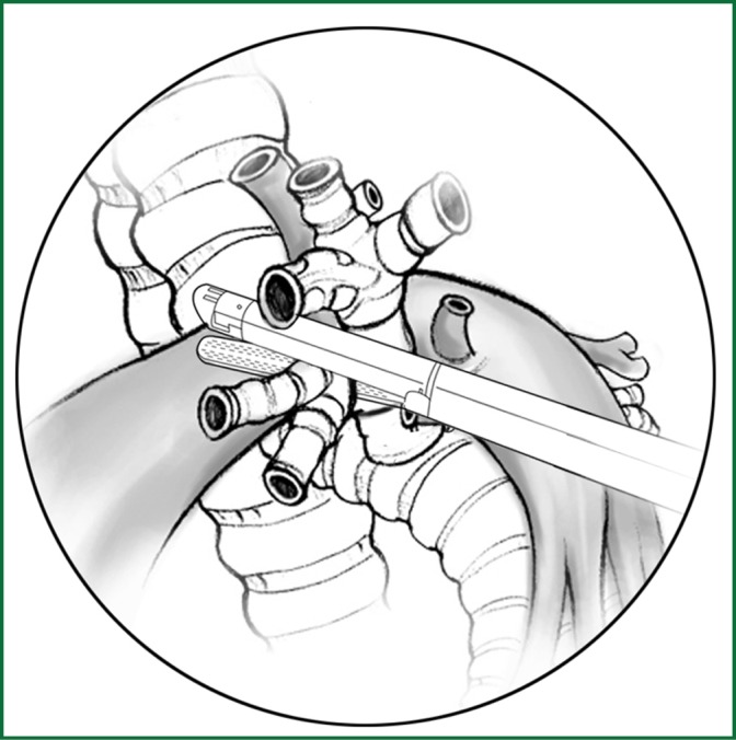
The location of various devices when the blood vessel is severed using an endoscopic cutting stapler.
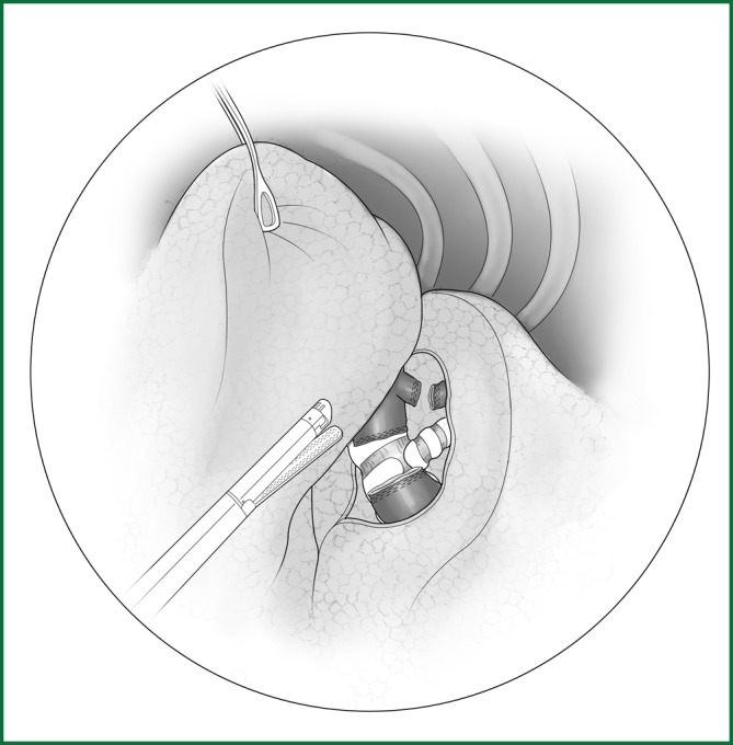
Sever the right upper lobar bronchus.
The reinforcement of the bronchus stump with a continuous suture.
The location of various devices while severing the right upper lobar bronchus.
Severing of the middle vein using an endoscopic cutting stapler.

The location of the various devices.

Ligation of the right middle pulmonary artery.
Severing of the middle lobar bronchus using an endoscopic cutting stapler.
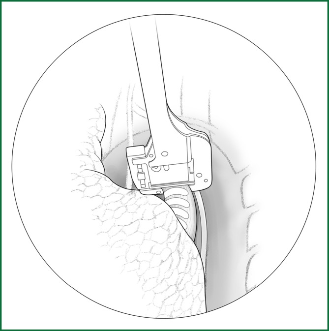
Severing of the right lower pulmonary vein using the endoscopic cutting stapler.
The location of various devices. Incision A is used for thoracoscopic viewing; the ring forceps is used to lift and pull the lobe via incisions B and C; and the endoscopic cutting stapler is used to sever the right lower pulmonary vein via incision B.
(upper right). Severing of the right lower pulmonary artery.
(left). Severing of the right lower lobar bronchus.
The locations of the various devices.

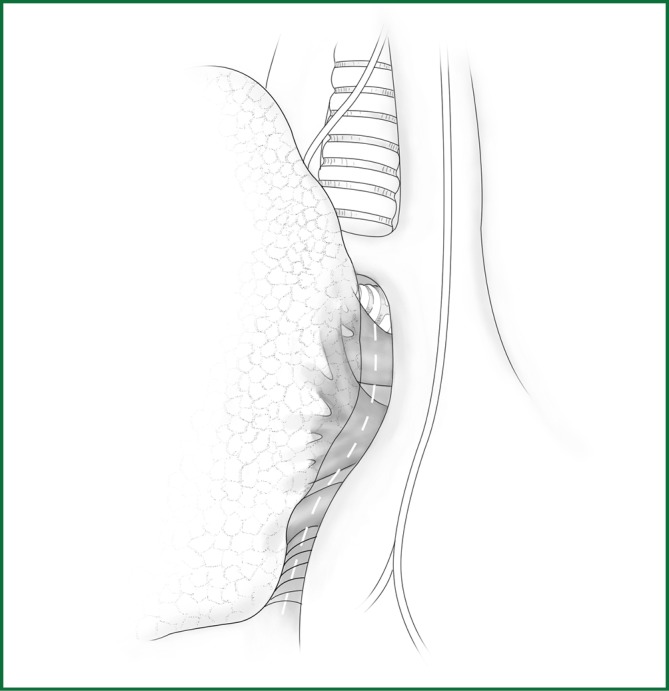
Incision of the mediastinal pleura. The dotted line indicated by the arrow is the cutting line along the pulmonary edge.
Severing of the left upper pulmonary vein using an endoscopic cutting stapler.
The location of the various devices.
Severing of each branch of the upper left pulmonary artery using an endoscopic cutting stapler.
Severing of the upper left lobar bronchus using an endoscopic cutting stapler.
After the left lower pulmonary vein is freed, it is severed using an endoscopic cutting stapler.
Severing of the left lower pulmonary artery trunk using an endoscopic cutting stapler.

Severing of the left lower lobar bronchus using an endoscopic cutting stapler.
Anatomical schematic of the bronchus and arteries of the lingual segment.
The dissection of the oblique fissure of the left lung using a cutting stapler.
The lingual segmental artery of the left upper pulmonary artery is clamped with a vascular clip and then severed.
The lingual segmental vein of the upper pulmonary vein is freed and then severed using a cutting stapler.
Severing of the lingual segmental bronchus using an endoscopic cutting stapler.
The lingual segment without inflation expansion is continuously severed using an endoscopic cutting stapler.
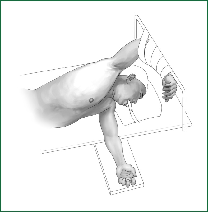
Patient’s position.
Cutting the mediastinal pleura.
Severing of the right lower pulmonary vein.

Severing of the right upper pulmonary vein.
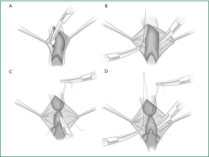
Handling of the right pulmonary artery trunk. (A) A stapler is used to clamp the right pulmonary artery trunk and (B) the stapler is used to clamp the first branch of the right pulmonary artery trunk.
The clamping of the right main pulmonary bronchus with a stapler.
Cutting the hilar mediastinal pleura of the left lung.
Severing of the left lower pulmonary vein.

Severing of the left upper pulmonary vein.

Severing of the left pulmonary artery trunk.
Severing of the left main pulmonary bronchus.
LinkOut - more resources
Full Text Sources