. 2012 Apr 1;2(2):70-78.
doi: 10.4161/bact.20632.

Mobilization of pathogenicity islands by Staphylococcus aureus strain Newman bacteriophages

Affiliations

Mobilization of pathogenicity islands by Staphylococcus aureus strain Newman bacteriophages

Altaira D Dearborn et al. Bacteriophage. .

Abstract

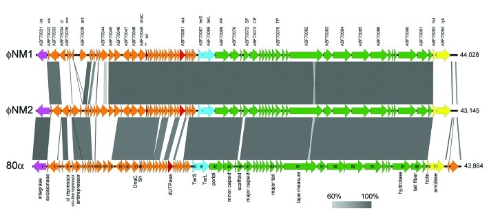
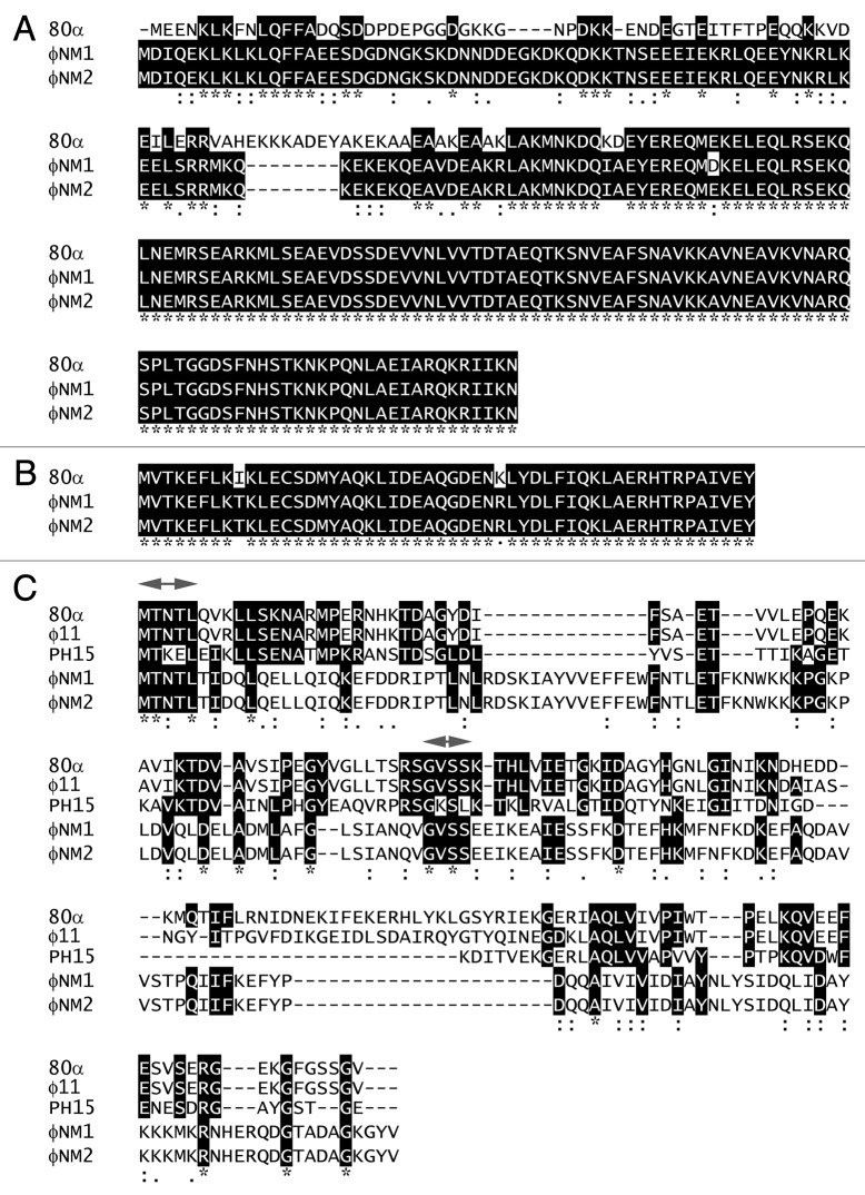
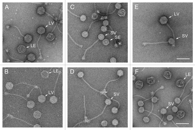
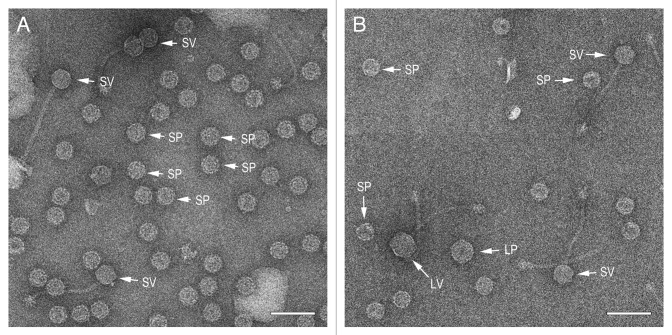
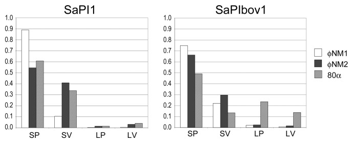
Staphylococcus aureus pathogenicity islands (SaPIs) are mobile genetic elements that encode virulence factors and depend on helper phages for their mobilization. Such mobilization is specific and depends on the ability of a phage protein to inactivate the SaPI repressor Stl. Phage 80α can mobilize several SaPIs, including SaPI1 and SaPIbov1, via its Sri and Dut proteins, respectively. In many cases, the capsids formed in the presence of the SaPI are smaller than those normally produced by the phage. Two SaPI-encoded proteins, CpmA and CpmB, are involved in this size determination process. S. aureus strain Newman contains four prophages, named φNM1 through φNM4. Phages φNM1 and φNM2 are very similar to phage 80α in the structural genes, and encode almost identical Sri proteins, while their Dut proteins are highly divergent. We show that φNM1 and φNM2 are able to mobilize both SaPI1 and SaPIbov1 and yield infectious transducing particles. The majority of the capsids formed in all cases are small, showing that both SaPIs can redirect the capsid size of both φNM1 and φNM2.


Figures

None
Figure 1. Genomic sequence alignments of φNM1 (GenBank entry DQ530359), φNM2 (DQ530360) and 80α (DQ517338), displayed using Easyfig v2.0. The arrows represent the ORFs annotated in the entries, whereas the shading between them represent the percentage identity (BLASTn) from 60% (light gray) to 100% (dark gray). The labels above the φNM1 sequence correspond to the protein identifiers from the GenBank file followed by the name of the protein product, where available (Table 1). The sri gene (corresponding to orf22 in 80α, which was not annotated in the φNM1 GenBank entry, is indicated with a double asterisk (**). The ORFs in 80α are numbered according to Christie et al. with protein functions shown below. Genes encoding structural proteins are colored green; terminase, light blue; lysis genes, yellow; integrase, magenta; Sri, dark red; Dut, light red.
None
Figure 2. Multiple protein sequence alignments of pertinent gene products from 80α, φNM1 and φNM2, including SP (A), Sri (B), and Dut (C). (See Table 1 for protein identifiers.) In (C), corresponding proteins from φ11 and S. epidermidis phage PH15 are also included, and the conserved MTNTL and GVSS motifs are indicated (arrows) above the alignment. Residues that are identical between three or more sequences are highlighted. Conservation between all five sequences is indicated underneath the alignment. The alignments were generated with Clustal Omega.
None
Figure 3. Electron micrographs of negatively stained, CsCl gradient-purified formed by induction of the φNM1 lysogen TB25 (A) and the φNM2 lysogen TB26 (B). Negatively stained transducing particles formed by mobilization of SaPI1 with φNM1 (C) and φNM2 (D), and by mobilization of SaPIbov1 with φNM1 (E) and φNM2 (F). Examples of small virions (SV), large virions (LV) and large empty capsids (LE) are indicated. Scale bars equal 100 nm.
None
Figure 4. Electron micrographs of negatively stained, partially purified lysates from the double lysogens AD1 (φNM1, SaPI1) (A) and AD5 (80α, SaPIbov1) (B). Examples of particles corresponding to small procapsids (SP), small virions (SV), large procapsids (LP) and large virions (LV) are indicated in each panel. Scale bars equal 100 nm.
None
Figure 5. Particle distribution histograms for the mobilization of SaPI1 and SaPIbov1 by phages φNM1 (white), φNM2 (black) and 80α (hatched). Particles were scored as small procapsids (SP), small virions (SV), large procapsids (LP) and large virions (LV) and plotted as a fraction of the total number of particles scored in each sample.

References

    1. Gordon RJ, Lowy FD. Pathogenesis of methicillin-resistant Staphylococcus aureus infection. Clin Infect Dis. 2008;46(Suppl 5):S350–9. doi: 10.1086/533591. - DOI - PMC - PubMed
    1. Otto M. Basis of virulence in community-associated methicillin-resistant Staphylococcus aureus. Annu Rev Microbiol. 2010;64:143–62. doi: 10.1146/annurev.micro.112408.134309. - DOI - PubMed
    1. Ziebuhr W, Ohlsen K, Karch H, Korhonen T, Hacker J. Evolution of bacterial pathogenesis. Cell Mol Life Sci. 1999;56:719–28. doi: 10.1007/s000180050018. - DOI - PMC - PubMed
    1. Malachowa N, DeLeo FR. Mobile genetic elements of Staphylococcus aureus. Cell Mol Life Sci. 2010;67:3057–71. doi: 10.1007/s00018-010-0389-4. - DOI - PMC - PubMed
    1. Novick RP, Christie GE, Penadés JR. The phage-related chromosomal islands of Gram-positive bacteria. Nat Rev Microbiol. 2010;8:541–51. doi: 10.1038/nrmicro2393. - DOI - PMC - PubMed

LinkOut - more resources