doi: 10.4103/1947-2714.100998.
Western blot: technique, theory, and trouble shooting
Affiliations
- PMID: 23050259
- PMCID: PMC3456489
- DOI: 10.4103/1947-2714.100998
Item in Clipboard
Western blot: technique, theory, and trouble shooting
N Am J Med Sci.
2012 Sep.
Abstract
Western blotting is an important technique used in cell and molecular biology. By using a western blot, researchers are able to identify specific proteins from a complex mixture of proteins extracted from cells. The technique uses three elements to accomplish this task: (1) separation by size, (2) transfer to a solid support, and (3) marking target protein using a proper primary and secondary antibody to visualize. This paper will attempt to explain the technique and theory behind western blot, and offer some ways to troubleshoot.
Keywords: Bio-medical research; protein; western blot.
Conflict of interest statement
Figures
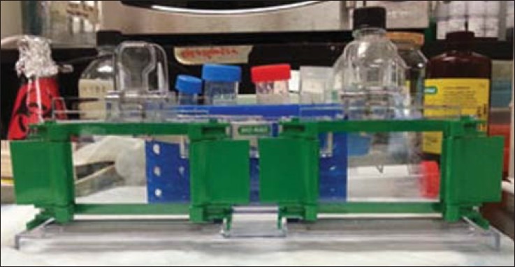
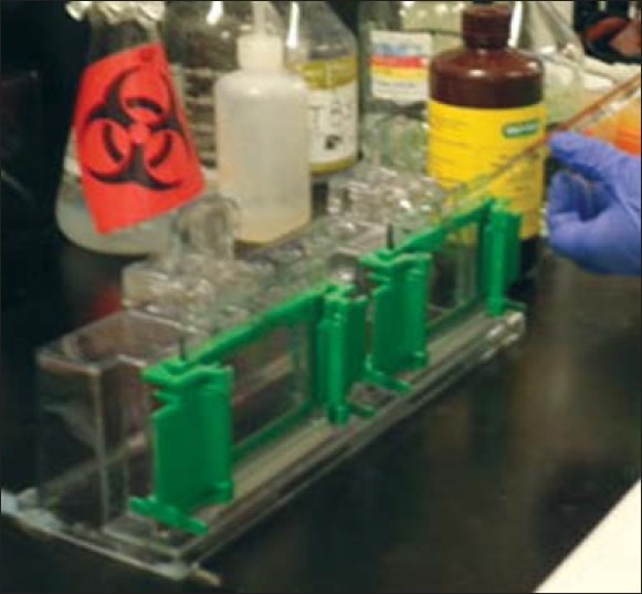
Assembled rack for gel solidification
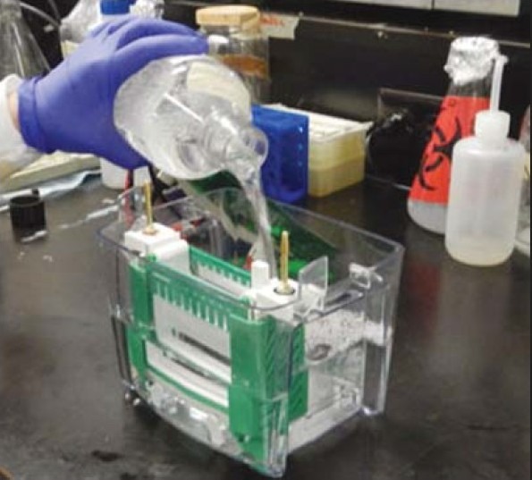
Add gel solution using a transfer pipette
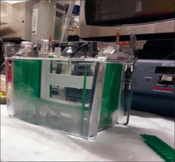
Add running buffer to the electrophorator
Add samples and molecular marker to the gel, after removing the combs
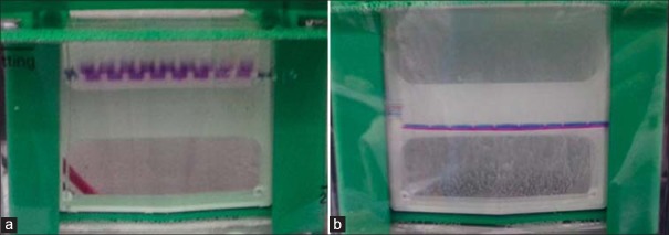
(a) Samples running through the stacking gel (lower voltage). (b): Samples running through the separating gel (higher voltage)
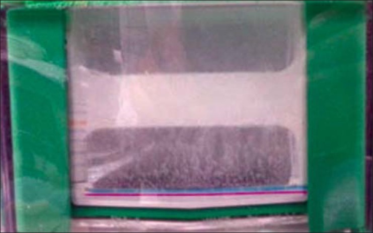
Run the gel to the bottom of the electrophorator
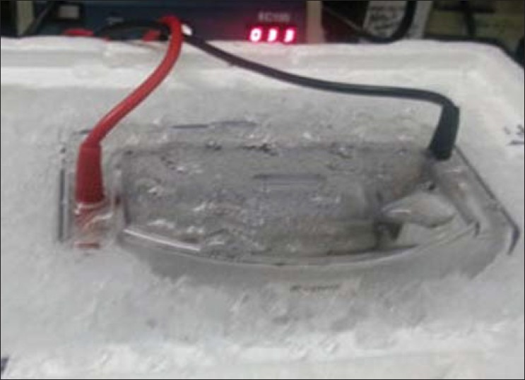
Transfer should be done on ice
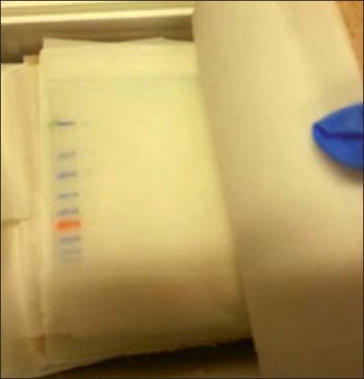
Membrane after transfer
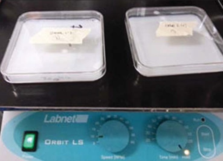
Use a shaker to incubate the membrane with antibody
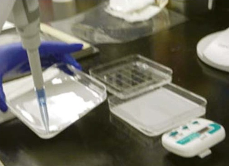
Incubate the membrane with ECL mix using a 1000 μL pipette to help the process
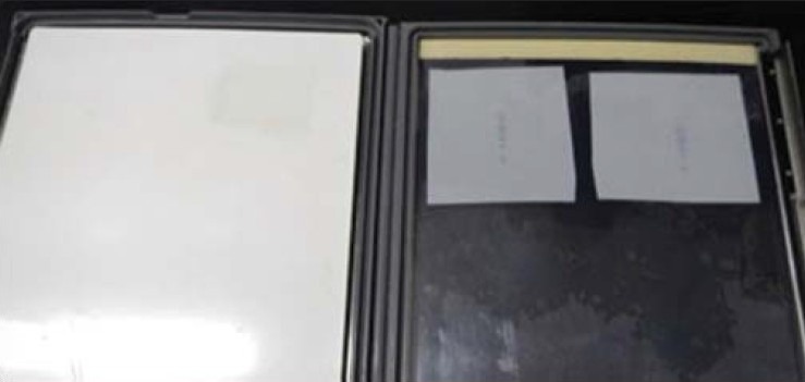
Use the cassette to expose the membrane in the dark room
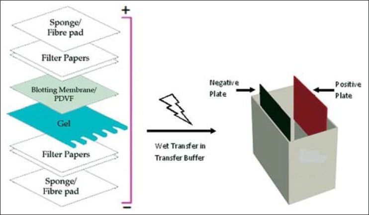
Assembly of a sandwich in western Blot
LinkOut - more resources
Full Text Sources
Other Literature Sources