. 2012 Nov;123 Suppl 2(Suppl 2):86-97.
doi: 10.1111/j.1471-4159.2012.07947.x.

The inhibitory effect of S-nitrosoglutathione on blood-brain barrier disruption and peroxynitrite formation in a rat model of experimental stroke

Affiliations

The inhibitory effect of S-nitrosoglutathione on blood-brain barrier disruption and peroxynitrite formation in a rat model of experimental stroke

Mushfiquddin Khan et al. J Neurochem. 2012 Nov.

Abstract

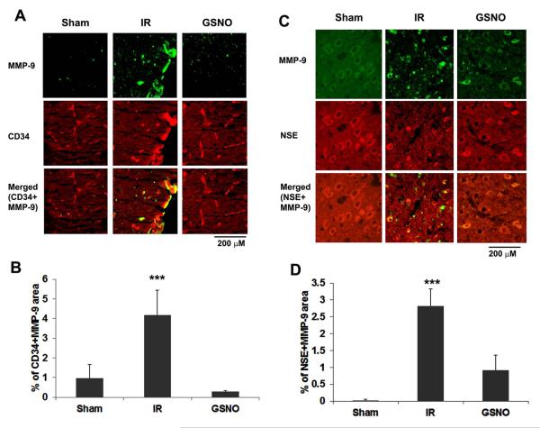
The hallmark of stroke injury is endothelial dysfunction leading to blood-brain barrier (BBB) leakage and edema. Among the causative factors of BBB disruption are accelerating peroxynitrite formation and the resultant decreased bioavailability of nitric oxide (NO). S-nitrosoglutathione (GSNO), an S-nitrosylating agent, was found not only to reduce the levels of peroxynitrite but also to protect the integrity of BBB in a rat model of cerebral ischemia and reperfusion (IR). A treatment with GSNO (3 μmol/kg) after IR reduced 3-nitrotyrosine levels in and around vessels and maintained NO levels in brain. This mechanism protected endothelial function by reducing BBB leakage, increasing the expression of Zonula occludens-1 (ZO-1), decreasing edema, and reducing the expression of matrix metalloproteinase-9 and E-selectin in the neurovascular unit. An administration of the peroxynitrite-forming agent 3-morpholino sydnonimine (3 μmol/kg) at reperfusion increased BBB leakage and decreased the expression of ZO-1, supporting the involvement of peroxynitrite in BBB disruption and edema. Mechanistically, the endothelium-protecting action of GSNO was invoked by reducing the activity of nuclear factor kappa B and increasing the expression of S-nitrosylated proteins. Taken together, the results support the ability of GSNO to improve endothelial function by reducing nitroxidative stress in stroke.


Figures

Figure 1
Figure 1
Effect of GSNO and SIN-1 on BBB leakage and edema after IR. Representative photographs showing extravasation of Evan’s blue (EB) in brain starting at 4 h and measured at 48 h after IR (A). Spectrofluorimetric estimation of EB was performed in ipsilateral hemisphere (B). Edema was measured at 24 h after IR (C). Significant EB leakage was observed in IR brain, whereas EB extravasations were not observed in sham brain. While SIN-1-treatment increased, the treatment with GSNO of IR decreased the EB extravasation in ipsilateral brain up to 3 h after the injury. GSNO treatment of IR also decreased edema. Data are expressed as means ±SD from triplicate determinations of five different samples in each group. *** p<0.001 and **p<0.01 vs. IR and SIN-1, ##p<0.05 vs. IR.
Figure 2
Figure 2
Effect of GSNO and SIN-1 on the expression of ZO-1 in ipsilateral (penumbra) rat brain. Western blot (A) and densitometry (B) of ZO-1 in the penumbra at 24 h after IR. Representative western blot (2 samples) from three different sets of experiments showed reduced expression of ZO-1 in brain from IR and SIN-1-treated animals. GSNO-treated IR animals had an expression of ZO-1 similar to the sham animals. *p<0.05 vs. sham and GSNO.
Figure 3
Figure 3
Effect of GSNO on the expression of 3-nitrotyrosine (3-NT) in ipsilateral (penumbra) rat brain (A, western analysis; B, densitometry; C, IHC analysis). Representative western blot (1 from Sham and 2 each from GSNO and IR) from three different sets of experiments at 4 h after IR showed enhanced reactivity of 3-NT in brain from IR animals. GSNO-treated animals had reduced expression of 3-NT. IHC of 3-NT (green) and smooth muscle actin (SMA, red) in ipsilateral (penumbra) rat brain at 4 h after IR. a-d) Sham-operated animal. e-h ) GSNO-treated IR animal. i-l) Veh-treated IR animal. i.i-l.i) Magnified view of i-l. Green fluorescence in IR sections indicates higher immunoreactivity for 3-NT (i, i-i) in vessels labeled with SMA (red fluorescence, j, j,i). Yellowish color in k and k.i sections indicates the colocalization of 3-NT with SMA. In the magnified sections, asterisk indicates lumen of vessel while arrow points to vessel. Sham- and GSNO-treated animals did not show any significant staining of 3-NT. The photomicrograph is representative of 3 samples in each group. *p<0.05 vs. GSNO.
Figure 4
Figure 4
Effect of GSNO on the expression of E-selectin in ipsilateral (penumbra) rat brain at 4 h after IR. Photomicrographs (A) and graph determining E-selectin positive area (B). Photomicrographs show enhanced reaction of E-selectin in the penumbra from IR compared with GSNO group. Sham brain does not show significant staining of E-selectin. Symbol V on sections indicates vessel. Photomicrographs are representative of 3 samples in each group. **p<0.01 vs. sham and GSNO.
Figure 5
Figure 5
Effect of GSNO on the expression of MMP-9 in ipsilateral (penumbra) rat brain at 4 h after IR. Photomicrographs of MMP-9/CD34 (A) and MMP-9/NSE (C), and graphs determining MMP-9/CD34 (B) and MMP-9/NSE (D) positive areas. Photomicrographs of IHC show enhanced reaction of MMP-9 (green color) in IR compared with GSNO group (A, B). Sham brain does not show MMP-9 positive cells. Colocalization (yellowish, merged) of MMP-9 (green color) with either endothelial cell marker CD34 (red) (A) or neuronal marker NSE (red) indicates that both neurons and endothelial cells have significantly increased expression of MMP-9 even at 4 h after IR. Photomicrographs are representative of 3 samples in each group. **p<0.01 vs. sham and GSNO.
Figure 6
Figure 6
Effect of GSNO on the expression of p65 of NF-κB and activity of NF-κB in ipsilateral (penumbra) rat brain at 4 h after IR. Photomicrographs (A), graph determining p65 positive area (B) and NF-κB activity (C). While the expression of P65 in sham was localized mainly in cytosol, both IR and GSNO groups had expression present mainly in nucleus (A). NF-κB activity in the ipsilateral brain tissue was measured as described in Methods. The activity was significantly high in IR compared with sham and GSNO groups. Values are mean + SD of triplicate determinations from three different experiments. ***p<0.001 vs. Sham and GSNO.
Figure 7
Figure 7
Effect of GSNO on the activity of constitutive NOS (A) and endothelial NOS (B) at 1 h and the levels of NO (C in ipsilateral (penumbra) brain) at 4h after IR. While activity of constitutive NOS is increased, endothelial NOS is decreased in the IR group. NO levels are also decreased in the IR group. GSNO and sham groups show similar activity of constitutive NOS and levels of NO. However, eNOS activity is decreased in the GSNO group compared with both sham and IR groups. The activity of cNOS and eNOS is expressed as pmol/mg protein/min and data are presented as mean+SD, n=3. ***p<0.001,**p<0.01 vs GSNO and Sham,+p<0.05 vs Sham, ++<0.05 vs IR.
Figure 8
Figure 8
Effect of GSNO on the expression of S-nitrosocysteine in ipsilateral (penumbra) rat brain at 4 h after IR. Photomicrographs (A) and graph determining S-nitrosocysteine positive area (B). The expression of S-nitrosocysteine was determined using IHC. Photomicrographs show reduced reaction of S-nitrosocysteine in the IR compared with sham and GSNO groups. Photomicrographs are representative of n=5 in each group (magnification x400).
Figure 9
Figure 9
Schematic showing the vicious cycle of peroxynitrite production in endothelial cell in an animal model of IR. GSNO was hypothesized to protect against neuroinflammatory secondary injury in IR by reducing peroxynitrite, thereby blocking the vicious superoxide/aberrant eNOS/peroxynitrite cycle.

References

    1. Barone FC, Feuerstein GZ. Inflammatory mediators and stroke: new opportunities for novel therapeutics. J Cereb Blood Flow Metab. 1999;19:819–834. - PubMed
    1. Barr TL, Latour LL, Lee KY, et al. Blood-brain barrier disruption in humans is independently associated with increased matrix metalloproteinase-9. Stroke. 2010;41:e123–128. - PMC - PubMed
    1. Berti R, Williams AJ, Moffett JR, Hale SL, Velarde LC, Elliott PJ, Yao C, Dave JR, Tortella FC. Quantitative real-time RT-PCR analysis of inflammatory gene expression associated with ischemia-reperfusion brain injury. J Cereb Blood Flow Metab. 2002;22:1068–1079. - PubMed
    1. Chen H, Yoshioka H, Kim GS, et al. Oxidative stress in ischemic brain damage: mechanisms of cell death and potential molecular targets for neuroprotection. Antioxid Redox Signal. 2011;14:1505–1517. - PMC - PubMed
    1. Chen HY, Chen TY, Lee MY, Chen ST, Hsu YS, Kuo YL, Chang GL, Wu TS, Lee EJ. Melatonin decreases neurovascular oxidative/nitrosative damage and protects against early increases in the blood-brain barrier permeability after transient focal cerebral ischemia in mice. J Pineal Res. 2006;41:175–182. - PubMed

Publication types

MeSH terms