. 2013 Feb 20;18(6):603-21.
doi: 10.1089/ars.2012.4623. Epub 2012 Dec 6.

Studies of mitochondrial and nonmitochondrial sources implicate nicotinamide adenine dinucleotide phosphate oxidase(s) in the increased skeletal muscle superoxide generation that occurs during contractile activity

Affiliations

Studies of mitochondrial and nonmitochondrial sources implicate nicotinamide adenine dinucleotide phosphate oxidase(s) in the increased skeletal muscle superoxide generation that occurs during contractile activity

Giorgos Konstantinos Sakellariou et al. Antioxid Redox Signal. .

Abstract

Aims: The sources of cytosolic superoxide in skeletal muscle have not been defined. This study examined the subcellular sites that contribute to cytosolic superoxide in mature single muscle fibers at rest and during contractile activity.

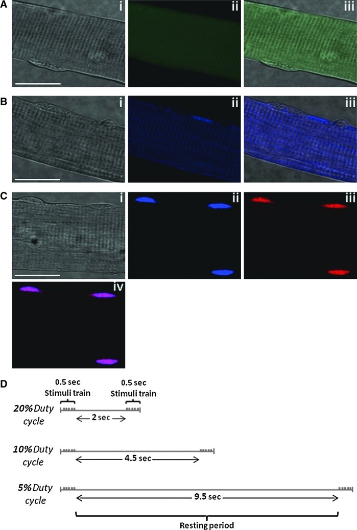
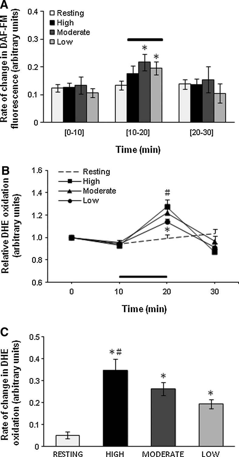
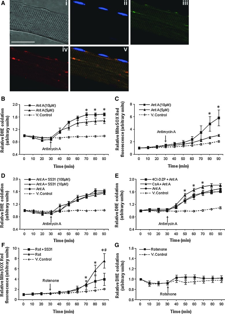
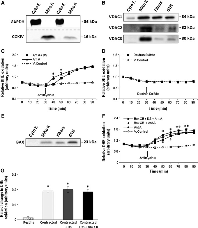
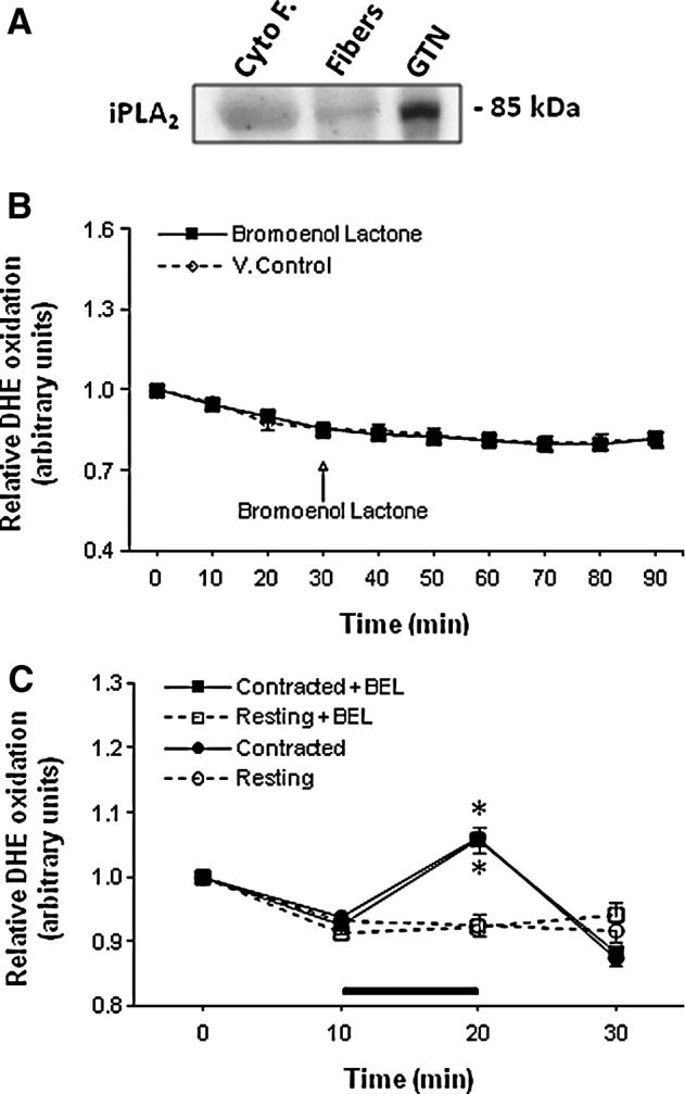
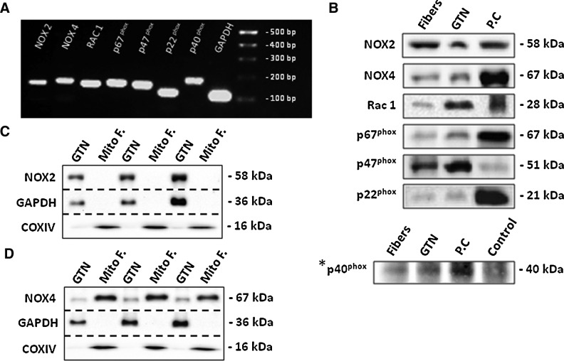
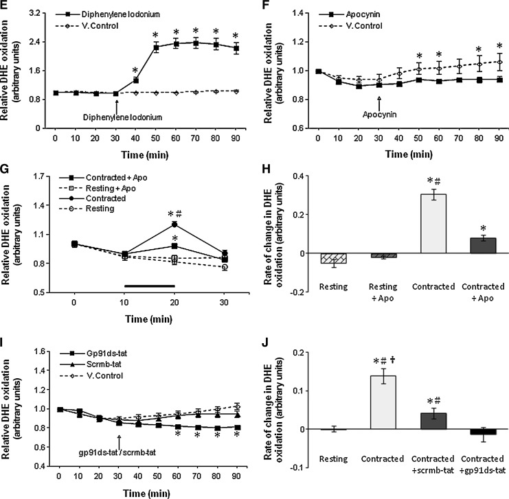
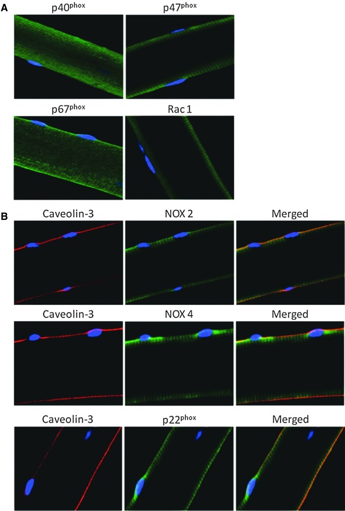
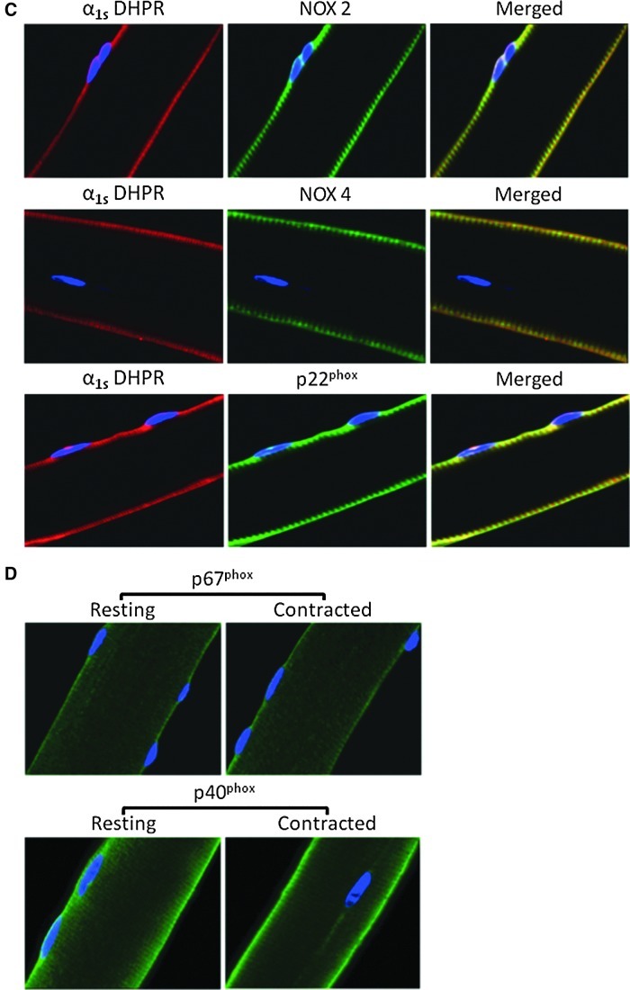
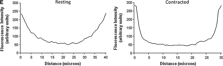
Results: Isolated fibers from mouse flexor digitorum brevis loaded with superoxide and nitric-oxide-sensitive fluorescent probes, specific pathway inhibitors and immunolocalization techniques were used to identify subcellular sites contributing to cytosolic superoxide. Treatment with the electron transport chain complex III inhibitor, antimycin A, but not the complex I inhibitor, rotenone, caused increased cytosolic superoxide through release from the mitochondrial intermembrane space via voltage-dependent anion or Bax channels, but inhibition of these channels did not affect contraction-induced increases in cytosolic superoxide. Nicotinamide adenine dinucleotide phosphate (NADPH) oxidase inhibitors decreased cytosolic superoxide at rest and following contractions. Protein and mRNA expression of NADPH oxidase subunits was demonstrated in single fibers. NOX2, NOX4, and p22(phox) subunits localized to the sarcolemma and transverse tubules; NOX4 was additionally expressed in mitochondria. Regulatory p40(phox) and p67(phox) proteins were found in the cytoplasm of resting fibers, but following contractions, p40(phox) appeared to translocate to the sarcolemma.

Innovation: Superoxide and other reactive oxygen species generated by skeletal muscle are important regulators of muscle force production and adaptations to contractions. This study has defined the relative contribution of mitochondrial and cytosolic sources of superoxide within the cytosol of single muscle fibers at rest and during contractions.

Conclusion: Muscle mitochondria do not modulate cytosolic superoxide in skeletal muscle but NADPH oxidase is a major contributor both at rest and during contractions.


Figures

FIG. 1.
FIG. 1.
DAF-FM DA and DHE to assess changes in NO and superoxide at rest and following contractile activity. (A) Confocal images of a single isolated fiber from the FDB muscle after 16 h in culture under bright field (i); fluorescent image following loading with DAF-FM DA (ii); merged image of (i) and (ii)(iii). (B) Confocal images of a single isolated fiber under bright field (i); fluorescent image showing blue fluorescence from nonoxidized DHE (ii); merged image of (i) and (ii)(iii). (C) Confocal images of a single isolated fiber under bright field (i); fluorescent image showing DAPI staining (ii); fluorescent image showing ethidium, the oxidized form of DHE (iii), and a merged image of (ii) and (iii)(iv). Original magnification: 60×(scale bar=30 μm). (D) Schematic illustration of the protocols for electrical stimulations at different intensities. The stimuli train was constant in all contraction protocols. The time at rest between repetitions varied according to the intensity of each protocol. DAF-FM DA, 4-amino-5-methylamino-2′,7′-difluorofluorescein diacetate; DAPI, 4′,6-diamidino-2-phenylindole dihydrochloride; DHE, dihydroethidium; FDB, flexor digitorum brevis.
FIG. 2.
FIG. 2.
Changes in DAF-FM fluorescence and DHE oxidation following contractile activity. (A) Rate of change in DAF-FM fluorescence from resting FDB fibers and fibers subjected to contractile activity of different intensities over the 10–20 min period. *p<0.05 compared with values from the same group prior to contractions 7–10 fibers in each group). (B) Relative change in DHE oxidation from resting FDB fibers and fibers subjected to contractile activity of different intensities over the 10–20-min period. *p<0.05 compared with fibers from the contracted groups at the corresponding time point. #p<0.05 compared with fibers subjected to the Low contraction protocol (n=7–8 fibers in each group). (C) Rate of change in DHE oxidation from resting FDB fibers and fibers subjected to a 10-min period of contractile activity with different intensities. *p<0.05 compared with fibers from the resting group. #p<0.05 compared with fibers subjected to the Low contractile activity protocol (n=7–8 fibers in each group).
FIG. 3.
FIG. 3.
Release of superoxide from intact mitochondria following treatment with Ant A does not occur through the mPTP or iMAC. (A) Confocal images of a single isolated fiber under bright field (i); fluorescent image following loading with DAPI (ii); fluorescence from MitoTracker Green FM (15 nM) (iii); fluorescent image from MitoSOX Red (iv); and a merged image of (ii)(iv)(v). Original magnification: 60×(scale bar=30 μm). (B) Relative change in DHE oxidation from isolated FDB fibers either untreated or treated with Ant A (5 or 10 μM) at 30 min. *p<0.05 compared with values from fibers treated with 5 μM Ant A or control vehicle-treated (V.control) fibers at the same time point (n=7–8 fibers in each group). (C) Relative change in MitoSOX Red fluorescence from isolated FDB fibers either untreated or treated with Ant A (5 or 10 μM) at 30 min. *p<0.05 compared with values from fibers treated with 5 μM Ant A and control vehicle-treated (V.control) fibers at the same time point (n=6–8 fibers in each group). (D) Relative change in DHE oxidation from control fibers and fibers loaded with Ant A (5 μM) at 30 min. Fibers were either treated with vehicle only (V.control) or treated with the mitochondrial-targeted SS-31 peptide at 10 or 100 μM (n=6 fibers in each group). (E) Relative change in DHE oxidation from control fibers and fibers loaded with Ant A (5 μM) at 30 min. Fibers were either treated with vehicle only (V.control) or treated with the mPTP (CsA) or iMAC (4Cl-DZP) inhibitors. *p<0.05 compared with values from fibers in all other Ant A–treated groups and control vehicle-treated (V.control) fibers at the corresponding time points (n=6–8 fibers in each group). (F) Relative change in MitoSOX Red fluorescence from control fibers and fibers treated with Rot at 30 min. A group of fibers was also treated with SS-31 peptide (10 μM). *p<0.05 compared with values from control vehicle-treated (V.control) fibers at the same time point. #p<0.05 compared with values from Rot-treated fibers incubated in the presence of SS-31 peptide at the corresponding time point (n=6–9 fibers in each group). (G) Relative change in DHE oxidation from control fibers and fibers treated with Rot at 30 min. 4Cl-DZP, 4′-chlorodiazepam; Ant A, antimycin A; CsA, cyclosporin A; iMAC, innermembrane anion channel; mPTP, mitochondrial permeability transition pore; Rot, rotenone.
FIG. 4.
FIG. 4.
Channels of the OMM mediate the diffusion of superoxide from the MIS to the cytosol of fibers following treatment with Ant A. (A) Example of Western blots for GAPDH and cytochrome oxidase IV (COXIV) to illustrate the purity of the extracted Mito F and Cyto F fractions obtained from the GTN muscle. (B) Representative Western blots of VDAC1, VDAC2, and VDAC3 proteins in Cyto F and Mito F fractions from GTN muscle, in lysate from single isolated fibers from the FDB muscle (fibers), and in whole GTN muscle. (C) Relative change in DHE oxidation from control fibers and fibers loaded with Ant A (5 μM) at 30 min. Fibers were either treated with vehicle alone or with DS. *p<0.05 compared with fibers treated with DS at the corresponding time point (n=7 fibers in each group). (D) Relative change in DHE oxidation from isolated FDB fibers either untreated or treated with DS at 30 min (n=7–8 fibers in each group). (E) Representative Western blot of Bax protein in cytosolic (Cyto F) and mitochondrial (Mito F) fractions from GTN muscle, in lysate from single isolated FDB muscle fibers, and in whole GTN muscle. (F) Relative change in DHE oxidation from control fibers and fibers loaded with Ant A (5 μM) at 30 min. Fibers were either untreated or treated with Bax CB or with Bax CB and DS. *p<0.05 compared with values from Ant A–treated fibers preincubated with Bax CB and DS at the same time point. #p<0.05 compared with values from fibers treated with Ant A in the presence of Bax CB at the same time point (n=6–7 fibers in each group). (G) Rate of change in DHE oxidation from resting fibers and fibers subjected to the Moderate contraction protocol for a period of 10 min. Contracted fibers were either untreated, treated with DS, or treated with DS and Bax CB. *p<0.05 compared with values from fibers at rest (n=9–11 fibers in each group). Bax CB, Bax channel blocker; Cyto F, cytosolic fractions; DS, dextran sulfate; GTN, gastrocnemius; Mito F, mitochondrial fractions; VDACs, voltage-dependent anion channels.
FIG. 5.
FIG. 5.
Effect of IPLA2 inhibition on cytosolic superoxide following contractile activity. (A) Representative Western blot of iPLA2 protein in Cyto F from GTN muscle, in lysate from single isolated FDB muscle fibers, and in whole GTN muscle. (B) Relative change in DHE oxidation from isolated FDB fibers either untreated or treated with BEL (0.6 μM) at 30 min (n=7–10 fibers in each group). (C) Relative change in DHE oxidation from resting FDB fibers and fibers subjected to the Moderate contraction protocol over the 10–20-min period. Fibers were either untreated or treated with BEL (0.6 μM). *p<0.05 compared with values from fibers of the same group prior to contractions (n=7–8 fibers in each group). BEL, bromoenol lactone; iPLA2, calcium-independent phospholipase A2.
FIG. 6.
FIG. 6.
NADPH oxidase is expressed in skeletal muscle fibers and regulates changes in superoxide following contractile activity. (A) RT-PCR amplification of NOX2, NOX4, Rac1, p67phox, p47phox, p22phox, p40phox, and GAPDH transcripts in single isolated fibers from the FDB muscle. The PCR products correspond to the amplicon sizes shown in Table 1. (B) Representative Western blots of NOX2, NOX4, Rac1, p67phox, p47phox, p22phox, and p40phox proteins in lysate from single isolated FDB muscle fibers and whole GTN muscle. Appropriate positive controls (PC) are shown: lysate from mouse heart for NOX2, NOX4, and p40phox; lysate from mouse liver for p67phox and p22phox; human platelet extract for Rac1 and Raw macrophage 264.7 whole cell lysate for p47phox. *p40phox was immunoprecipitated; see the section “Materials and Methods” for details. (C, D) Representative Western blots to show detection of NOX4, but not NOX2 proteins in Mito F from GTN muscle compared with lysate from whole GTN muscle. (E) Relative change in DHE oxidation from isolated FDB fibers either untreated or treated with DPI at 30 min. *p<0.05 compared with control-untreated fibers at the same time points (n=6–8 fibers in each group). (F) Relative change in DHE oxidation from isolated FDB fibers either untreated or treated with APO at 30 min. *p<0.05 compared with control vehicle-treated (V.control) fibers at the same time points (n=9 fibers in each group). (G) Relative change in DHE oxidation from resting FDB fibers and fibers subjected to the Moderate contraction protocol over the 10–20-min period. Fibers were either untreated or treated with APO. *p<0.05 compared with values from fibers of the same group prior to contractions. #p<0.05 compared with contracted fibers treated with APO at the corresponding time point (n=14–17 fibers in each group). (H) Rate of change in DHE oxidation from resting fibers and fibers subjected to a 10-min period of contractile activity. Fibers were either untreated or treated with APO. *p<0.05 compared with fibers from both resting groups. #p<0.05 compared with contracted fibers treated with APO (n=14–17 fibers in each group). (I) Relative change in DHE oxidation from isolated FDB fibers either untreated or treated with gp91ds-tat or scrmb-tat at 30 min. *p<0.05 compared with vehicle-treated (V.control) fibers and fibers treated with scrmb-tat at the corresponding time points (n=6–7 fibers in each group). (J) Rate of change in DHE oxidation from resting FDB fibers and fibers subjected to the Moderate contraction protocol for a period of 10 min. Contracted fibers were either untreated or treated with gp91ds-tat or scrmb-tat. *p<0.05 compared with values from fibers of the resting group. #p<0.05 compared with contracted fibers treated with gp91ds-tat. p<0.05 compared with values from contracted fibers treated with scrmb-tat (n=7 fibers in each group). APO, apocynin; DPI, diphenyleneiodonium chloride; RT-PCR, real-time–polymerase chain reaction.
FIG. 6.
FIG. 6.
NADPH oxidase is expressed in skeletal muscle fibers and regulates changes in superoxide following contractile activity. (A) RT-PCR amplification of NOX2, NOX4, Rac1, p67phox, p47phox, p22phox, p40phox, and GAPDH transcripts in single isolated fibers from the FDB muscle. The PCR products correspond to the amplicon sizes shown in Table 1. (B) Representative Western blots of NOX2, NOX4, Rac1, p67phox, p47phox, p22phox, and p40phox proteins in lysate from single isolated FDB muscle fibers and whole GTN muscle. Appropriate positive controls (PC) are shown: lysate from mouse heart for NOX2, NOX4, and p40phox; lysate from mouse liver for p67phox and p22phox; human platelet extract for Rac1 and Raw macrophage 264.7 whole cell lysate for p47phox. *p40phox was immunoprecipitated; see the section “Materials and Methods” for details. (C, D) Representative Western blots to show detection of NOX4, but not NOX2 proteins in Mito F from GTN muscle compared with lysate from whole GTN muscle. (E) Relative change in DHE oxidation from isolated FDB fibers either untreated or treated with DPI at 30 min. *p<0.05 compared with control-untreated fibers at the same time points (n=6–8 fibers in each group). (F) Relative change in DHE oxidation from isolated FDB fibers either untreated or treated with APO at 30 min. *p<0.05 compared with control vehicle-treated (V.control) fibers at the same time points (n=9 fibers in each group). (G) Relative change in DHE oxidation from resting FDB fibers and fibers subjected to the Moderate contraction protocol over the 10–20-min period. Fibers were either untreated or treated with APO. *p<0.05 compared with values from fibers of the same group prior to contractions. #p<0.05 compared with contracted fibers treated with APO at the corresponding time point (n=14–17 fibers in each group). (H) Rate of change in DHE oxidation from resting fibers and fibers subjected to a 10-min period of contractile activity. Fibers were either untreated or treated with APO. *p<0.05 compared with fibers from both resting groups. #p<0.05 compared with contracted fibers treated with APO (n=14–17 fibers in each group). (I) Relative change in DHE oxidation from isolated FDB fibers either untreated or treated with gp91ds-tat or scrmb-tat at 30 min. *p<0.05 compared with vehicle-treated (V.control) fibers and fibers treated with scrmb-tat at the corresponding time points (n=6–7 fibers in each group). (J) Rate of change in DHE oxidation from resting FDB fibers and fibers subjected to the Moderate contraction protocol for a period of 10 min. Contracted fibers were either untreated or treated with gp91ds-tat or scrmb-tat. *p<0.05 compared with values from fibers of the resting group. #p<0.05 compared with contracted fibers treated with gp91ds-tat. p<0.05 compared with values from contracted fibers treated with scrmb-tat (n=7 fibers in each group). APO, apocynin; DPI, diphenyleneiodonium chloride; RT-PCR, real-time–polymerase chain reaction.
FIG. 7.
FIG. 7.
NOX-catalytic subunits are located on the plasma membrane and T-tubules of skeletal muscle fibers. (A) Immunocytochemistry of single isolated fibers from the FDB muscle showing the expression of p40phox, p47phox, p67phox and Rac1 subunits of the NADPH oxidase complex. (B) Immunocytochemistry for the NOX2, NOX4 and p22phox NADPH oxidase components. Fibers were co-immunostained with an antibody to Caveolin-3 (red staining) to demonstrate sarcolemmal colocalization (yellow staining). (C) Fibers were immunostained using antibodies against NOX2, NOX4 and p22phox and co-immunostained with an antibody to α1s DHPR (red staining) to demonstrate T-tubular colocalization (yellow staining). (D) Immunocytochemistry of single isolated fibers showing the subcellular location of the cytosolic NADPH oxidase subunits p40phox and p67phox at rest and following a 10-min period of moderate contractions. Nuclei (blue staining) were stained with DAPI. (E) Profile of the distribution of fluorescence from immunostaining for p40phox across the single resting and contracted fibers shown in (D). α1s, subunit of dihydropyridine receptor; T-tubules, transverse tubules.
FIG. 7.
FIG. 7.
NOX-catalytic subunits are located on the plasma membrane and T-tubules of skeletal muscle fibers. (A) Immunocytochemistry of single isolated fibers from the FDB muscle showing the expression of p40phox, p47phox, p67phox and Rac1 subunits of the NADPH oxidase complex. (B) Immunocytochemistry for the NOX2, NOX4 and p22phox NADPH oxidase components. Fibers were co-immunostained with an antibody to Caveolin-3 (red staining) to demonstrate sarcolemmal colocalization (yellow staining). (C) Fibers were immunostained using antibodies against NOX2, NOX4 and p22phox and co-immunostained with an antibody to α1s DHPR (red staining) to demonstrate T-tubular colocalization (yellow staining). (D) Immunocytochemistry of single isolated fibers showing the subcellular location of the cytosolic NADPH oxidase subunits p40phox and p67phox at rest and following a 10-min period of moderate contractions. Nuclei (blue staining) were stained with DAPI. (E) Profile of the distribution of fluorescence from immunostaining for p40phox across the single resting and contracted fibers shown in (D). α1s, subunit of dihydropyridine receptor; T-tubules, transverse tubules.

References

    1. Abou-Sleiman PM. Muqit MM. Wood NW. Expanding insights of mitochondrial dysfunction in Parkinson's disease. Nat Rev Neurosci. 2006;7:207–219. - PubMed
    1. Ackermann EJ. Conde-Frieboes K. Dennis EA. Inhibition of macrophage Ca(2+)-independent phospholipase A2 by bromoenol lactone and trifluoromethyl ketones. J Biol Chem. 1995;270:445–450. - PubMed
    1. Ago T. Kuroda J. Pain J. Fu C. Li H. Sadoshima J. Upregulation of Nox4 by hypertrophic stimuli promotes apoptosis and mitochondrial dysfunction in cardiac myocytes. Circ Res. 2010;106:1253–1264. - PMC - PubMed
    1. Aydin J. Andersson DC. Hanninen SL. Wredenberg A. Tavi P. Park CB. Larsson NG. Bruton JD. Westerblad H. Increased mitochondrial Ca2+ and decreased sarcoplasmic reticulum Ca2+ in mitochondrial myopathy. Hum Mol Genet. 2009;18:278–288. - PubMed
    1. Balcerczyk A. Soszynski M. Rybaczek D. Przygodzki T. Karowicz-Bilinska A. Maszewski J. Bartosz G. Induction of apoptosis and modulation of production of reactive oxygen species in human endothelial cells by diphenyleneiodonium. Biochem Pharmacol. 2005;69:1263–1273. - PubMed

Publication types

MeSH terms