. 2012 Oct;1271(1):97-103.
doi: 10.1111/j.1749-6632.2012.06726.x.

Diversification and conservation of the extraembryonic tissues in mediating nutrient uptake during amniote development

Affiliations
Free PMC article

Diversification and conservation of the extraembryonic tissues in mediating nutrient uptake during amniote development

Guojun Sheng et al. Ann N Y Acad Sci. 2012 Oct.
Free PMC article

Abstract

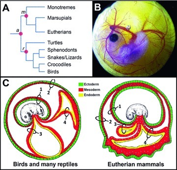
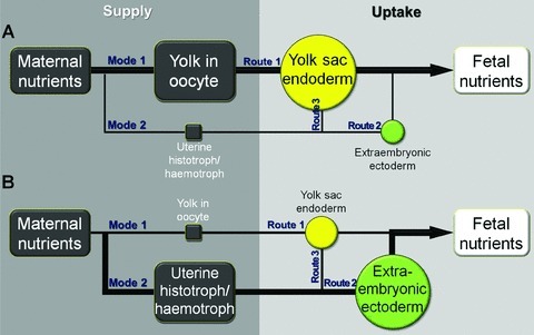
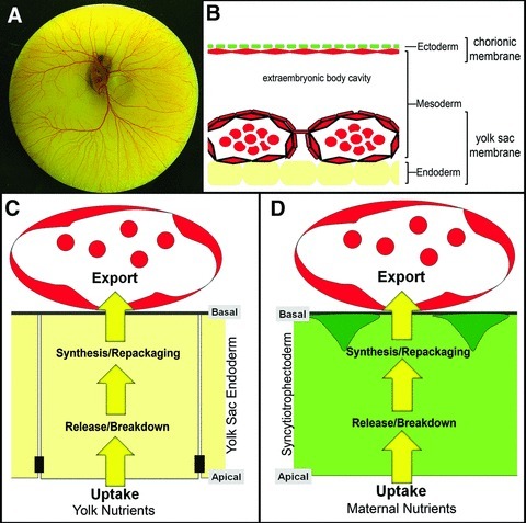
The transfer of nutrients from the mother through the chorioallantoic placenta meets the nutritional needs of the embryo during human prenatal development. Although all amniotes start with a similar "tool kit" of extraembryonic tissues, an enormous diversity of extraembryonic tissue formation has evolved to accommodate embryological and physiological constraints unique to their developmental programs. A comparative knowledge of these extraembryonic tissues and their role in nutrient uptake during development is required to fully appreciate the adaptive changes in placental mammals. Here, we offer a comparative embryological perspective and propose that there are three conserved nutrient transfer routes among the amniotes. We highlight the importance of the yolk sac endoderm, thought to be a vestigial remnant of our amniote lineage, in mediating nutrient uptake during early human development. We also draw attention to the similarity between yolk sac endoderm-mediated and trophectoderm-mediated nutrient uptake.


Figures

Figure 1
Figure 1
Extraembryonic tissues during amniote development. (A) A simplified phylogenetic tree of the extant amniotes. a, amniotes; m, mammals; r, reptiles (including birds). (B) A developing turtle (Pelodiscus sinensis) embryo, showing the embryo proper together with all associated extraembryonic tissues. (C) Two examples of how an amniote embryo organizes its extraembryonic tissues. Modified after Ferner and Mess, with permission from Elsevier. Left: schematic view of the organization in birds and many reptiles. Right: schematic view of the organization in eutherian mammals. 1, amnion; 2, chorion; 3, yolk sac; 4, allantois. Germ layers are color-coded (green, ectoderm; red, mesoderm; yellow, endoderm).
Figure 2
Figure 2
Variations in maternal nutrient supply and embryonic nutrient uptake. On the maternal side, nutrients are either deposited in the oocyte internally during its maturation (vitellogenesis) (mode 1) or provided to the developing embryo externally from the oviduct/uterus through either simple apposition of fetal and maternal tissues or complex placentation (collectively termed uterine histotroph/hemotroph here) (mode 2). Nutrients in the oocyte are taken up by the embryo through the yolk sac endoderm (route 1), and nutrients in the uterine histotroph/hemotroph are taken up by either the extraembryonic ectoderm (chorionic ectoderm or trophectoderm) (route 2) or the yolk sac endoderm (route 3). (A) In avian embryos, mode 1 and route 1 are dominant. (B) In eutherian embryos, mode 2 and route 2 are dominant. But in all amniote embryos, both maternal supply modes and all three nutrient uptake routes are operational, and the importance of route 3 transfer in mammalian embryos has been underappreciated.
Figure 3
Figure 3
Maternal-to-fetal nutrient transfer is an active and complex process. (A) A developing chicken embryo is overshadowed by the massive yolk sac and its associated yolk sac vasculature. (B) A schematic view of the germ-layer and cell-layer organization in the extraembryonic region of a developing chicken embryo. Ectoderm, green; mesoderm, red; endoderm, yellow. Yolk, located below the endoderm, is omitted. (C) The yolk sac endoderm is an epithelium with its apical side facing the yolk and its basal side facing the extraembryonic vasculature (smooth muscle cells omitted). The endoderm cells break down and modify lipids taken up from the yolk, break down yolk proteins, and synthesize de novo all major serum proteins and minor regulatory molecules to be released into circulation. (D) A schematic view of the extraembryonic ectoderm- (syncytiotrophectoderm-) mediated nutrient uptake in humans in comparison with the yolk sac endoderm-mediated nutrient uptake shown in C. Light green, syncytiotrophectoderm; dark green, cytotrophectoderm cells. Despite differences in germ-layer origin and nutrient composition, there are remarkable cellular and biochemical similarities between these two processes.

References

    1. Blair JE, Hedges SB. Molecular phylogeny and divergence times of deuterostome animals. Mol. Biol. Evol. 2005;22:2275–2284. - PubMed
    1. Selwood L, Johnson MH. Trophoblast and hypoblast in the monotreme, marsupial and eutherian mammal: evolution and origins. Bioessays. 2006;28:128–145. - PubMed
    1. Ferner K, Mess A. Evolution and development of fetal membranes and placentation in amniote vertebrates. Respir. Physiol. Neurobiol. 2011;178:39–50. - PubMed
    1. Hughes RL, Hall LS. Early development and embryology of the platypus. Philos. Trans R. Soc. Lond. B. Biol. Sci. 1998;353:1101–1114. - PMC - PubMed
    1. Enders AC, Carter AM. Comparative placentation: some interesting modifications for histotrophic nutrition—a review. Placenta. 2006;27(Suppl A):S11–S16. - PubMed