. 2012 Oct 25;490(7421):508-13.
doi: 10.1038/nature11558. Epub 2012 Oct 10.

Structure of the agonist-bound neurotensin receptor

Affiliations

Structure of the agonist-bound neurotensin receptor

Jim F White et al. Nature. .

Abstract

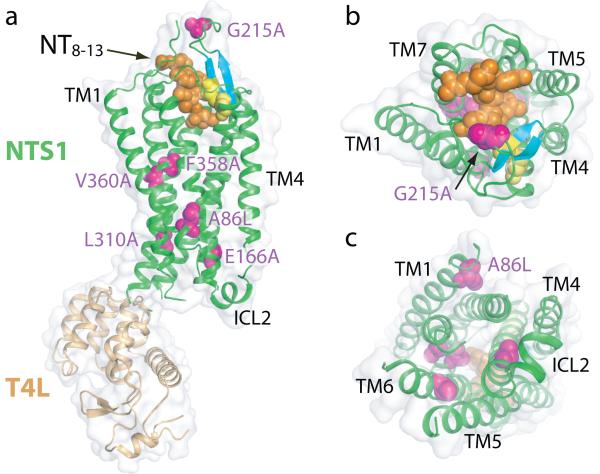
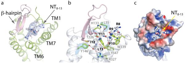
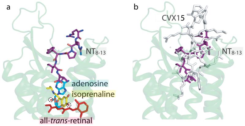
Neurotensin (NTS) is a 13-amino-acid peptide that functions as both a neurotransmitter and a hormone through the activation of the neurotensin receptor NTSR1, a G-protein-coupled receptor (GPCR). In the brain, NTS modulates the activity of dopaminergic systems, opioid-independent analgesia, and the inhibition of food intake; in the gut, NTS regulates a range of digestive processes. Here we present the structure at 2.8 Å resolution of Rattus norvegicus NTSR1 in an active-like state, bound to NTS(8-13), the carboxy-terminal portion of NTS responsible for agonist-induced activation of the receptor. The peptide agonist binds to NTSR1 in an extended conformation nearly perpendicular to the membrane plane, with the C terminus oriented towards the receptor core. Our findings provide, to our knowledge, the first insight into the binding mode of a peptide agonist to a GPCR and may support the development of non-peptide ligands that could be useful in the treatment of neurological disorders, cancer and obesity.


Figures

Fig. 1
Fig. 1. Overview of the NTS1 structure bound to the peptide agonist NT8-13
Cartoon representation of NTS1-GW5-T4L; (a) side view, (b) extracellular view, (c) intracellular view. Space filling models are used to depict the agonist NT8-13 (orange), the side chains of thermostabilizing mutations (purple) and the disulphide bond (yellow) between the conserved residues C142 and C225. Also shown are the β-hairpin in extracellular loop 2 (blue-green) and the π-helix in intracellular loop 2 (ICL2). T4L has been omitted from the intracellular view for clarity.
Fig. 2
Fig. 2. NTS1-GW5-T4L is in an active-like conformation
(a) Alignment of NTS1-GW5 (green) with the inactive state of the nociceptin receptor (NOP) (red, PDB code 4EA3). NTS1 was most similar to NOP (root mean squared deviation = 2.1 Å for Cα atoms in the TM domains) upon alignment of the TM domains of NTS1 with other peptide receptors-. (b) Alignment of the inactive state of rhodopsin (light brown, PDB code 1GZM), its active state Meta-II (ref.) (blue-green, PDB code 3PQR) and NTS1-GW5 (green). (c) Alignment of the inactive state of β2AR (pale mauve, PDB code 2RH1) and its active state (pale grey, PDB code 3SN6) and NTS1-GW5 (green). All views are from the intracellular surface. The arrows indicate the apparent displacement of TM5 and TM6 of NTS1-GW5 relative to the corresponding helix positions in the inactive receptor structures.
Fig. 3
Fig. 3. The conserved D/ERY and NPxxY motifs in NTS1-GW5
(a) Comparison of the side chain orientations of R3.50 and Y/N5.58 of NTS1-GW5 and the inactive nociceptin receptor (NOP) (PDB code 4EA3). NTS1-GW5 residues (R167, N257, labeled) in green; corresponding NOP residues (R148, Y235, unlabeled) in red. D1473.49 of NOP and A1663.49 of NTS1 are also shown. (b) In an inactive GPCR, R3.50 interacts with E3.49 as shown here for rhodopsin (pale brown, PDB code 1GZM). This salt bridge is broken upon activation allowing R3.50 to contact residue5.58 as depicted for active rhodopsin (ref.) (blue-green, PDB code 3PQR) (residues E134, R135 and Y223). (c) The same comparison for inactive β2AR (pale mauve, PDB code 2RH1) and active β2AR (pale grey, PDB code 3SN6) (residues E130, R131 and Y219). (d) Y3697.53 of NTS1 (green) occupies space as seen for Y3067.53 in active rhodopsin (blue-green, PDB code 3PQR). The orientation of Y3067.53 in the inactive rhodopsin (pale brown, PDB code 1GZM) and the inactive NOP (red, PDB code 4EA3) is shown for comparison.
Fig. 4
Fig. 4. The NTS1 agonist binding pocket
All views are from the extracellular side with NT8-13 shown as a stick model. (a) Cartoon representation of the ligand binding pocket with the ECL2 β-hairpin (pink). The quality of the electron density of NT8-13 is depicted as blue isosurface (2Fo-Fc map contoured at 1σ). (b) Key NTS1 residues (green residues with grey labels) in contact with the peptide ligand (grey residues with bold black labels). Hydrogen bonds and salt bridges are indicated by dashed lines (black). The electron density maps of selected NTS1 residues (L55, Y146, T226, R327) are shown as blue isosurface (2Fo-Fc map contoured at 1σ). (c) The charge complementarity between NT8-13 and its binding pocket is depicted with the NTS1 surface colored according to its electrostatic potential (−4 kT/e to +4 kT/e; red, negative; blue, positive). The positively charged arginine side chains of the ligand face the electronegative rim of the binding pocket whereas the negatively charged carboxylate of L13 resides in an electropositive environment. See also Supplementary Information.
Fig. 5
Fig. 5. A new paradigm for peptide agonist binding
Compared to small endogenous agonists, the NT8-13 binding cavity is located near the receptor surface. (a) The structures of the agonist-bound adenosine A2A receptor (PDB ID 2YDO), β1AR (PDB ID 2Y03) and meta-rhodopsin II (ref.) (PDB code 3PQR) were aligned in PyMOL. Only the cartoon representation for NTS1-GW5 is shown (pale green) with NT8-13 depicted in purple. The agonists adenosine (blue-green), isoprenaline (yellow; an isopropyl derivative of the endogenous ligand noradrenaline) and all-trans-retinal (red) are labeled. The chemical groups in adenosine and isoprenaline that make agonist-specific contacts to the receptor are circled (dashed line). (b) The cyclic antagonist peptide CVX15 binds to the CXCR4 receptor (grey, PDB ID 3OE0) in a similar fashion to NT8-13 (purple) in NTS1-GW5 (pale green cartoon).

Comment in

References

    1. Carraway R, Leeman SE. The isolation of a new hypotensive peptide, neurotensin, from bovine hypothalami. J. Biol. Chem. 1973;248:6854–6861. - PubMed
    1. Bissette G, Nemeroff CB, Loosen PT, Prange AJ, Jr., Lipton MA. Hypothermia and intolerance to cold induced by intracisternal administration of the hypothalamic peptide neurotensin. Nature. 1976;262:607–609. - PubMed
    1. Carraway RE, Plona AM. Involvement of neurotensin in cancer growth: evidence, mechanisms and development of diagnostic tools. Peptides. 2006;27:2445–2460. - PubMed
    1. Griebel G, Holsboer F. Neuropeptide receptor ligands as drugs for psychiatric diseases: the end of the beginning? Nat. Rev. Drug Discov. 2012;11:462–478. - PubMed
    1. Kitabgi P. Targeting neurotensin receptors with agonists and antagonists for therapeutic purposes. Curr. Opin. Drug Discov. Devel. 2002;5:764–776. - PubMed

Publication types

Associated data