. 2012 Nov 1;491(7422):66-71.
doi: 10.1038/nature11525. Epub 2012 Oct 10.

Generation of functional thyroid from embryonic stem cells

Affiliations

Generation of functional thyroid from embryonic stem cells

Francesco Antonica et al. Nature. .

Abstract

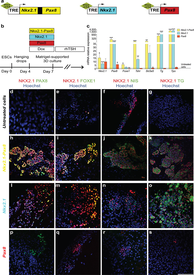
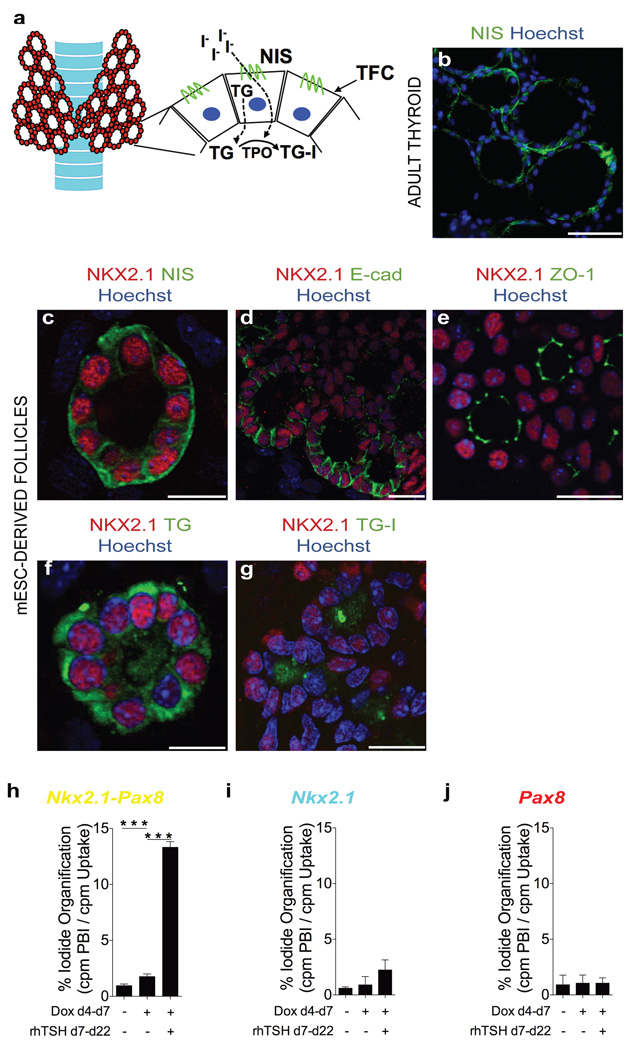
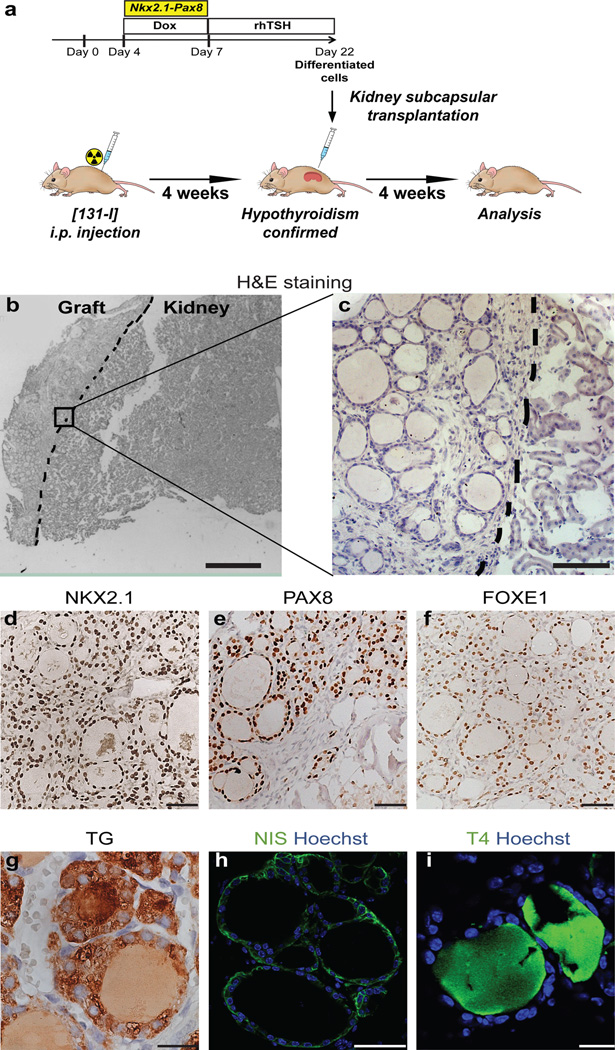
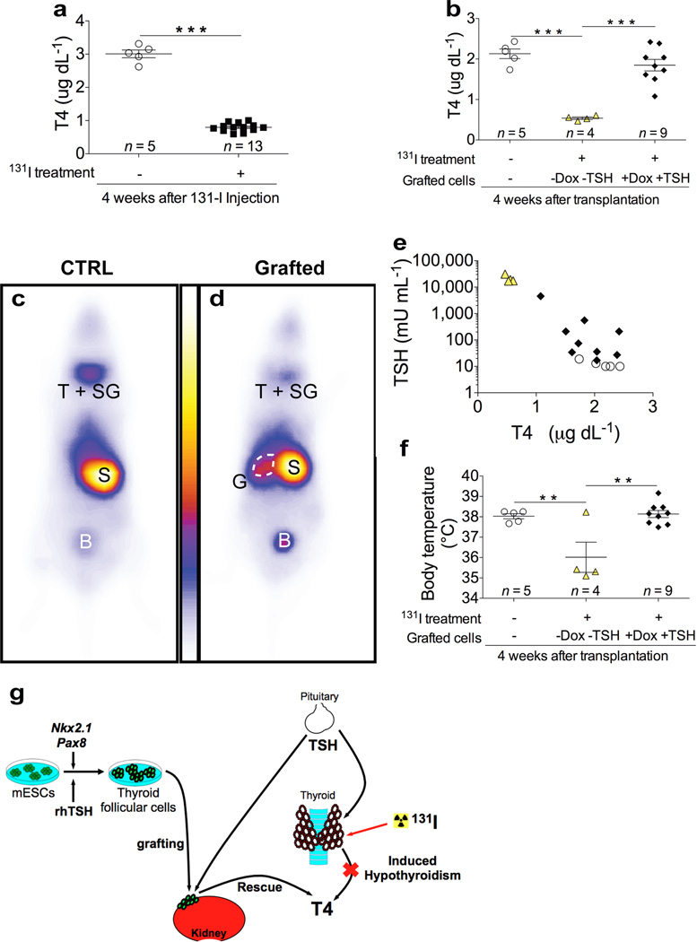
The primary function of the thyroid gland is to metabolize iodide by synthesizing thyroid hormones, which are critical regulators of growth, development and metabolism in almost all tissues. So far, research on thyroid morphogenesis has been missing an efficient stem-cell model system that allows for the in vitro recapitulation of the molecular and morphogenic events regulating thyroid follicular-cell differentiation and subsequent assembly into functional thyroid follicles. Here we report that a transient overexpression of the transcription factors NKX2-1 and PAX8 is sufficient to direct mouse embryonic stem-cell differentiation into thyroid follicular cells that organize into three-dimensional follicular structures when treated with thyrotropin. These in vitro-derived follicles showed appreciable iodide organification activity. Importantly, when grafted in vivo into athyroid mice, these follicles rescued thyroid hormone plasma levels and promoted subsequent symptomatic recovery. Thus, mouse embryonic stem cells can be induced to differentiate into thyroid follicular cells in vitro and generate functional thyroid tissue.


Figures

Figure 1
Figure 1. Ectopic expression of Nkx2.1 and Pax8 promotes the differentiation of mESCs into thyroid follicles
a, Schematic representation of tetracycline-inducible mESC lines. b, Schematic diagram of thyroid follicle differentiation protocol from mESCs. c, Expression of endogenous Nkx2.1 and Pax8, Foxe1, Tshr, Slc5a5, Tg and Tpo at day 22 in cells differentiated after Dox-mediated induction of Nkx2.1-Pax8 (yellow columns), Nkx2.1 (cyan columns) and Pax8 (red columns). Relative expression of each transcript is presented as fold change compared to untreated cells at day 22 as mean ± s.e.m. (n = 6). Unpaired t-test was used for statistical analysis. *P < 0.05, **P < 0.01, ***P < 0.001. d–s, Immunostaining at day 22 of untreated cells (d–g) and after Dox-mediated induction of Nkx2.1-Pax8 (h–k), Nkx2.1 (l–o) and Pax8 (p–s) for NKX2.1/PAX8 (d,h,l,p), NKX2.1/FOXE1 (e,i,m,q), NKX2.1/NIS (f,j,n,r) and NKX2.1/TG (g,k,o,s). Scale bar, 100 µm (d–s).
Figure 2
Figure 2. mESC-derived thyroid cells show full morphological and functional maturation
a, Schematic diagram of thyroid gland organised in follicles. b, Immunostaining of NIS in adult thyroid tissue. c–f, Immunofluorescence at day 22 of thyroid follicles derived from mESCs upon ectopic expression of Nkx2.1 and Pax8 for NKX2.1/NIS (c), NKX2.1/E-cad (d), NKX2.1/ZO-1 (e) and NKX2.1/TG (f). g, Immunodetection of iodinated TG (TG-I) in the luminal compartment of NKX2.1-positive follicles. h–j, Iodide organification assay in cells differentiated after Dox-induction of Nkx2.1-Pax8 (h), Nkx2.1 (i) and Pax8 (j). Histograms show the organification percentage of iodine-125 at day 22 in cells differentiated without Dox and rhTSH (left column), in presence of Dox only (central column) and upon Dox and rhTSH treatment (right column). Data are presented as mean ± s.e.m. (n = 3). Tukey’s Multiple Comparison Test was used for statistical analysis. ***P < 0.001. Scale bars, 200 µm (b) and 20 µm (c–g).
Figure 3
Figure 3. Grafting of mESC-derived thyroid follicles in mice
a, Schematic diagram of protocol for mESC-derived thyroid follicles transplantation in the renal capsule of mice with radio-ablated thyroid (hypothyroid mice). b–i, Histological analysis of kidneys sections 4 weeks after grafting. Hematoxylin and eosin staining (H&E) on OCT-embedded grafted kidney showed: - localization of transplanted tissue in the cortical area of the host organ (left side) (b) - and single cuboidal epithelium organization of transplanted tissue (c); Immunohistochemistry of NKX2.1 (d), PAX8 (e), FOXE1 (f), TG (g), and immunofluorescence of NIS (h) and T4 (i) in grafted tissue. Scale bars 300 µm (b), 100 µm (c), 50 µm (d,e,f,h) and 20 µm (g,i).
Figure 4
Figure 4. Rescue of experimentally induced hypothyroidism by transplanting mESC-derived thyroid follicles
a, Plasma total T4 level 4 weeks after injection in untreated (open circles) and iodine-131 treated (black squares) mice. b, Plasma total T4 level 4 weeks after transplantation of differentiated cells in iodine-131 treated mice. c–d, Whole-body images of mice 30 minutes after the injection of 99mTc-pertechnate. 4 weeks after grafting, body scan was performed on untreated control mice (CTRL) (c) or iodine-131 treated mice grafted with mESC-derived follicles (Grafted) (d); T+SG, thyroid and salivary glands; S, stomach; B, bladder; G, grafted mESC-derived follicles (G). e, Relationship between plasma TSH and T4 levels 4 weeks after grafting. f, Body temperature measurements 4 weeks after grafting. In b,e,f, open circles show iodine-131 untreated and ungrafted mice; yellow triangles show mice treated with iodine-131 and grafted with cells differentiated without Dox and rhTSH (-Dox-TSH); black diamonds show mice treated with iodine-131 and grafted with cells differentiated with Dox and rhTSH (+Dox+TSH). The values are shown as dot plot (a,b,f) or scatter plot (e) and as mean ± s.e.m.. Unpaired t-test (a) and Tukey’s Multiple Comparison Test (b,f) were used for statistical analysis. **P < 0.01, ***P < 0.001. g, Graphical summary: Nkx2.1 and Pax8 co-expression in combination with rhTSH treatment leads to differentiation of mESC into fully functional thyroid follicles that promote in vivo hormonal and symptomatic recovery of hypothyroid state.

Comment in

References

    1. De Felice M. Thyroid Development and Its Disorders: Genetics and Molecular Mechanisms. Endocrine Reviews. 2004;25:722–746. - PubMed
    1. De Felice M, Di Lauro R. Minireview: Intrinsic and Extrinsic Factors in Thyroid Gland Development: An Update. Endocrinology. 2011;152:2948–2956. - PubMed
    1. Mauchamp J, Mirrione A, Alquier C, Andre F. Follicle-like structure and polarized monolayer: role of the extracellular matrix on thyroid cell organization in primary culture. Biol Cell. 1998;90:369–380. - PubMed
    1. Nunez J, Pommier J. Formation of thyroid hormones. Vitam Horm. 1982;39:175–229. - PubMed
    1. Kimura S, et al. The T/ebp null mouse: thyroid-specific enhancer-binding protein is essential for the organogenesis of the thyroid, lung, ventral forebrain, and pituitary. Genes Dev. 1996;10:60–69. - PubMed

Publication types

MeSH terms