Review
. 2013 Jul;70(14):2425-41.
doi: 10.1007/s00018-012-1173-4. Epub 2012 Sep 28.

Stress-induced self-cannibalism: on the regulation of autophagy by endoplasmic reticulum stress

Affiliations
Review

Stress-induced self-cannibalism: on the regulation of autophagy by endoplasmic reticulum stress

Shane Deegan et al. Cell Mol Life Sci. 2013 Jul.

Abstract

Macroautophagy (autophagy) is a cellular catabolic process which can be described as a self-cannibalism. It serves as an essential protective response during conditions of endoplasmic reticulum (ER) stress through the bulk removal and degradation of unfolded proteins and damaged organelles; in particular, mitochondria (mitophagy) and ER (reticulophagy). Autophagy is genetically regulated and the autophagic machinery facilitates removal of damaged cell components and proteins; however, if the cell stress is acute or irreversible, cell death ensues. Despite these advances in the field, very little is known about how autophagy is initiated and how the autophagy machinery is transcriptionally regulated in response to ER stress. Some three dozen autophagy genes have been shown to be required for the correct assembly and function of the autophagic machinery; however; very little is known about how these genes are regulated by cellular stress. Here, we will review current knowledge regarding how ER stress and the unfolded protein response (UPR) induce autophagy, including description of the different autophagy-related genes which are regulated by the UPR.


Figures

Fig. 1
Fig. 1
The accumulation of unfolded protein in the ER lumen results in the dissociation of Grp78 from the three UPR sensors PERK, ATF6, and IRE1. Following Grp78 dissociation, PERK dimerizes and autophosphorylates, activating its cytosolic kinase domain. PERK phosphorylates EIF2α inhibiting general protein synthesis and facilitating/permitting non-canonical translation of ATF4 mRNA. Active PERK also phosphorylates NRF2 resulting in its dissociation from KEAP1, allowing NRF2 to translocate to the nucleus. Activation of ATF6 leads to its translocation to the Golgi where it is processed by site 1 and site 2 proteases (S1P and S2P) into an active transcription factor which results in the transcription of XBP1 mRNA. Activation of IRE1 results from its dimerization and autophosphorylation in a manner similar to PERK. IRE1 contains an endoribonuclease domain which processes unspliced XBP1 mRNA. Spliced XBP1 (XBP1s) mRNA is translated into an active transcription factor. IRE1 also possesses a kinase domain that recruits TRAF2 and ASK1 leading to the activation of JNK
Fig. 2
Fig. 2
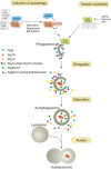
The autophagy pathway is divided into different phases; induction, vesicle nucleation, elongation, maturation, lysosomal fusion and degradation. Activation of the ULK1/2 complex requires mTORC1 inhibition and AMPK-mediated phosphorylation of ULK1. This complex is essential for the initial induction of the phagophore. The PI3K complex (see text and Fig. 3) is activated upon Bcl-2/Bcl-XL dissociation from Beclin’s BH3 domain. PI3K complex I is required for the induction and nucleation of the phagophore whereas PI3K complex II is involved in the expansion and curvature of the autophagosomal membrane (see text for details). The elongation phase of the autophagsome requires the conversion of LC3I to LC3II and the formation of the ATG12–ATG5–ATG16 complex. LC3II and ATG12–5–16 complex are required for substrate specificity and scaffolding roles on the autophagosome. Upon maturation of the autophagosome, ATG12–5–16 and the outer membrane bound LC3II are recycled back in the cytosol. The mature autophagosome fuses with a lysosome where it is degraded by resident cathepsins
Fig. 3
Fig. 3
The UPR can regulate autophagy at different stages in the process, induction, vesicle nucleation, and elongation and maturation. Left hand panel induction of autophagy by the UPR can occur through multiple pathways. Ca2+ release from the ER lumen via the inositol 1,4,5-trisphosphate receptor (IP3R) can activate calcium calmodulin kinase II (CaMKII). CaMKII can subsequently phosphorylate and activate AMP kinase (AMPK) which in turn phosphorylates and activates the tuberous sclerosis complex (TSC). TSC inhibits mTORC1 and subsequently relieves mTORC1 inhibition on the ULK1/2 complex. PERK activation results in the non-canonical translation of the transcription factor ATF4. ATF4 can transcriptionally upregulate REDD1 which results in the activation of TSC and subsequent inhibition of mTORC1. ATF4 can also transcriptionally upregulate another transcription factor known as CHOP. CHOP targets expression tribbles-related protein 3 (TRB3). TRB3 can directly inhibit Akt which relieves Akt’s inhibitory effects on TSC, resulting in mTORC1 inhibition and subsequent activation of ULK1/2 complex. CHOP also transcriptionally upregulates ERO1-α. ERO1-α has been shown to stimulate IP3R-mediated Ca2+ release from the ER lumen resulting in activation of CaMKII–AMPK–TSC arm leading to mTORC1 inhibition and subsequent activation of ULK1/2 complex. Middle panel the activation of the PI3K complex is an essential step for the induction, nucleation and curvature of the phagophore. Multiple players are involved in the activation of the PI3K complex in response to ER stress. DAPK1 remains in a phosphorylated inactive state under resting conditions. In response to ER stress DAPK1 is dephosphorylated resulting in the activation of its kinase domain. DAPK1 can phosphorylate Beclin’s BH3 domain preventing the inhibitory association of Bcl-2/Bcl-XL. The PERK–ATF4–CHOP arm can also promote the activation of the PI3K complex. CHOP has been reported to transcriptionally upregulate BH3-only proteins. BH3-only proteins can bind to Bcl-2/Bcl-XL and displace them from Beclin’s BH3 domain. IRE1-mediated activation of JNK can also result in activation of the PI3K complex. JNK has been shown to phosphorylate Bcl-2/Bcl-XL and inhibit their association with Beclin1. Right hand panel elongation and maturation of the phagophore requires two important processes to occur, the conversion of LC3I to LC3II and the formation of the ATG12–5–16 complex (see text for details). ATF4 can transcriptionally upregulate LC3 and ATG12, while CHOP can transcriptionally upregulate ATG5. The transcriptional upregulation of these three proteins is essential for the formation of the autophagosome

References

    1. McLaughlin M, Vandenbroeck K. The endoplasmic reticulum protein folding factory and its chaperones: new targets for drug discovery? Br J Pharmacol. 2011;162:328–345. - PMC - PubMed
    1. Gotoh T, Endo M, Oike Y (2011) Endoplasmic reticulum stress-related inflammation and cardiovascular diseases. Int J Inflam 2011, Article ID 259462 - PMC - PubMed
    1. Gorman AM, Healy SJ, Jager R, Samali A (2012) Stress management at the ER: regulators of ER stress-induced apoptosis. Pharmacol Ther. doi:10.1016/j.pharmthera.2012.02.003 - PubMed
    1. Cawley K, Deegan S, Samali A, Gupta S (2011) In: Michael Conn P (ed) Methods in enzymology, vol 490. Academic, London, p 31–51 - PubMed
    1. Szegezdi E, Logue SE, Gorman AM, Samali A. Mediators of endoplasmic reticulum stress-induced apoptosis. EMBO Rep. 2006;7:880–885. doi: 10.1038/sj.embor.7400779. - DOI - PMC - PubMed

Publication types

Substances

LinkOut - more resources