. 2012 Dec 15;18(24):6599-608.
doi: 10.1158/1078-0432.CCR-12-0838. Epub 2012 Oct 10.

Identification of KIF5B-RET and GOPC-ROS1 fusions in lung adenocarcinomas through a comprehensive mRNA-based screen for tyrosine kinase fusions

Affiliations

Identification of KIF5B-RET and GOPC-ROS1 fusions in lung adenocarcinomas through a comprehensive mRNA-based screen for tyrosine kinase fusions

Yoshiyuki Suehara et al. Clin Cancer Res. .

Abstract

Background: The mutually exclusive pattern of the major driver oncogenes in lung cancer suggests that other mutually exclusive oncogenes exist. We conducted a systematic search for tyrosine kinase fusions by screening all tyrosine kinases for aberrantly high RNA expression levels of the 3' kinase domain (KD) exons relative to more 5' exons.

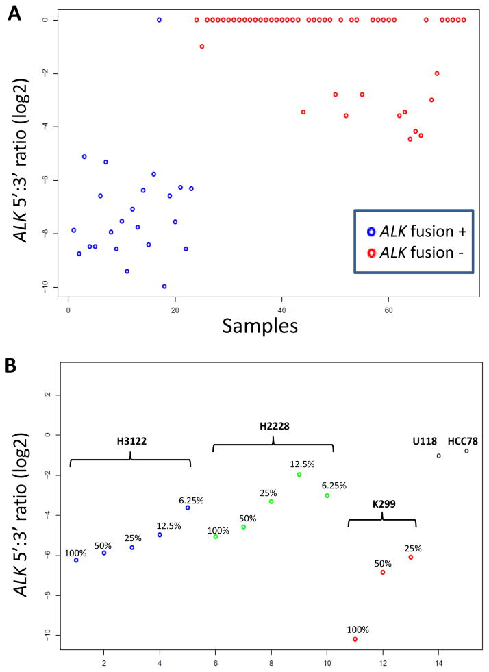
Methods: We studied 69 patients (including five never smokers and 64 current or former smokers) with lung adenocarcinoma negative for all major mutations in KRAS, EGFR, BRAF, MEK1, HER2, and for ALK fusions (termed "pan-negative"). A NanoString-based assay was designed to query the transcripts of 90 tyrosine kinases at two points: 5' to the KD and within the KD or 3' to it. Tumor RNAs were hybridized to the NanoString probes and analyzed for outlier 3' to 5' expression ratios. Presumed novel fusion events were studied by rapid amplification of cDNA ends (RACE) and confirmatory reverse transcriptase PCR (RT-PCR) and FISH.

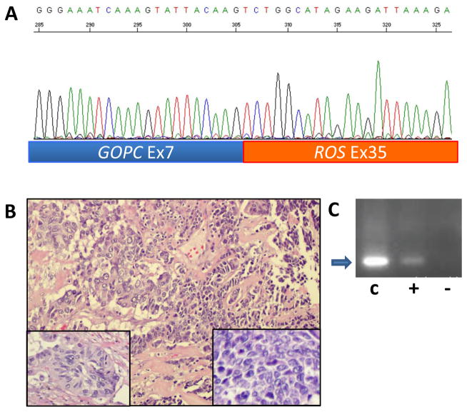
Results: We identified one case each of aberrant 3' to 5' ratios in ROS1 and RET. RACE isolated a GOPC-ROS1 (FIG-ROS1) fusion in the former and a KIF5B-RET fusion in the latter, both confirmed by RT-PCR. The RET rearrangement was also confirmed by FISH. The KIF5B-RET patient was one of only five never smokers in this cohort.

Conclusion: The KIF5B-RET fusion defines an additional subset of lung cancer with a potentially targetable driver oncogene enriched in never smokers with "pan-negative" lung adenocarcinomas. We also report in lung cancer the GOPC-ROS1 fusion originally discovered and characterized in a glioma cell line.


Conflict of interest statement

Potential conflicts of interest: none to declare

Figures

Figure 1
Figure 1. Overall strategy for identification of “pan-negative” tumors for discovery of novel tyrosine kinase fusions
Note that samples with PIK3CA mutations were not excluded from further analysis because of their known frequent overlap with other major driver mutations. See Materials and Methods for further details.
Figure 2
Figure 2. Principle of NanoString assay for evidence of tyrosine kinase fusions
As functional tyrosine kinase fusions invariably occur upstream of the exons encoding the kinase domain, probes were designed to measure the expression at two regions for each gene transcript, a 5′ probe pair located far upstream of the kinase domain exons, and the second located within those exons or further 3′. Kinase fusions often cause an imbalance in the RNA expression level of these two regions attributable to stronger activation of the promoter of the fusion partner gene and/or, in some cases, loss of the non-oncogenic, reciprocal fusion gene due to an unbalanced translocation event. The 3′/5′ expression ratios are calculated for each kinase gene in the assay and samples with outlier ratios were visualized on log scale plots.
Figure 3
Figure 3. NanoString assay validation
A. Detection of ALK fusions in lung adenocarcinoma samples using ALK 3′ to 5′ expression ratios. The lone discordant case had lower RNA quality and quantity compared to other samples. B. Serial dilutions of RNAs from cell lines with known ALK fusions. The U118 and HCC78 cell lines are shown as negative controls. Based on a cutoff of log2 ratio of −4, samples with at least 25% ALK-fusion positive tumor cell content should generally be detectable. e: exon.
Figure 4
Figure 4. NanoString Assay results for ROS1 and RET
A & B. For both genes, the locations of the 5′ and 3′ probes are shown schematically, with the kinase domain indicated in red. Samples with outlier negative 5′:3′ ratios for ROS1 and RET are indicated by the arrows. These samples were subjected to 5′RACE leading to the identification of GOPC-ROS1 and KIF5B-RET fusions, respectively. The U118 and HCC78 cell lines are included in the ROS1 plot as positive controls. Neg: pan-negative lung adenocarcinomas (see text); KRAS: KRAS-mutated lung adenocarcinomas (n=17); EGFR: EGFR-mutated lung adenocarcinomas (n=11); normal: non-neoplastic lung tissue (n=37).
Figure 5
Figure 5. Identification of GOPC-ROS1 fusion
A. Sequence of product of 5′RACE shows an in-frame fusion of GOPC exon 7 to ROS1 exon 35 in this cancer from a 68 year old white female with a 82 pack-year smoking history. B. Histology shows a combined small cell and adenocarcinoma. The adenocarcinoma component (left portion of field) is poorly differentiated with solid and acinar growth patterns (left inset). The small cell component of the tumor (right portion of field) is composed of organoid nests and trabeculae of densely packed cells with scant cytoplasm (right inset). The nuclei are spindle and angulated with finely granular chromatin and inconspicuous nucleoli (right inset). There is marked tumor necrosis and brisk mitotic activity in both components. C. RT-PCR confirmation of GOPC-ROS1 fusion. Lane C shows the positive control product (arrow) in the U188 cell line RNA. Lane “+” shows the same product in the tumor RNA. The RT-PCR product was also sequence-verified. Lane “−” is a negative control showing a lack of product in the tumor RNA when the RT is omitted.
Figure 6
Figure 6. Identification of KIF5B-RET fusion
A. Sequence of product of 5′RACE shows an in-frame fusion of KIF5B exon 15 to RET exon 12 in this cancer from a 60 year old never smoker white female. B. Histology shows lung adenocarcinoma with predominantly papillary and acinar growth patterns (left). Nuclei are pleomorphic and display prominent intranuclear pseudoinclusions (right). C. FISH analysis shows splitting of green (5′probe) and red (3′probe) signals of RET breakapart FISH assay. Inset: normal bone marrow cells showing fused or overlapping red and green signals only.

References

    1. Pao W, Iafrate AJ, Su Z. Genetically informed lung cancer medicine. J Pathol. 2011;223:230–40. - PubMed
    1. Pao W, Girard N. New driver mutations in non-small-cell lung cancer. Lancet Oncol. 2011;12:175–80. - PubMed
    1. Johnson BE, Kris MG, Kwiatkowski DJ, Iafrate AJ, Wistuba II, Shyr Y, et al. Clinical characteristics of planned 1000 patients with adenocarcinoma of lung (ACL) undergoing genomic characterization in the US Lung Cancer Mutation Consortium (LCMC) J Thorac Oncol. 2011;6:S346.
    1. Arcila ME, Lau C, Jhanwar SC, Zakowski MF, Kris MG, Ladanyi M. Comprehensive analysis for clinically relevant oncogenic driver mutations in 1131 consecutive lung adenocarcinomas. Mod Pathol. 2011;24s1:404A.
    1. Kris MG, Arcila M, Lau C, Rekhtman N, Brzostowski E, Pilloff M, et al. Two year results of LC-MAP: an institutional program to routinely profile tumor specimens for the presence of mutations in targetable pathways in all patients with non-small cell lung cancers. J Thorac Oncol. 2011;6:S347.

Publication types

MeSH terms