Clinical Trial
. 2012 Oct 10;4(155):155ra137.
doi: 10.1126/scitranslmed.3004373.

Neural stem cell engraftment and myelination in the human brain

Affiliations
Clinical Trial

Neural stem cell engraftment and myelination in the human brain

Nalin Gupta et al. Sci Transl Med. .

Erratum in

  • Sci Transl Med. 2012 Dec 19;4(165):165er8

Abstract

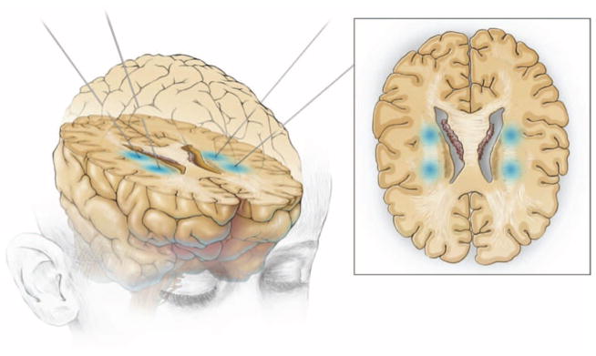
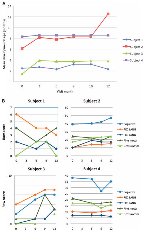
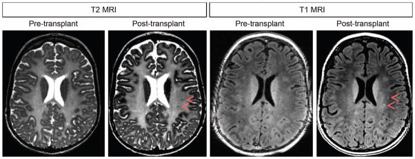
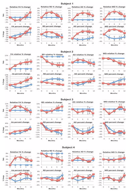
Pelizaeus-Merzbacher disease (PMD) is a rare leukodystrophy caused by mutation of the proteolipid protein 1 gene. Defective oligodendrocytes in PMD fail to myelinate axons, causing global neurological dysfunction. Human central nervous system stem cells (HuCNS-SCs) can develop into oligodendrocytes and confer structurally normal myelin when transplanted into a hypomyelinating mouse model. A 1-year, open-label phase-1 study was undertaken to evaluate safety and to detect evidence of myelin formation after HuCNS-SC transplantation. Allogeneic HuCNS-SCs were surgically implanted into the frontal lobe white matter in four male subjects with an early-onset severe form of PMD. Immunosuppression was administered for 9 months. Serial neurological evaluations, developmental assessments, and cranial magnetic resonance imaging (MRI) and MR spectroscopy, including high-angular resolution diffusion tensor imaging (DTI), were performed at baseline and after transplantation. The neurosurgical procedure, immunosuppression regimen, and HuCNS-SC transplantation were well tolerated. Modest gains in neurological function were observed in three of the four subjects. No clinical or radiological adverse effects were directly attributed to the donor cells. Reduced T1 and T2 relaxation times were observed in the regions of transplantation 9 months after the procedure in the three subjects. Normalized DTI showed increasing fractional anisotropy and reduced radial diffusivity, consistent with myelination, in the region of transplantation compared to control white matter regions remote to the transplant sites. These phase 1 findings indicate a favorable safety profile for HuCNS-SCs in subjects with PMD. The MRI results suggest durable cell engraftment and donor-derived myelin in the transplanted host white matter.


Conflict of interest statement

Competing interests: D.H.R. has nothing to disclose. S.L.H. is employed by the sponsor StemCells Inc., and other primary study personnel (N.G., R.G.H., J.S., S.-M.K., T.R., R.P., R.J.J., M.U., and A.J.B.) received partial salary support from the sponsor. HuCNS-SC for use in PMD is claimed by a host of U.S. patents issued to StemCells Inc.: U.S. patent nos. 5,968,829 (“Human CNS neural stem cells”); 7,361,505 (“Multipotent neural stem cell compositions”); 7,153,686 (“Compositions of enriched central nervous system stem cell and progenitor cell populations”); 6,777,233 (“Cultures of human CNS neural stem cells”); 5,851,832 (“In vitro growth and proliferation of multipotent neural stem cells and their progeny”); 6,497,872 (“Neural transplantation using proliferated multipotent neural stem cells and their progeny”); and 7,166,277 (“Remyelination of neurons using multipotent neural stem cell progeny”).

Figures

Fig. 1
Fig. 1
Sites of neural stem cell direct implantation. Under MRI guidance, HuCNS-SCs were transplanted into the deep white matter, the corona radiata, of the frontal white matter in two sites in each hemisphere of the brain of four PMD patients.
Fig. 2
Fig. 2
The CAS of child development and the Bayley-III. (A) Results for CAS showed overall stability of mean developmental age (in months) for all subjects, with the exception of subject 2, who demonstrated an overall gain from 6 to 12 months. (B) Bayley-III cognitive, language [receptive (REC) and expressive (EXP)], and motor (fine and gross) subtest raw scores at screening and 6, 9, and 12 months after transplant for all subjects. Small increases in select subtest scores were noted for subjects 2 and 4. Subjects 1 and 3 posttransplant scores, although variable, remained within the extreme lower end of the test range. Note difference in scale of the raw score axis used for each subject.
Fig. 3
Fig. 3
DTI measurements at control and implantation site ROIs in subject 2. T1-weighted MR images in orthogonal planes with boxes showing ROIs used for DTI analyses. ROIs adjacent to transplant sites are shown in green, and ROIs used to acquire control data are shown in aqua. Red arrowheads highlight the location of ROIs.
Fig. 4
Fig. 4
MRI images at baseline (pretransplant) and 12 months after transplant for subject 2. Small ovoid areas of T2 hypointensity and T1 hyperintensity (red arrowheads) were not present before transplantation and then gradually became visible after transplant. These areas do not represent small cysts because decreasing T2 hyperintensity is observed.
Fig. 5
Fig. 5
Absolute and relative diffusion metrics versus time. Zero is the starting point for each value on the preimplantation MRI study. Absolute values show percentage of change in each value at each imaging time point (red boxes for the implant site and blue diamonds for control sites). Relative values show control values as baseline (zero) and values at implant site as percentage of change from control. FA, fractional anisotropy; MD, mean diffusivity; RD, radial diffusivity; AD, axial diffusivity.

Comment in

References

    1. Gencic S, Abuelo D, Ambler M, Hudson LD. Pelizaeus-Merzbacher disease: An X-linked neurologic disorder of myelin metabolism with a novel mutation in the gene encoding proteolipid protein. Am J Hum Genet. 1989;45:435–442. - PMC - PubMed
    1. Schiffmann R, van der Knaap MS. Invited article: An MRI-based approach to the diagnosis of white matter disorders. Neurology. 2009;72:750–759. - PMC - PubMed
    1. Garbern JY. Pelizaeus-Merzbacher disease: Genetic and cellular pathogenesis. Cell Mol Life Sci. 2007;64:50–65. - PMC - PubMed
    1. Schneider A, Montague P, Griffiths I, Fanarraga M, Kennedy P, Brophy P, Nave KA. Uncoupling of hypomyelination and glial cell death by a mutation in the proteolipid protein gene. Nature. 1992;358:758–761. - PubMed
    1. Dhaunchak AS, Colman DR, Nave KA. Misalignment of PLP/DM20 transmembrane domains determines protein misfolding in Pelizaeus–Merzbacher disease. J Neurosci. 2011;31:14961–14971. - PMC - PubMed

Publication types