

Tau pathology in frontotemporal lobar degeneration with C9ORF72 hexanucleotide repeat expansion
- PMID: 23053135
- PMCID: PMC3551994
- DOI: 10.1007/s00401-012-1048-7
Tau pathology in frontotemporal lobar degeneration with C9ORF72 hexanucleotide repeat expansion
Abstract
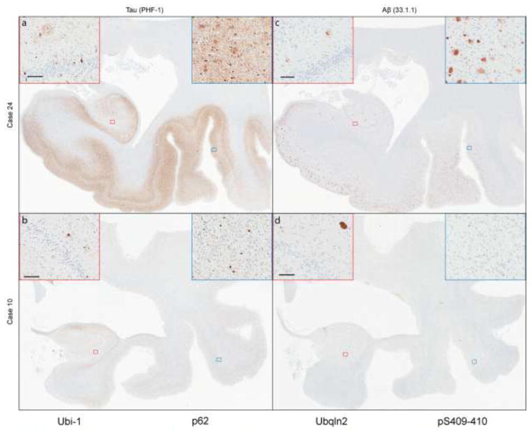
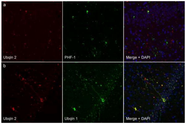
An expanded GGGGCC hexanucleotide repeat in C9ORF72 is the most common genetic cause of amyotrophic lateral sclerosis and frontotemporal lobar degeneration associated with TDP-43 pathology (FTLD-TDP). In addition to TDP-43-positive neuronal and glial inclusions, C9ORF72-linked FTLD-TDP has characteristic TDP-43-negative neuronal cytoplasmic and intranuclear inclusions as well as dystrophic neurites in the hippocampus and cerebellum. These lesions are immunopositive for ubiquitin and ubiquitin-binding proteins, such as sequestosome-1/p62 and ubiquilin-2. Studies examining the frequency of the C9ORF72 mutation in clinically probable Alzheimer's disease (AD) have found a small proportion of AD cases with the mutation. This prompted us to systematically explore the frequency of Alzheimer-type pathology in a series of 17 FTLD-TDP cases with mutations in C9ORF72 (FTLD-C9ORF72). We identified four cases with sufficient Alzheimer-type pathology to meet criteria for intermediate-to-high-likelihood AD. We compared AD pathology in the 17 FTLD-C9ORF72 to 13 cases of FTLD-TDP linked to mutations in the gene for progranulin (FTLD-GRN) and 36 cases of sporadic FTLD (sFTLD). FTLD-C9ORF72 cases had higher Braak neurofibrillary tangle stage than FTLD-GRN. Increased tau pathology in FTLD-C9ORF72 was assessed with thioflavin-S fluorescent microscopy-based neurofibrillary tangle counts and with image analysis of tau burden in temporal cortex and hippocampus. FTLD-C9ORF72 had significantly more neurofibrillary tangles and higher tau burden compared with FTLD-GRN. The differences were most marked in limbic regions. On the other hand, sFTLD and FTLD-C9ORF72 had a similar burden of tau pathology. These results suggest FTLD-C9ORF72 has increased propensity for tau pathology compared to FTLD-GRN, but not sFTLD. The accumulation of tau as well as lesions immunoreactive for ubiquitin and ubiquitin-binding proteins (p62 and ubiquilin-2) suggests that mutations in C9ORF72 may involve disrupted protein degradation that favors accumulation of multiple different proteins.
Figures
References
-
- Al-Sarraj S, King A, Troakes C, et al. p62 positive, TDP-43 negative, neuronal cytoplasmic and intranuclear inclusions in the cerebellum and hippocampus define the pathology of C9orf72-linked FTLD and MND/ALS. Acta Neuropathologica. 2011;122:691–702. - PubMed
-
- Arai T, Mackenzie IR, Hasegawa M, et al. Phosphorylated TDP-43 in Alzheimer’s disease and dementia with Lewy bodies. Acta Neuropathologica. 2009;117:125–136. - PubMed
-
- Babu JR, Geetha T, Wooten MW. Sequestosome 1/p62 shuttles polyubiquitinated tau for proteasomal degradation. J Neurochem. 2005;94:192–203. - PubMed
-
- Behrens MI, Mukherjee O, Tu PH, et al. Neuropathologic heterogeneity in HDDD1: a familial frontotemporal lobar degeneration with ubiquitin-positive inclusions and progranulin mutation. Alzheimer Dis Assoc Disord. 2007;21:1–7. - PubMed
Publication types
MeSH terms
Substances
Grants and funding
- P50 AG016574/AG/NIA NIH HHS/United States
- R01-AG37491/AG/NIA NIH HHS/United States
- P50-AG16574/AG/NIA NIH HHS/United States
- R01-AG26251/AG/NIA NIH HHS/United States
- R01 AG026251/AG/NIA NIH HHS/United States
- R01 NS080882/NS/NINDS NIH HHS/United States
- P50-NS72187/NS/NINDS NIH HHS/United States
- P50 NS072187/NS/NINDS NIH HHS/United States
- R01 NS065782/NS/NINDS NIH HHS/United States
- R01-NS65782/NS/NINDS NIH HHS/United States
- R01 AG037491/AG/NIA NIH HHS/United States
- P01 AG003949/AG/NIA NIH HHS/United States
- P01-AG03949/AG/NIA NIH HHS/United States
LinkOut - more resources
Full Text Sources
Other Literature Sources
Miscellaneous
