. 2012 Oct;404(6-7):1789-802.
doi: 10.1007/s00216-012-6246-6. Epub 2012 Jul 22.

The toxicity outcome of silica nanoparticles (Ludox®) is influenced by testing techniques and treatment modalities

Affiliations

The toxicity outcome of silica nanoparticles (Ludox®) is influenced by testing techniques and treatment modalities

Caterina Fede et al. Anal Bioanal Chem. 2012 Oct.

Abstract

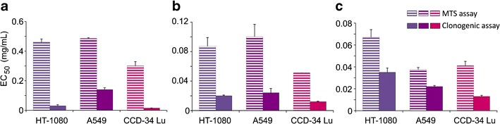
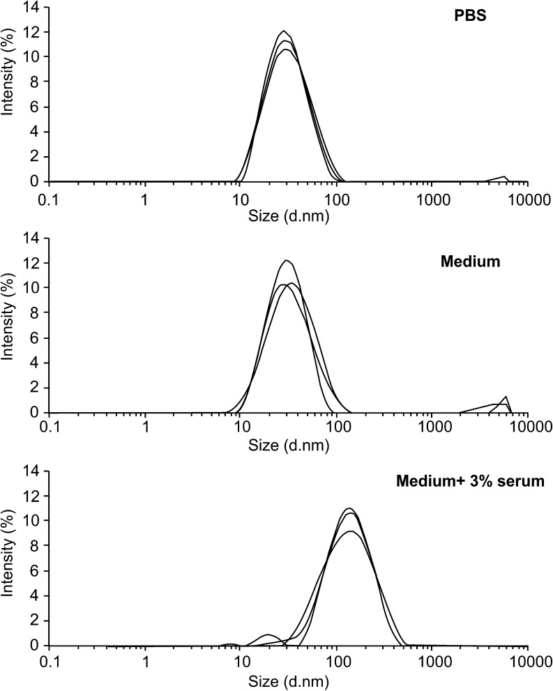
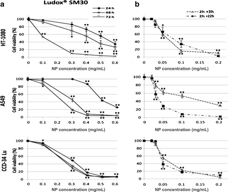
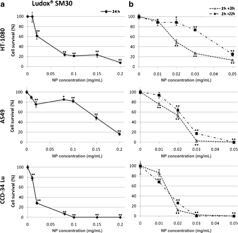
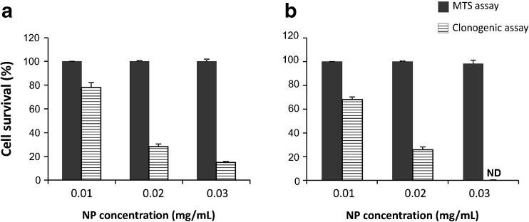
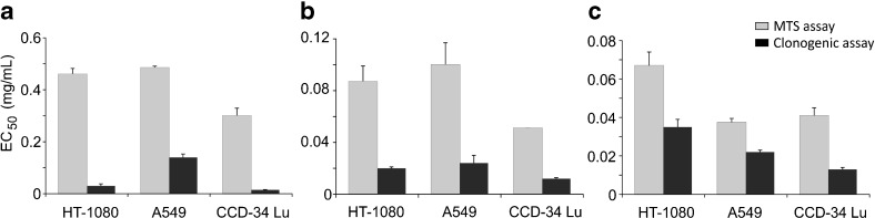
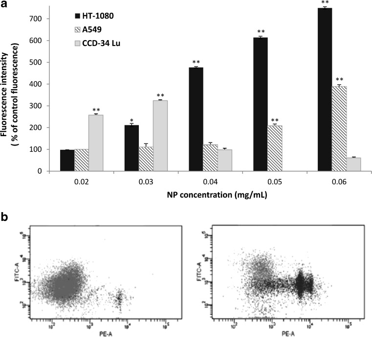
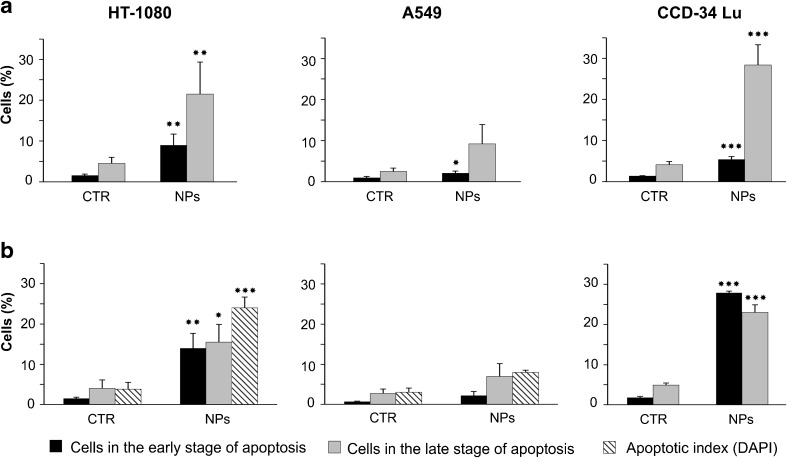
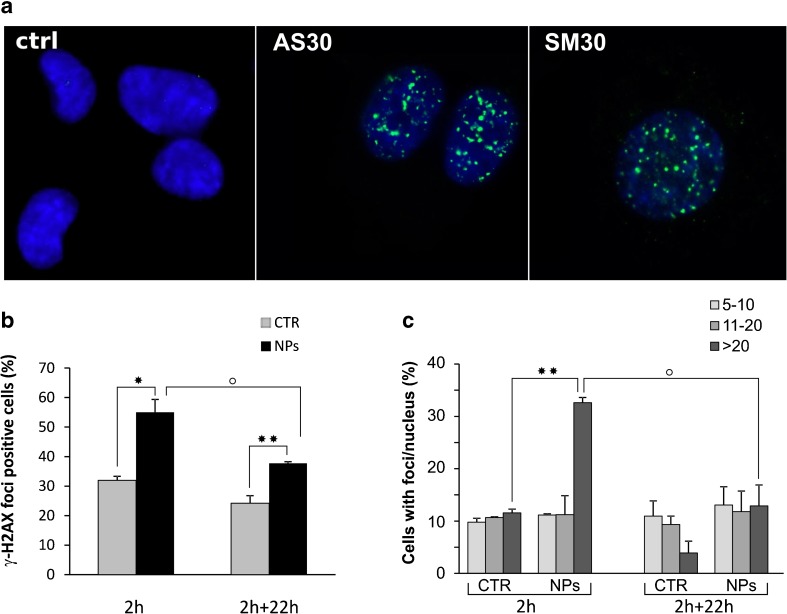
We analyzed the influence of the kind of cytotoxicity test and its application modality in defining the level of hazard of the in vitro exposures to nanostructures. We assessed the cytotoxicity induced by two different Ludox® silica nanoparticles (NPs), AS30 and SM30, on three human cell lines, CCD-34Lu, A549, and HT-1080. Dynamic light scattering measurements showed particle agglomeration when NPs are diluted in culture medium supplemented with fetal calf serum. We examined the impact of such particle aggregation on the cytotoxicity by exposing the cells to NPs under different treatment modalities: short incubation (2 h) in serum-free medium or long incubation (24-72 h) in serum-containing medium. Under this last modality, NP suspensions tended to form aggregates and were toxic at concentrations five- to tenfold higher than in serum-free medium. The results of cell survival varied considerably when the long-term clonogenic assay was performed to validate the data of the short-term MTS assay. Indeed, the half maximum effective concentrations (EC(50)) in all the three cell lines were four- to fivefold lower when calculated from the data of clonogenic assay than of MTS. Moreover, the mechanisms of NP toxicity were cell-type-specific, showing that CCD-34Lu are prone to the induction of plasma membrane damages and HT-1080 are prone to DNA double-strand break and apoptosis induction. Taken together, our results demonstrate that the choice of testing strategy and treatment conditions plays an important role in assessing the in vitro toxicity of NPs.


Figures

Figure
Figure
Fig. 1
Fig. 1
Dynamic light scattering (DLS) particle size distribution of Ludox® NPs SM30 (1 mg/mL) suspended in PBS, in culture medium, and in medium with 3 % of serum. The measures were performed for each sample after 2 h incubation at 37 °C
Fig. 2
Fig. 2
Cell viability measured by MTS assay in HT-1080, A549, and CCD-34 Lu cells treated with increasing concentrations of Ludox® NPs SM30 in medium with 3 % of serum (a) or without serum, followed by a recovery for 3 or 22 h in complete medium (10 % of serum) (b). The data represent mean ± SD (3 ≤ n ≤ 15). *p < 0.05, **p < 0.01 (t test; treated vs. control cells)
Fig. 3
Fig. 3
Cell survival measured by clonogenic assay in HT-1080, A549, and CCD-34Lu cells treated with increasing concentrations of Ludox® NPs SM30. Cell cloning was performed after a 24-h treatment with NPs in medium containing 3 % of serum (a), or after a 2 h treatment in serum-free medium followed by a recovery for 3 or 22 h in complete medium (b). The data represent mean ± SD (3 ≤ n ≤ 12). *p < 0.05, **p < 0.01 (t test; treated vs. control cells)
Fig. 4
Fig. 4
Cell survival of CCD-34 Lu assessed by MTS and clonogenic assays. The cells were incubated with Ludox® SM30 for 24 h in medium with 3 % of serum (a) or for 2 h in serum-free medium followed by a recovery of 22 h in complete medium (b). The data represent mean ± SD from four independent experiments. Cell survival determined by clonogenic assay was significantly lower than that determined by MTS for all the three tested doses (p < 0.001, t test, clonogenic vs MTS). ND = not detectable
Fig. 5
Fig. 5
Cytotoxicity of SM30 NPs expressed as half-maximum effective concentration (EC50 value in milligrams per milliliter), as assessed by MTS and clonogenic assays. a Treatment of 24 h in medium with 3 % of serum; b treatment of 2 h in serum-free medium, followed by recovery of 3 h in complete medium (serum 10 %) or of 22 h in complete medium (c). The data represent mean values of EC50± SD (3 ≤ n ≤ 15). In all treatment conditions and in all cell lines, the values of EC50 derived from clonogenic assay were significantly lower than those determined by MTS (p < 0.001, t test)
Fig. 6
Fig. 6
Reactive oxygen species (ROS) generation in cells treated with Ludox SM30 for 2 h in serum-free medium. a The mean florescence intensity of the ROS probe (carboxy-DCF) is expressed as percentage of control fluorescence. The data represent mean ± SE (n = 3). *p < 0.05, **p < 0.01 (t test; treated vs. control cells). b Dot plots obtained from representative experiment, showing propidium iodide (PE) versus carboxy-DCF fluorescence (FITC) in CCD-34Lu cells; left panel: untreated cells; right panel: cells treated with SM30 (0.03 mg/mL) for 2 h in serum-free medium
Fig. 7
Fig. 7
Apoptosis induction in cells treated with Ludox® SM30 (0.04 mg/mL) for 2 h in serum-free medium, followed by a recovery of 3 (a) or 22 h (b) in complete medium. After the recovery, the cells were double-stained with Annexin V–FITC/propidium iodide and analyzed by flow cytometry to detect cells in the early or in the late stage of apoptosis. Data represent means ± SD (n = 3). *p < 0.05, **p < 0.01, ***p < 0.001 (t test; treated vs. control cells). The presence of apoptotic bodies (apoptotic index) was checked by DAPI staining at the end of 2 + 22 h treatment (***p < 0.01, t test, treated vs. control cells)
Fig. 8
Fig. 8
Induction of DNA double-strand breaks in HT-1080 cells. a Immunofluorescence of γ-H2AX foci in HT-1080 treated with Ludox ® AS30 and SM30 NPs in serum-free medium. b Percentage of HT-1080 cells positive for γ-H2AX foci after 2 h incubation with 0.04 mg/mL of SM30 NPs in serum-free medium. Cells were fixed at the end of treatment (2 h) and after a recovery of 22 h in complete medium (2 + 22 h). c Positive cells for γ-H2AX foci were categorized on the basis of the number of foci/nucleus (5–10, 11–20, >20 foci). Data represent means ± SD (2 ≤ n ≤ 4). *p < 0.05, **p < 0.01, t test, treated versus control cells; ° p < 0.05, t test, (2 h) versus (2 + 22 h) treated cells

References

    1. Sanvincens N, Marco MP. Multifunctional nanoparticles—properties and prospects for their use in human medicine. Trends Biotechnol. 2008;26:425–433. doi: 10.1016/j.tibtech.2008.04.005. - DOI - PubMed
    1. Abu-Salah KM, Ansari AA, Alrokayan SA. DNA-based applications in nanobiotechnology. J Biomed Biotechnol. 2010;2010:715295. doi: 10.1155/2010/715295. - DOI - PMC - PubMed
    1. Karn B, Kuiken T, Otto M. Nanotechnology and in situ remediation: a review of the benefits and potential risks. Environ Health Perspect. 2009;117:1813–1831. - PMC - PubMed
    1. Liu D, Gu N. Nanomaterials for fresh-kipping and sterilization in food preservation. Recent Pat Food Nutr Agric. 2009;1:149–154. doi: 10.2174/1876142910901020149. - DOI - PubMed
    1. Santra S, Zhang P, Wang K, Tapec R, Tan W. Conjugation of biomolecules with luminophore-doped silica nanoparticles for photostable biomarkers. Anal Chem. 2001;73:4988–4993. doi: 10.1021/ac010406+. - DOI - PubMed

Publication types

Substances

LinkOut - more resources