. 2012 Dec 6;87(6):132.
doi: 10.1095/biolreprod.112.103366. Print 2012 Jun.

Microbial products alter the expression of membrane-associated mucin and antimicrobial peptides in a three-dimensional human endocervical epithelial cell model

Affiliations

Microbial products alter the expression of membrane-associated mucin and antimicrobial peptides in a three-dimensional human endocervical epithelial cell model

Andrea L Radtke et al. Biol Reprod. .

Abstract

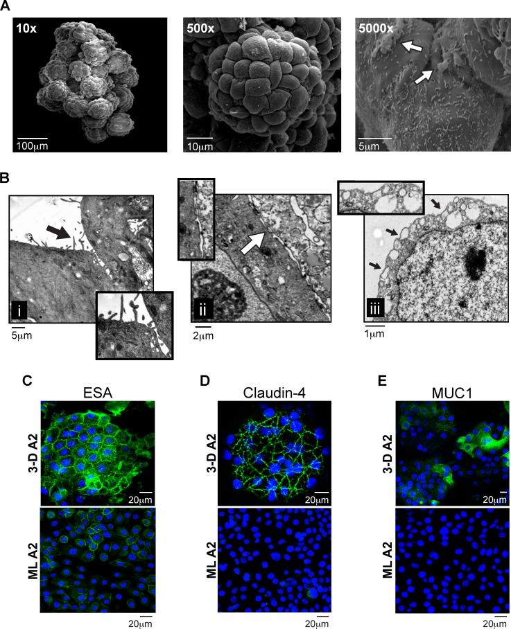
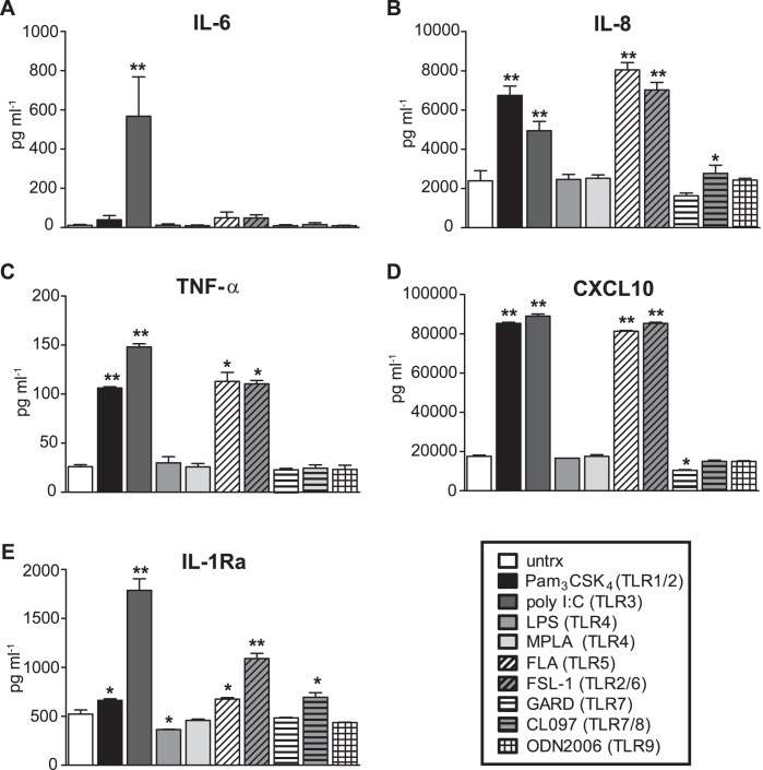
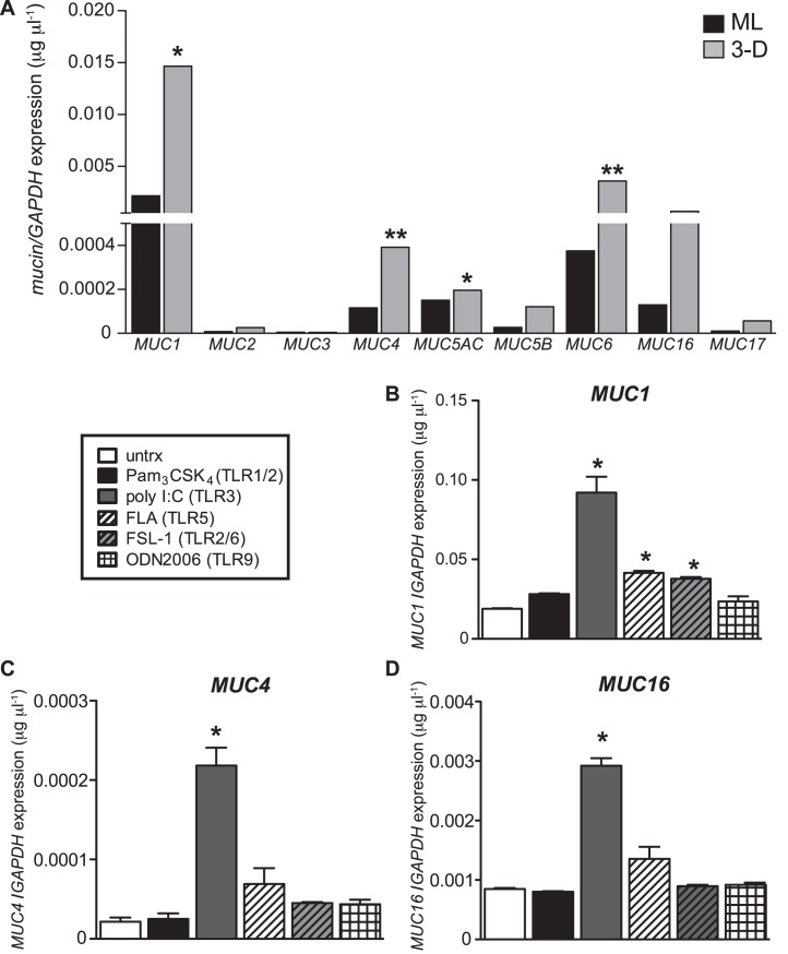
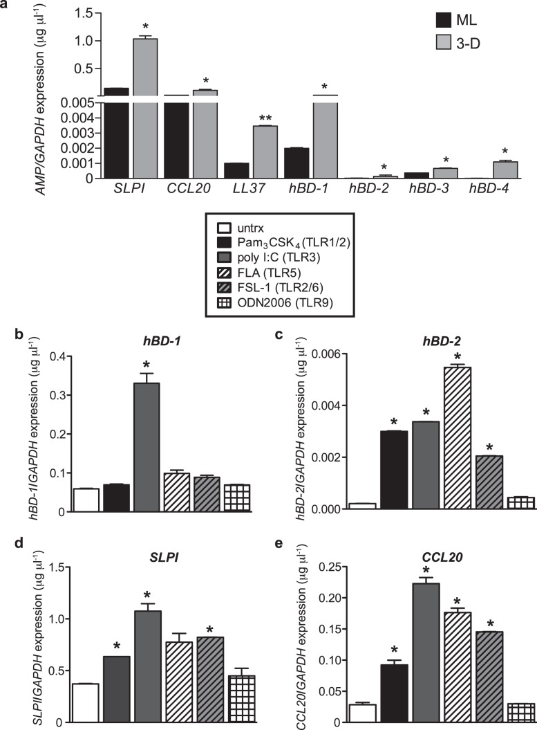
Our understanding of the mechanisms that regulate tissue-specific mucosal defense can be limited by the lack of appropriate human in vitro models. The endocervix lies between the microbe-rich vaginal cavity and the relatively sterile endometrium and is a major portal of entry for Chlamydia trachomatis, Neisseria gonorrhoeae, Mycoplasma genitalium, human immunodeficiency virus (HIV), and herpes simplex virus (HSV) infection in women. The endocervix is lined with a simple epithelium, and these cells produce mucus, which plays a key role in immune defense and reproduction. Here we describe the development of a human three-dimensional endocervical epithelial cell model generated by rotating wall vessel bioreactor technology. The model is composed of cellular aggregates that recapitulate major structural and barrier properties essential for the function and protection of the endocervix, including junctional complexes, microvilli, innate immune receptors, antimicrobial peptides, and mucins, the major structural component of mucus. Using this model, we also report, for the first time, that the membrane-associated mucin genes MUC1, MUC4, and MUC16 are differentially regulated in these aggregates by different bacterial and viral products. Differential induction of antimicrobial peptides was also observed with these products. Together these data define unique and flexible innate endocervical immune signatures that follow exposure to microbial products and that likely play a critical role in the outcome of pathogen challenge at this site.


Figures

FIG. 1
FIG. 1
Morphology and structural characteristics of 3-D endocervical EC. A) SEM images of a 3-D human endocervical EC aggregate at magnifications ×10, ×500, and ×5000. White arrow points to areas of mucus. B) TEM images of a 3-D endocervical EC aggregate. Insets highlight i) microvilli (black arrow), ii) tight junctional complexes (white arrow), and iii) secretory vesicles (black arrows). Confocal immunofluorescence microscopy images of 3-D endocervical EC (top panel) and confluent endocervical EC ML (bottom panel) were labeled with anti-ESA (C), anti-claudin-4 (D), and anti-MUC1 (E) antibodies and counterstained with DAPI to label nuclei.
FIG. 2
FIG. 2
Barrier capability of the tight junctional complexes in ML and 3-D endocervical EC. ML (gray lines) and 3-D (black) endocervical EC viability/active metabolism were determined by MTT assay following N-9 treatment for 4 h (solid lines) or 24 h (dashed lines). Points shown are representative mean ± SD values from triplicate samples from three independent experiments. Statistical comparisons were made between ML and 3-D EC at 4 and 24 h post-N-9 treatment for each concentration by using the Student t-test. **P < 0.01 represents (black) 24-h treatment (tx) and (gray) 4-h.
FIG. 3
FIG. 3
Cytokine production from 3-D endocervical EC following microbial product exposure. Cytokine levels produced by 3-D endocervical EC 24 h poststimulation with a customized panel of TLR agonists were compared to untreated (untrx) EC. IL-6 (A), IL-8 (B), TNF-α (C), CXCL10 (D), and IL-1Ra (E). Cytokine and chemokine values shown are representative mean ± SD levels from duplicate samples from three independent experiments. Statistical comparisons were made between TLR agonist treated EC versus untreated EC for each cytokine measured by using the Student t-test. *P < 0.05; **P < 0.01.
FIG. 4
FIG. 4
Mucin expression in 3-D endocervical EC after microbial product stimulation. A) ML and 3-D endocervical EC were profiled for mucins by qRT-PCR analysis. Statistical comparisons were made between ML and 3-D endocervical EC for each gene, using the Student t-test. 3-D endocervical EC MUC1 (B), MUC4 (C), and MUC16 (D) expression 24 h postexposure with TLR agonists. All gene expression was normalized to that of GAPDH. Expression levels shown are representative mean ± SD values from duplicate samples from at least three independent experiments. Statistical comparisons were made between untreated (untrx) and TLR agonist-treated EC by using the Student t-test. *P < 0.05; **P < 0.01.
FIG. 5
FIG. 5
Elevated expression of AMPs in 3-D endocervical EC after microbial product stimulation. A) ML and 3-D endocervical EC aggregates were profiled for mucins by qRT-PCR analysis. Statistical comparisons were made between ML and 3-D endocervical EC for each gene using the Student t-test. 3-D endocervical EC hBD-1 (B), hBD-2 (C), SLPI (D), and CCL20 (E) expression at 24 h postexposure with TLR agonists. All gene expression was normalized to that of GAPDH. Representative expression levels shown are the mean ± SD values from duplicate samples from at least three independent experiments. Statistical comparisons were made between untreated (untrx) and TLR agonist-treated EC by using the Student t-test. *P < 0.05; **P < 0.01.

References

    1. Hickey DK, Patel MV, Fahey JV, Wira CR. Innate and adaptive immunity at mucosal surfaces of the female reproductive tract: stratification and integration of immune protection against the transmission of sexually transmitted infections. J Reprod Immunol 2011; 88: 185 194. - PMC - PubMed
    1. Hafez ESE. Structural and ultrastructural parameters of the uterine cervix. Obstet Gynecol Surv 1982; 37: 507 516. - PubMed
    1. Ludwig H, Metzger H. The Human Female Reproductive Tract: A Scanning Electron Microscopy Atlas. New York: Springer-Verlag; 1979.
    1. Blaskewicz CD, Pudney J, Anderson DJ. Structure and function of intercellular junctions in human cervical and vaginal mucosal epithelia. Biol Reprod 2011; 85: 97 104. - PMC - PubMed
    1. Kaushic C, Nazli A, Ferreira VH, Kafka JK. Primary human epithelial cell culture system for studying interactions between female upper genital tract and sexually transmitted viruses, HSV-2 and HIV-1. Methods 2011; 55: 114 121. - PubMed

Publication types

MeSH terms