. 2012 Nov 15;189(10):4909-20.
doi: 10.4049/jimmunol.1201622. Epub 2012 Oct 10.

Development of a lipopolysaccharide-targeted peptide mimic vaccine against Q fever

Affiliations

Development of a lipopolysaccharide-targeted peptide mimic vaccine against Q fever

Ying Peng et al. J Immunol. .

Abstract

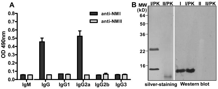
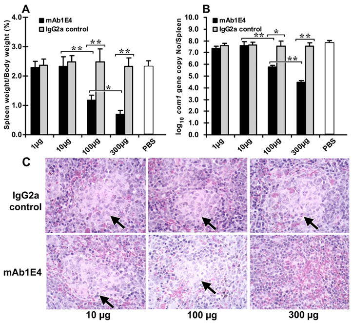
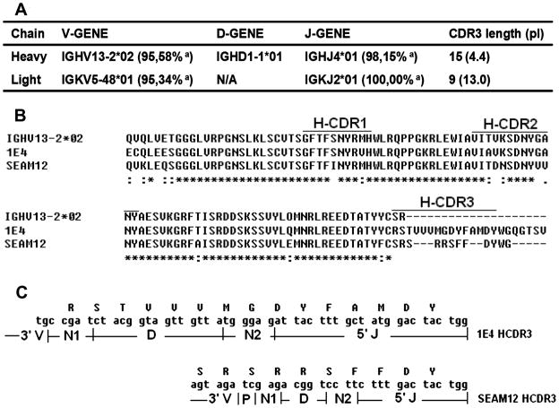
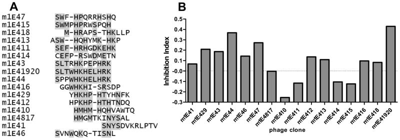
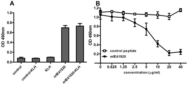
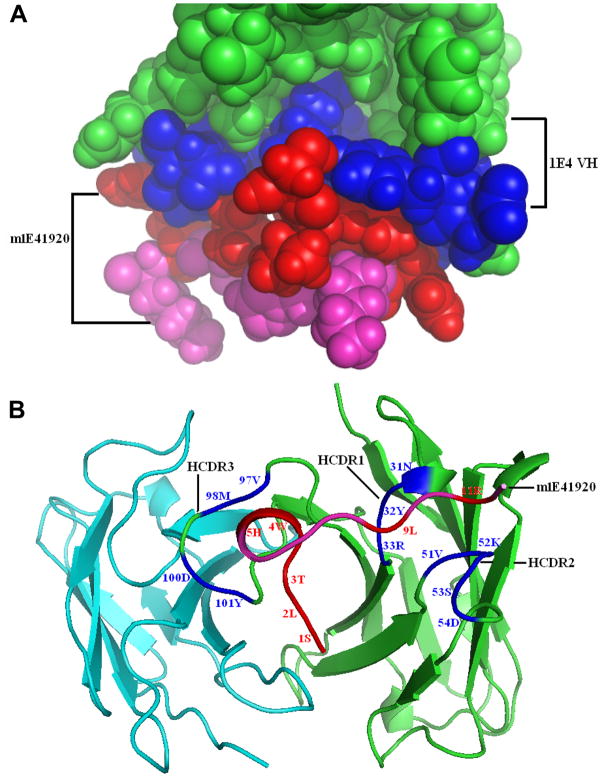
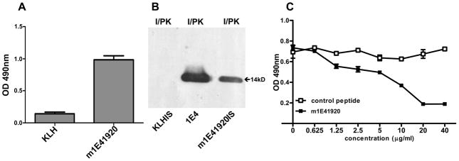
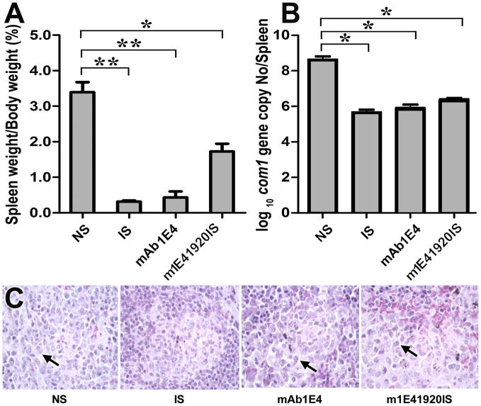
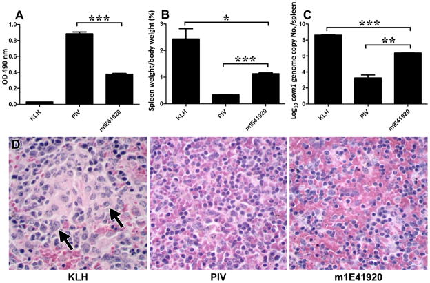
Coxiella burnetii is a Gram-negative bacterium that causes acute and chronic Q fever in humans. Creation of a safe and effective new generation vaccine to prevent Q fever remains an important public health goal. Previous studies suggested that Ab-mediated immunity to C. burnetii phase I LPS (PI-LPS) is protective. To identify the potential peptides that can mimic the protective epitopes on PI-LPS, a PI-LPS-specific mAb 1E4 was generated, characterized, and used to screen a phage display library. Interestingly, our results indicate that 1E4 was able to inhibit C. burnetii infection in vivo, suggesting that 1E4 is a protective mAb. After three rounds of biopanning by 1E4 from the phage display library, a mimetic peptide, m1E41920, was identified, chemically synthesized, and conjugated to keyhole limpet hemocyanin (KLH) for examining its immunogenicity. The results indicate that the synthetic peptide m1E41920 was able to inhibit the binding of 1E4 to PI Ag, suggesting m1E41920 shares the same binding site of 1E4 with the epitopes of PI Ag. In addition, m1E41920-KLH elicited a specific IgG response to PI Ag, and immune sera from m1E41920-KLH-immunized mice was able to inhibit C. burnetii infection in vivo, suggesting that m1E41920 may specifically mimic the protective epitope of PI-LPS. Furthermore, m1E41920-KLH was able to confer significant protection against C. burnetii challenge. Thus, m1E41920-KLH is a protective Ag and may be useful for developing a safe and effective vaccine against Q fever. This study demonstrates the feasibility of developing a peptide mimic vaccine against Q fever.


Figures

Figure 1
Figure 1
Characterization of 1E4 by ELISA and Western blotting. Panel A, isotyping of 1E4 by ELISA with PI antigen. A 96-well microtiter plate was coated with inactivated PI or PII antigen and incubated with the hybridoma supernatant from cloned hybridoma 1E4. The reactivity of 1E4 with anti-IgM, -IgG, -IgG1, -IgG2a -IgG2b and -IgG3 was measured by OD 490. The values represent the average absorbance at 490 nm of duplicate wells. Panel B, the reactivity of 1E4 with PI and PII antigens, and proteinase K digested PI and PII antigens in Western blotting. I: PI antigen; I/PK, proteinase K-treated PI antigen; II: PII antigen; II/PK, proteinase K-treated PII antigen.
Figure 2
Figure 2
Evaluation of the ability of 1E4 to inhibit C. burnetii infection in vivo. The inhibition of C. burnetii was performed by incubation of 1×10⁷ virulent C. burnetii NMI with 1, 10, 100 and 300 μg of purified 1E4, or mouse IgG2a isotype control at 4 °C overnight. Six week-old BALB/c mice were infected by i.p. injection with 1×10⁷ of 1E4 or control IgG2a-treated C. burnetii NMI. Mice infected with 1×10⁷ of PBS-treated C. burnetii NMI were used as negative controls. Splenomegaly and bacterial burden in the spleen were measured at 14 days post infection and used as indicators to evaluate the ability of 1E4 to inhibit C. burnetii infection in BALB/c mice. Panel A, splenomegaly was measured by spleen weight as a percentage of body weight. Panel B, bacterial burden in the spleen was determined by real time-PCR and reported as log10 of C. burnetii com1 gene copy numbers. Panel C, Pathological changes in the spleen at 14 days post challenge. The data presented in each group is the average with standard deviation of four mice. *, P<0.05; **, P<0.01.
Figure 3
Figure 3
Analysis of VH and VL gene usage and CDR sequence of 1E4. Panel A, analysis of 1E4 VH and VL gene usage by IMGT/V-QUEST. a: identity to germline genes. Panel B, comparison of amino acid sequence of 1E4 VH gene with the anti-Neisseria meningitidis group B polysaccharide mAb SEAM12 and murine germline family IGHV13-2 by ClustalW program. A star (*) indicates the same amino acid residue identity in a position, while a dot (:) indicates a different residue, and a dash (-) indicates inserted spaces placed in the sequence to provide maximum identity. Panel C, comparison of the HCDR3 conjunction between 1E4 and SEAM12. N: nucleotide addition mutation; P: P deletion mutation.
Figure 4
Figure 4
Identification of 1E4-specific phage clones. Panel A, amino acid sequence alignment of nineteen 1E4 recognized phagotopes. The consensus residues are shadowed. Panel B, the reactivity of recombinant phage clones with 1E4 was analyzed by ELISA. A 96-well microtiter plate was coated with individual 1010 of purified phage particles and incubated with 5μg/ml of 1E4 with or without 2μg/ml of PI antigen. The Inhibition of 1E4 binding by PI antigen was measured by the inhibition index {(A490 without inhibitor-A490 with inhibitor)/A490 without inhibitor}.
Figure 5
Figure 5
Evaluation of the binding ability of the synthetic mimetic peptide m1E41920 and m1E41920-KLH conjugate with 1E4 by ELISA and competitive ELISA. Panel A, binding activity of m1E41920 and m1E41920-KLH conjugate with 1E4 was measured by ELISA. A 96-well microtiter plate was coated with m1E41920, m1E41920-KLH, control peptide, control-KLH or KLH and incubated with 5μg/ml of 1E4. The values represent the average absorbance at 490 nm of duplicate wells. Panel B, competitive inhibition ELISA analysis of the ability of synthetic peptide m1E41920 to inhibit 1E4 binding with PI antigen. A 96-well microtiter plate was coated with inactivated PI antigen and incubated with 5μg/ml of 1E4 mixed with different concentrations of m1E41920 or control peptide. Points plotted represent the average absorbance at 490 nm of duplicate wells.
Figure 6
Figure 6
Models of 1E4-m1E41920 complex established by the molecular modeling and docking procedures. Panel A, the images were generated using Pymol and the 1E4-m1E41920 interaction model was showed as a spacefill graph. The directly contacted m1E41920 and 1E4 VH residues in direct contact were identified and displayed as red and blue spacefill graph, respectively. The contacted residues of the 1E4 VH paratopes in complex with m1E41920 are shown in blue and the remainders are shown in green. The peptide mimic m1E41920 fits well into the binding site of the parent mAb 1E4 VH chain. Panel B, the model docking structure of the 1E4-m1E41920 complex was displayed as a cartoon graph. The contacted residues and amino acid abbreviations are shown in the corresponding color described in panel A. The peptide mimic m1E41920 fits into the heavy chain groove with the middle four residue domain (WHKH) in a helix turn whereas it directly contacts HCDR1, HCDR2 and HCDR3 in an extended conformation through the N terminal domain SLTWH and C terminal domain -L-R-, respectively.
Figure 7
Figure 7
Analysis of m1E41920-KLH elicited antibody response to PI antigen. Panel A, m1E41920-KLH elicited IgG response to PI antigen. Immune sera from m1E41920-KLH-immunized mice were tested for IgG response to PI antigen by ELISA. A 96-well microtiter plate was coated with inactivated PI antigen and incubated with immune sera from KLH-or m1E41920-KLH-immunized mice at 1:400 dilutions. The reactivity of immune sera from KLH- or m1E41920-KLH-immunized mice with anti-IgG was measured by OD 490. The values represent the average absorbance at 490 nm of duplicate wells. Panel B, Western blotting analysis of the reactivity of 1E4 and immune sera from KLH- or m1E41920-KLH-immunized mice with proteinase K digested PI antigens. Panel C, evaluation of the binding ability of immune sera from m1E41920-KLH-immunized mice with PI antigen by the competitive inhibition ELISA. A 96-well microtiter plate was coated with inactivated PI antigen and incubated with 1:400 diluted pooled immune sera from m1E41920-KLH-immunized mice mixed with different concentrations of synthetic peptide m1E41920 or control peptide. Points plotted represent the average absorbance at 490 nm of duplicate wells.
Figure 8
Figure 8
Evaluation of the ability of immune sera from m1E41920-KLH-immunized mice to inhibit C. burnetii infection in BALB/c mice. The inhibition of C. burnetii was performed by incubation of 1×10⁷ virulent C. burnetii with 30 μl of normal mouse sera or immune sera from m1E41920-KLH-immunized mice at 4 °C overnight. In addition, 1×10⁷ virulent C. burnetii NMI was treated with 30 μl of immune sera from PIV-vaccinated BALB/c mice or 300 μg of purified 1E4 in the same manner and used as positive controls. Six week-old BALB/c mice were infected by i.p. injection with 1×10⁷ of normal mouse sera, immune sera and 1E4-treated C. burnetii, respectively. Splenomegaly and bacterial burden in the spleen were measured at 14 days post infection and used as indicators to evaluate the ability of immune sera from m1E41920-KLH-immunized mice to inhibit C. burnetii infection in BALB/c mice with negative and positive controls. Panel A, splenomegaly was measured by spleen weight as percentage of body weight. Panel B, bacterial burden in the spleen was determined by real time-PCR and reported as log10 of C. burnetii com1 gene copy numbers. Panel C, Pathological changes in the spleen at 14 days post challenge. The data presented in each group is the average with standard deviation of four mice. *, P<0.05; **, P<0.01.
Figure 9
Figure 9
Evaluation of the protective efficacy of m1E41920-KLH against C. burnetii infection in BALB/c mice. Panel A, m1E41920-KLH induced specific IgG response to PI antigen as measured by ELISA. A 96-well microtiter plate was coated with inactivated PI antigen and incubated with immune sera from KLH-, PIV- or m1E41920-KLH-immunized mice at 1:400 dilutions. The reactivity of immune sera with anti-IgG was measured by OD 490. The values represent the average absorbance at 490 nm of duplicate wells. Panel B, splenomegaly was measured by spleen weight as percentage of body weight. Panel C, bacterial burden in the spleen was determined by real time-PCR and reported as log10 of C. burnetii com1 gene copy numbers. Panel D, Pathological changes in the spleen at 14 days post challenge. The data presented in each group is the average with standard deviation of four mice. *, P<0.05; **, P<0.01; ***, P<0.001.

Similar articles

Cited by

References

    1. Angelakis E, Raoult D. Q Fever. Vet Microbiol. 2010;140:297–309. - PubMed
    1. Bell JF, Lackman DB, Meis A, Hadlow WJ. Recurrent Reaction of Site of Q Fever Vaccination in a Sensitized Person. Mil Med. 1964;129:591–5. - PubMed
    1. Marmion BP, Ormsbee RA, Kyrkou M, et al. Vaccine prophylaxis of abattoir-associated Q fever. Lancet. 1984;2:1411–4. - PubMed
    1. Ackland JR, Worswick DA, Marmion BP. Vaccine prophylaxis of Q fever. A follow-up study of the efficacy of Q-Vax (CSL) 1985–1990. Med J Aust. 1994;160:704–8. - PubMed
    1. Stoker MG, Fiset P. Phase variation of the Nine Mile and other strains of Rickettsia burnetii. Can J Microbiol. 1956;2:310–21. - PubMed

Publication types

MeSH terms