Comparative Study
. 2013 Jan;18(1):70-5.
doi: 10.1007/s00776-012-0312-y. Epub 2012 Sep 28.

Ultrasonographic observation of the healing process in the gap after a Ponseti-type Achilles tenotomy for idiopathic congenital clubfoot at two-year follow-up

Affiliations
Comparative Study

Ultrasonographic observation of the healing process in the gap after a Ponseti-type Achilles tenotomy for idiopathic congenital clubfoot at two-year follow-up

Hisateru Niki et al. J Orthop Sci. 2013 Jan.

Abstract

Background: Ponseti management usually requires Achilles tenotomy during the final stage of serial casting. However, we lack a good understanding of the sequential tendon healing process after tenotomy in the Ponseti bracing protocol. The purpose of this study was to clarify the ultrasonographic process of tendon healing in the gap for up to two years after Ponseti-type Achilles tenotomy in patients with clubfeet.

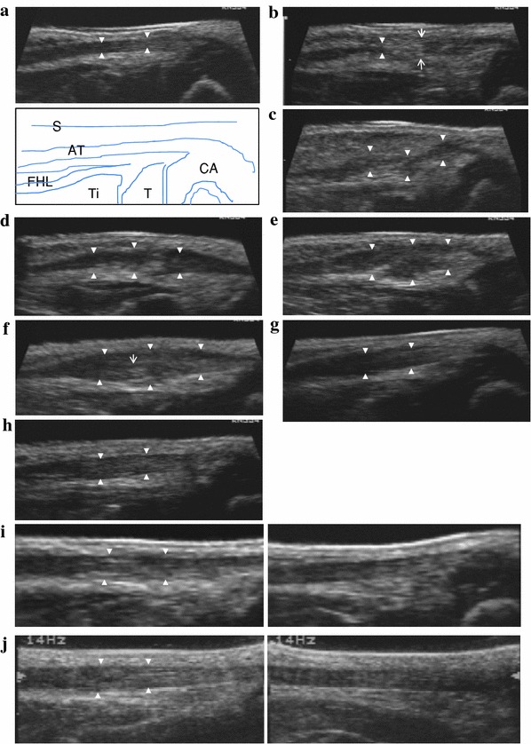
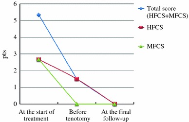
Methods: We conducted an ultrasonographic study to clarify the sequential changes in gap healing for up to two years after tenotomy. The subjects were 23 patients with 33 clubfeet. Achilles tenotomy was performed at mean 10.4 (8-16) weeks after birth. Dynamic and static ultrasonography was performed before tenotomy and at 1, 2, 3, 4, 6, 8, and 12 weeks as well as at 4, 6, 12, 18, and 24 months after tenotomy.

Results: Continuity and gliding were noted within four weeks. The united portion continued to thicken for up to three months after tenotomy. Starting from the fourth month, the healed portion began to lose its thickness, and this process continued into the sixth month. At one year, the thickness of the tendon did not differ much from that of the tendon on the opposing foot. In cases where patients had clubfoot on both feet and underwent simultaneous tenotomies, measurement of the tendons could not be accurately compared. At two years after tenotomy, slight irregularity of the internal structure persisted when compared with the unaffected foot. In addition, clinical and X-ray findings were evaluated simultaneously, and no recurrence was confirmed.

Conclusions: To our knowledge, our results are the first to describe the process of gap healing in the tendon after tenotomy up to and beyond two years, as recommended in the Ponseti bracing protocol. Level of evidence IV.


Figures

Fig. 1
Fig. 1
a Before tenotomy. Case 1 at two and a half months after birth. The Achilles tendon is clearly depicted as a low-echo region (solid white triangles). AT Achilles tendon, CA calcaneus, T talus, Ti tibia, FHL flexor hallucis longus, S skin. b 1–2 weeks after tenotomy. Case 1 at three months after birth and at 14 days after tenotomy. Solid white triangles show the proximal edge. White arrows show tenotomy portion. The gap zone is of mixed echogenicity. c 2–3 weeks after tenotomy. Case 1 at three months and one week after birth and at 21 days after tenotomy. Solid white triangles show that the lesion continued to be visible as a low echo. The gap is filled with irregular hypoechoic tissue. d 3–4 weeks after tenotomy. Case 1 at three and a half months after birth and at 28 days after tenotomy. Continuity of the tendon is identified. A bulbous appearance of the gap after tenotomy can be seen. e 4–6 weeks after tenotomy. Case 1 at three months and three weeks after birth and at 35 days after tenotomy. The united portion (solid white triangles) appears swollen. f 8–12 weeks after tenotomy. Case 1 at five months after birth and at 70 days after tenotomy. The united portion (solid white triangles) continues to thicken up to 8–12 weeks after tenotomy. The fibers are seen to display a linear configuration (narrow white arrow). g 14–16 weeks after tenotomy. Case 1 at six and a half months after birth and at 112 days after tenotomy. The healed portion (solid white triangles) begins to decrease in size. h Six months after tenotomy. Case 1 at eight and a half months after birth and at 169 days after tenotomy. The decease in size continued into the sixth month. Homogeneous tendon fibers are observed within the gap (white solid triangles). i One year after tenotomy. Case 1 at one year and two months after birth and at 358 days after tenotomy. Left shows the affected side and right the nonaffected side in the same case. The thickness does not differ much from that on the nonaffected side. The arrangement of tendon fibers within the gap is not similar at all to that on the normal side. j Two years after tenotomy. Case 1 at two years and three months after birth and at 719 days after tenotomy. Left shows the affected side and right the nonaffected side in the same case. Slight irregularity in the internal structure persists when compared with the nonaffected side
Fig. 2
Fig. 2
Modified Catterall/Pirani scores. HFCS hindfoot contracture, MFCS midfoot contracture. Total score (HFCS + MFCS): 0–6 pts, HFCS: 0–3 pts, MFCS: 0–3pts. Most abnormal: 6 pts, normal: 0 pts

References

    1. Herzenberg JE, Radler C, Bor N. Ponseti versus traditional methods of casting for idiopathic clubfoot. J Pediatr Orthop. 2002;22:517–521. - PubMed
    1. Ponseti IV. Congenital clubfoot: fundamentals of treatment. New York: Oxford University Press; 1996. p. 140.
    1. Colburn M, Williams M. Evaluation of the treatment of idiopathic clubfoot by using the Ponseti method. J Foot Ankle Surg. 2003;42:259–267. doi: 10.1016/S1067-2516(03)00312-0. - DOI - PubMed
    1. Scher DM, Feldman DS, van Bosse HJ, Sala DA, Lehman WB. Predicting the need for tenotomy in the Ponseti method for correction of clubfoot. J Pediatr Orthop. 2004;24:349–352. doi: 10.1097/01241398-200407000-00001. - DOI - PubMed
    1. Morcuende JA, Abbasi D, Dolan LA, Ponseti IV. Results of an accelerated Ponseti protocol for clubfoot. J Pediatr Orthop. 2005;25:623–626. doi: 10.1097/01.bpo.0000162015.44865.5e. - DOI - PubMed

Publication types