. 2013 Jan;18(1):57-76.
doi: 10.1007/s10495-012-0762-x.

In vivo knockdown of intersectin-1s alters endothelial cell phenotype and causes microvascular remodeling in the mouse lungs

Affiliations

In vivo knockdown of intersectin-1s alters endothelial cell phenotype and causes microvascular remodeling in the mouse lungs

Cristina Bardita et al. Apoptosis. 2013 Jan.

Abstract

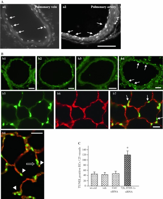
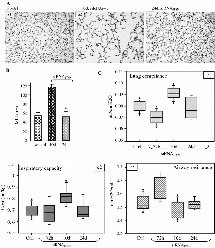
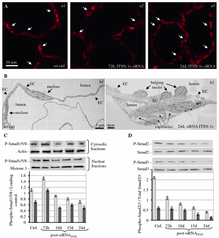
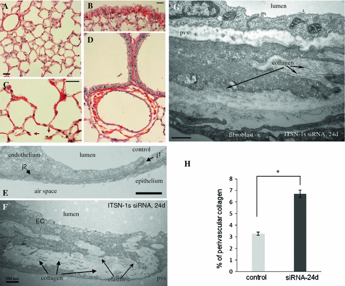
Intersectin-1s (ITSN-1s) is a general endocytic protein involved in regulating lung vascular permeability and endothelial cells (ECs) survival, via MEK/Erk1/2(MAPK) signaling. To investigate the in vivo effects of ITSN-1s deficiency and the resulting ECs apoptosis on pulmonary vasculature and lung homeostasis, we used an ITSN-1s knocked-down (KD(ITSN)) mouse generated by repeated delivery of a specific siRNA targeting ITSN-1 gene (siRNA(ITSN)). Biochemical and histological analyses as well as electron microscopy (EM) revealed that acute KD(ITSN) [3-days (3d) post-siRNA(ITSN) treatment] inhibited Erk1/2(MAPK) pro-survival signaling, causing significant ECs apoptosis and lung injury; at 10d of KD(ITSN), caspase-3 activation was at peak, terminal deoxynucleotidyl transferase dUTP nick-end labeling (TUNEL)-positive ECs showed 3.4-fold increase, the mean linear intercept (MLI) showed 48 % augment and pulmonary microvessel density as revealed by aquaporin-1 staining (AQP-1) decreased by 30 %, all compared to controls; pulmonary function was altered. Concomitantly, expression of several growth factors known to activate Erk1/2(MAPK) and suppress Bad pro-apoptotic activity increased. KD(ITSN) altered Smads activity, downstream of the transforming growth factor beta-receptor-1 (TβR1), as shown by subcellular fractionation and immunoblot analyses. Moreover, 24d post-siRNA(ITSN), surviving ECs became hyper-proliferative and apoptotic-resistant against ITSN-1s deficiency, as demonstrated by EM imaging, 5-bromo-deoxyuridine (BrdU) incorporation and Bad-Ser(112/155) phosphorylation, respectively, leading to increased microvessel density and repair of the injured lungs, as well as matrix deposition. In sum, ECs endocytic dysfunction and apoptotic death caused by KD(ITSN) contribute to the initial lung injury and microvascular loss, followed by endothelial phenotypic changes and microvascular remodeling in the remaining murine pulmonary microvascular bed.


Figures

Fig. 1
Fig. 1
Acute inhibition (72 h post-siRNAITSN) of ITSN-1s expression induces apoptosis in mouse lungs. A, B TUNEL demonstrates an increase in apoptotic ECs death in the lungs of mice with acute ITSN-1s inhibition (a1, a2, b4), by reference to controls, wild-type mice (b1), vehicle-injected mice (b2) or non-specific siRNA-treated mice (b3). TUNEL-positive ECs (arrows) were detected in large vessels—segments from a pulmonary vein (a1) and a pulmonary artery (a2) are shown, as well as in medium-sized (50–100 μm diameter) vessels (b4). Bars 30 μm (a1, a2), 20 μm (b1, b2). Lung sections of KDITSN mice (3d) were stained with TUNEL (b5) and CD31 Ab (b6) to evaluate the presence of apoptotic ECs within the microvessels of the alveolar septum (b7). Arrows in b7, indicate TUNEL-positive alveolar epithelial cells. The tips of damaged septal walls display TUNEL-positive ECs nuclei (b8, arrowheads) and alveolar epithelial cells nuclei (b8, open arrow) Bars 20 μm (b3–b7); 10 μm (b8). C Quantification of TUNEL-positive ECs in medium-sized vessels (50–100 μm) of controls and siRNAITSN-treated mice lungs; *P < 0.05. Results are representative for three different experiments, with 3–4 mice/experimental condition
Fig. 2
Fig. 2
Acute inhibition (72 h post-siRNAITSN) of ITSN-1s expression causes lung microvessel loss. A Micrographs of AQP-1staining of lung sections from controls (a1) and KDITSN mice, 3d, (a2), show decreased number of AQP-1 labeled microvessels (diameter or transverse diameter, respectively <9 μm) in siRNAITSN relative to control specimens. Arrows indicate microvessel profiles whose boundaries are labeled by AQP-1 Ab. Isotype-matched IgG staining of mouse lung sections of wt- (a3) and KDITSN mice (a4) produced no ECs staining. The NovaRed substrate produced a light-reddish background. Bars 10 μm (a1, a2), 15 μm (a3, a4). Results are representative for three different experiments, with 3–4 mice/experimental condition
Fig. 3
Fig. 3
KDITSN induces lung injury. A Representative histo-pathology of mouse lungs from wild-type control (a1), vehicle (a2), siRNActrl (a3), siRNAITSN (1 single dose) treated mice, at 72 h (a4) and 240 h (a5) post-siRNAITSN treatment. H&E staining of paraffin-embedded tissue shows expanded airway spaces (a4, asterisks) in mouse lungs with acute inhibition of ITSN-1s. Bar 60 μm. B MLI of control and siRNAITSN-treated mice. All results are representative for at least 3 independent experiments; 30 random power fields were counted from each time point, (n = 3 mice/group; *P < 0.05). C EM morphological analysis of KDITSN mouse lungs, 72 h post-siRNAITSN. Fragments of ECs of wild-type mice show normal mitochondria (m), (a1, arrows), flattened Golgi cisternae, situated in close apposition as stacks (circled area), caveolar profiles open to the lumen or apparently free in the cytosol, (inset a.1.1) and a healthy nucleus (a2). ECs of KDITSN show increased number of mitochondrial units (m; a3, a5), swollen Golgi apparatus (a3, circled area) and chromatin condensation (a4). Fragment of ECs showing numerous finger-like projections (a5, arrowheads). Note also the enlarged perivascular space (pvs) and the thick alveolar septum in the KDITSN by comparison to control (inset, c5.1). Bars 100 nm (c1.1); 200 nm (a1–a5, c5.1). Results are representative for 3 different experiments, with 3 mice/experimental condition
Fig. 4
Fig. 4
Chronic inhibition of ITSN-1s in mouse lung endothelium causes a peak in caspases-3 activation and significant ECs death at 10d of KDITSN. A Western blot analyses of lung lysates (70 μg proteins/lane) of siRNAITSN and control mice show augmented immunoreactivity for cleaved caspases-3 at 10d of KDITSN, while total caspase-3 expression is unchanged. b Densitometric analysis of representative HyBlot CL films indicates more than 3-fold increase in cleaved caspase-3 immunoreactivity at 10d of KDITSN, by reference to untreated mice. Densitometric values ± SEM are representative for three independent experiments. c Lung sections of KDITSN mice (10d) were stained with TUNEL (c1) and CD31 Ab/AlexaFluor594 (c2) to evaluate the presence of apoptotic ECs within the microvessels of the alveolar wall. The merged image revealed numerous apoptotic ECs (c3, arrows) as well as epithelial alveolar cells (c3, open arrows). Bar 20 μm. d Quantification of TUNEL-positive ECs in medium-sized vessels (50–100 μm) of controls and siRNAITSN-treated mice lungs; P < 0.05 for all time points, relative to controls. Results are representative for 3 different experiments, with 3–4 mice/experimental condition
Fig. 5
Fig. 5
Mouse lung chronically depleted of ITSN-1s show phenotypically-altered ECs. A BrdU/FITC Ab immunostaining (a1, a4, a5), followed by CD31/AlexaFluor594 (a2, a6, a7) shows BrdU positive ECs within alveolar wall, at 10d (a3, arrows) and 24d (a8, a9, arrows). Arrowheads (a8, a9) point towards BrdU positive alveolar epithelial cells. Bars: 20 μm, (a1–a9). B Representative Western blots of lung lysates of control, wt-mice (a), siRNActrl (b) and veh (c), 72 h post-injection as well as siRNAITSN-treated mice, at the time points indicated, using phospho-specific Erk1/2MAPK and total Erk1/2MAPK Abs. The graph shows densitometric analysis of Erk1/2MAPK activation (phospho-Erk1/2 relative to total Erk1/2) as mean ± SEM of 3 separate experiments. C Expression TGFβ, BMP-2/4 and VEGF-A, in mouse lung lysates of control and siRNAITSN-treated mice, at the time points indicated assessed by Western blot with specific anti-TGFβ, BMP-2/4 and VEGF-A Abs. Actin was used as loading control. D. Densitometric analysis of TGF-β, BMP-2/4 and VEGF-A immunoreactivity. Different Abs and detection conditions did not allow a quantitative assessment of the ratio between the growth factors. E. Evaluation of Bad phosphorylation by Western blotting with anti-phospho Ser112-Bad, Ser136-Bad and Ser155-Bad Abs. The blots were stripped and reprobed with an anti-Bad Ab. The Western blots C, E are representative for 3–4 different experiments; densitometric analyses B, D were applied on 3 different HyBlot CL films. Densitometric values ± SEM are representative for 3 independent experiments
Fig. 6
Fig. 6
KDITSN-1s induces mouse lung injury and alters pulmonary function tests. A Histology (H&E) of untreated (a1), siRNAITSN, 10d, (a2) and siRNAITSN, 24d, (a3) Bar 20 μm. B Morphometric analyses of MLI show no difference between the mice with chronic siRNAITSN (24d) and controls. 30 random high power fields were counted for each group. Data are shown as mean values ± SEM. *P < 0.05 (siRNAITSN 24d compared to siRNAITSN, 10d). C Pulmonary function tests—CL, IC and airway RL—measurements in KDITSN mouse. Lines within the boxes show medians; bounds of the boxes show 25th and the 75th percentiles of the data, respectively; the dark circles show outliers. All data are presented as mean ± SEM; P < 0.001. Results are representative for three different experiments, with 3–4 mice/experimental condition
Fig. 7
Fig. 7
Chronic KDITSN-1s in mouse lungs induces microvascular remodeling A. Micrographs of GS-1 lectin staining of paraffin-embedded sections show microvessel profiles (arrows), within the alveolar walls in control (a1), in mice treated with siRNAITSN for 3d (a2) and in mice treated with siRNAITSN for 24d (a3). Bar 10 μm. B Ultrastructural features of microvascular remodeling in KDITSN mouse lungs. Two vessels profiles in wt-mouse lung display elongated ECs nuclei (b1). Note the relatively uniform thickness of the ECs throughout the vessel perimeter. Segment of a mid-sized vessel in KDITSN mouse lung shows a distorted endothelium and several nuclei protruding into the lumen (b2, arrows). New pulmonary microvessels (dashed arrows) with narrow openings are abundant and located in very close proximity to each other. C Smad1/5/8 phosphorylation in mouse lung cytosolic (light grey bars) and nuclear fractions (dark grey bars) of controls and siRNAITSN-treated mice. Nuclei were isolated from mouse lungs as in [69] and extracts were prepared using NE-PER Nuclear Extraction Reagent (Pierce Biotechnology, Rockford IL). Actin and histone 3 were used as loading controls for the cytosolic and nuclear fractions respectively. D Smad2 (light grey bars) and Smad3 (dark grey bars) phosphorylation in total lung lysates of control and siRNAITSN-treated mice. Total Smad2/3 were used as loading controls. Results are representative for 3–5 different experiments
Fig. 8
Fig. 8
Chronic ITSN-1s deficiency leads to dense perivascular collagen depositions Representative micrographs of Picrosirius Red-stained lung sections of control (A) and siRNAITSN-treated mice for 24d (B–D), to assess collagen deposition. Short septal walls remnants were often noticed with collagen bundles at the tip (C, arrows). Bars 20 μm. Electron micrographs show segments of the alveolar septal wall in a control microvessel (E), as well as in a postcapillary venule (10–15 μm diameter) (F), and a precapillary arteriole (20–25 μm diameter) (G) of ITSN-1s deficient mice. Collagen fibrils (G, arrows), and fibrilar material (proteoglicans, elastic fibers, etc.), accumulated in the basement membrane between the ECs, smc and fibroblast. j1—interendothelial junction, j2—epithelial junction, EC—endothelial cell, pvs—perivascular space, smc—smooth muscle cell. Bar 500 nm. H Quantification of the amount of collagen encircling middle-sized lung vessels in control and ITSN-1s chronic inhibition mice. Results are representative for 3 different experiments, with 3 mice/experimental condition. Collagen layers were measured in 20–25 randomly chosen high power fields comprising 25 medium-sized blood vessels for each experimental condition. Data are expressed as mean ± SEM, *P < 0.05
Fig. 9
Fig. 9
Schematic representation of TGFβ-Smads/Erk1/2MAPK signaling switch in ECs of KDITSN TGFβ transmits its signal by binding to type II and type I receptors, resulting in activation of type I receptor and subsequent phosphorylation of R-Smads. ITSN-1s binds mSos [7] and thus, KDITSN may increase mSos availability for Grb2 interaction, and preferential formation of ALK5/mSos/Grb2 signaling complex. This may result in ineffective assembly of ALK5/Smad2/SARA complexes and subsequent alteration of the Smad2/3-Erk1/2 signaling balance toward persistent Ras/MEK/Erk1/2 activation. ALK5 functions to activate Ras/Erk1/2MAPK necessary for restoring pro-survival signaling, lost due to KDITSN. This signaling event may suppress Smad2/3 phosphorylation. Note that Smad2/3 phosphorylation can be inhibited not only upon Ras/Erk1/2 activation but also upon Smad1/5/8 activation. Smad1/5/8 phosphorylation downstream of BMPR2 is triggered while the receptor is at the plasma membrane; the transcriptional response is however, dependent on BMPR2 internalization [55]

References

    1. Hussain NK, Yamabhai M, Ramjaun AR, Guy AM, Baranes D, O’Bryan JP, et al. Splice variants of intersectin are components of the endocytic machinery in neurons and nonneuronal cells. J Biol Chem. 1999;274:15671–15677. doi: 10.1074/jbc.274.22.15671. - DOI - PubMed
    1. Predescu SA, Predescu DN, Timblin BK, Stan RV, Malik AB. Intersectin regulates fission and internalization of caveolae in endothelial cells. Mol Biol Cell. 2003;14:4997–5010. doi: 10.1091/mbc.E03-01-0041. - DOI - PMC - PubMed
    1. Knezevic I, Predescu D, Bardita C, Wang M, Sharma T, Keith B, et al. Regulation of dynamin-2 assembly-disassembly and function through the SH3A domain of intersectin-1s. J Cell Mol Med. 2011;15:2364–2376. doi: 10.1111/j.1582-4934.2010.01226.x. - DOI - PMC - PubMed
    1. Henne WM, Boucrot E, Meinecke M, Evergren E, Vallis Y, Mittal R, et al. FCHo proteins are nucleators of clathrin-mediated endocytosis. Science. 2010;328:1281–1284. doi: 10.1126/science.1188462. - DOI - PMC - PubMed
    1. Yamabhai M, Hoffman NG, Hardison NL, McPherson PS, Castagnoli L, Cesareni G, et al. Intersectin, a novel adaptor protein with two Eps15 homology and five Src homology 3 domains. J Biol Chem. 1998;273:31401–31407. doi: 10.1074/jbc.273.47.31401. - DOI - PubMed

Publication types

Substances