. 2013 Mar;8(1):94-117.
doi: 10.1007/s11481-012-9401-0. Epub 2012 Oct 10.

Microglial activation and antioxidant responses induced by the Parkinson's disease protein α-synuclein

Affiliations

Microglial activation and antioxidant responses induced by the Parkinson's disease protein α-synuclein

Dawn Béraud et al. J Neuroimmune Pharmacol. 2013 Mar.

Abstract

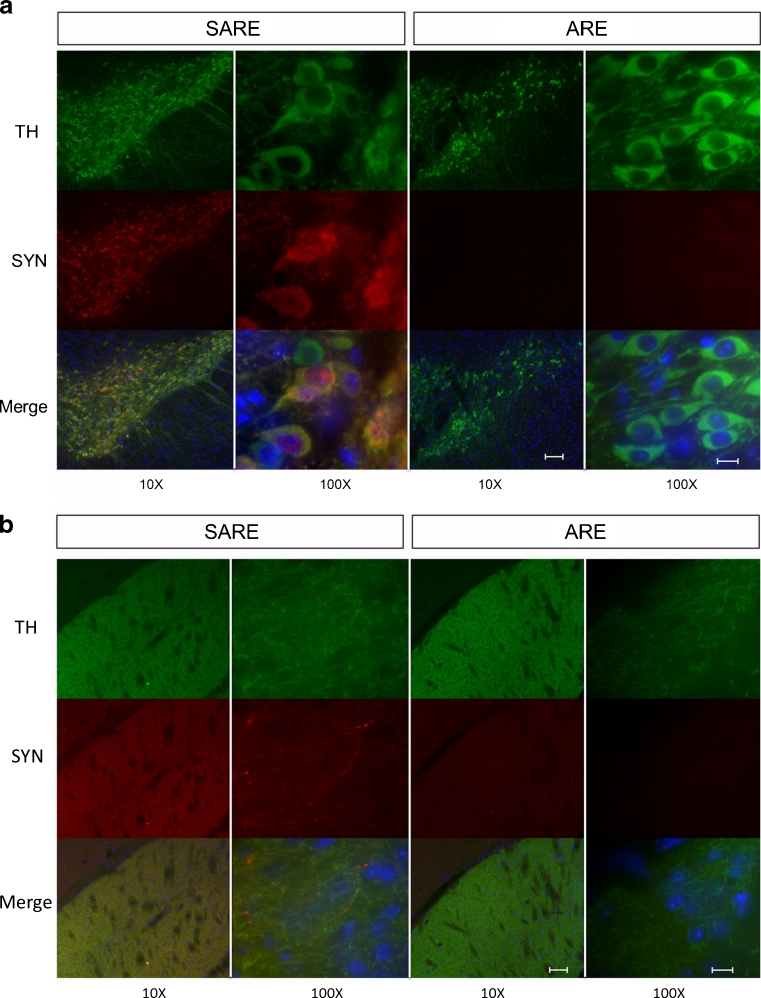
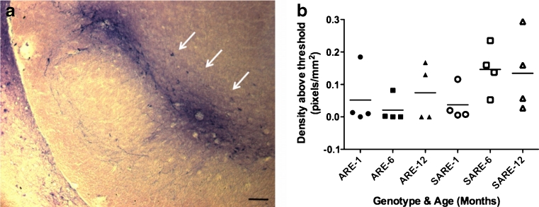
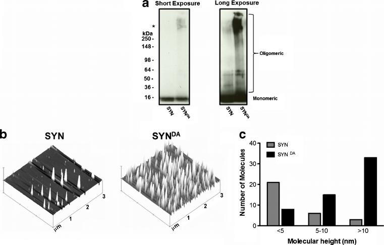
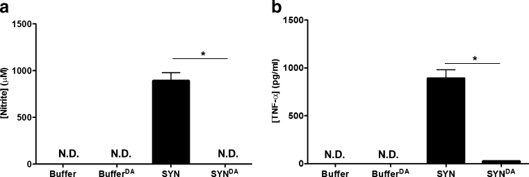
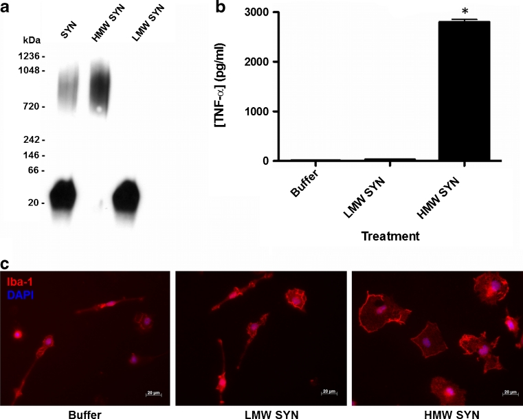
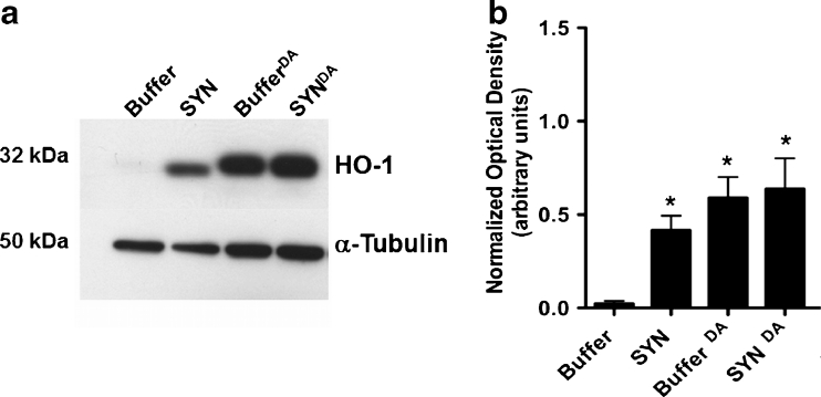
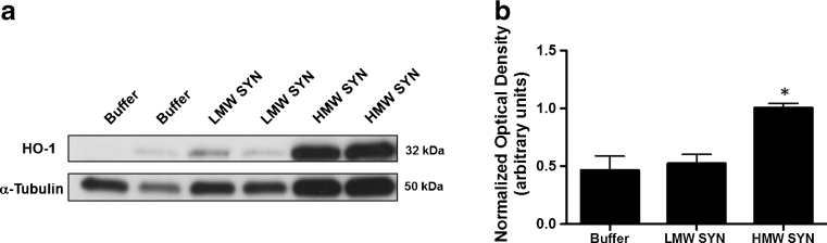
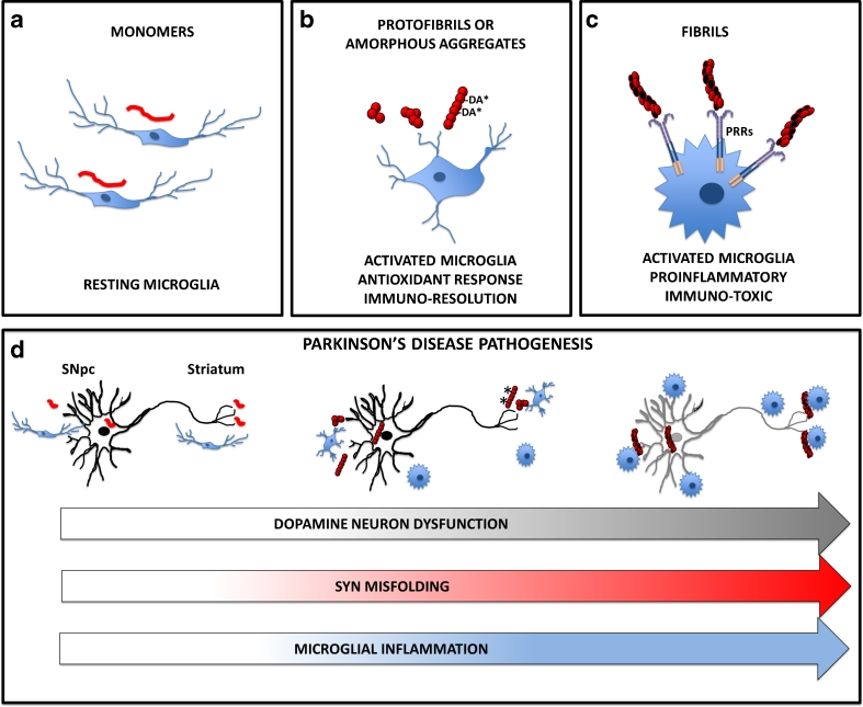
Parkinson's disease (PD) is the second most common age-related neurodegenerative disorder typified by tremor, rigidity, akinesia and postural instability due in part to the loss of dopamine within the nigrostriatal system. The pathologic features of this disorder include the loss of substantia nigra dopamine neurons and attendant striatal terminals, the presence of large protein-rich neuronal inclusions containing fibrillar α-synuclein and increased numbers of activated microglia. Evidence suggests that both misfolded α-synuclein and oxidative stress play an important role in the pathogenesis of sporadic PD. Here we review evidence that α-synuclein activates glia inducing inflammation and that Nrf2-directed phase-II antioxidant enzymes play an important role in PD. We also provide new evidence that the expression of antioxidant enzymes regulated in part by Nrf2 is increased in a mouse model of α-synuclein overexpression. We show that misfolded α-synuclein directly activates microglia inducing the production and release of the proinflammatory cytokine, TNF-α, and increasing antioxidant enzyme expression. Importantly, we demonstrate that the precise structure of α-synuclein is important for induction of this proinflammatory pathway. This complex α-synuclein-directed glial response highlights the importance of protein misfolding, oxidative stress and inflammation in PD and represents a potential locus for the development of novel therapeutics focused on induction of the Nrf2-directed antioxidant pathway and inhibition of protein misfolding.


Conflict of interest statement

The authors declare that they have no conflict of interest.

Figures

Fig. 1
Fig. 1
α-Synuclein and TH co-localize in the SNpc and striatum of SARE mice. Substantia nigra a and striatal b images from 1-month old SARE and ARE mice following co-immunohistochemistry for tyrosine hydroxylase (TH; green) and α-synuclein (SYN; red) demonstrating co-localization (Merge; yellow) of these proteins in mice that overexpress human α-synuclein under the control of a rat TH promoter (SARE). Images were taken at 10X (scale bar = 100 μm) and 100X (scale bar = 10 μm) magnifications
Fig. 2
Fig. 2
Antioxidant responses in vivo. The SNpc of 1-, 6- and 12-month old ARE and SARE mice (n = 4/genotype/age) were processed for hPLAP activity measurements using a BCIP/NBT staining protocol. a Image of phosphatase activity in the SNpc of an SARE mouse (12-months of age) demonstrating robust activity in this region of the brain (white arrows). Images were taken at 10X (scale bar = 100 μm) magnification. b The density of phosphatase activity was determined from the SNpc of BCIP/NBT stained tissue for all mice and reported as pixels/mm2. There is no statistically significant difference between α-synuclein overexpressing mice (SARE) and ARE mice in phosphatase activity
Fig. 3
Fig. 3
α-Synuclein overexpression increases the expression of antioxidant genes in the striatum. Quantitative RT-PCR was performed on cDNA obtained from 1-, 6- and 12-month old ARE and SARE mouse striata. a SARE mice had significantly higher expression of genes associated with the detoxification of hydrogen peroxide and quinones (HMOX1, NQO1, GPX1 and GPX4) at 1- and 6-months of age compared with age-matched ARE mice. b SARE mice had significantly higher expression of genes associated with glutathione metabolism (GCLM, GSS and GSR) at 1- and 6-months of age compared with age-matched ARE mice. Expression values were normalized to 18S rRNA as an internal control. All SARE measurements at 1- and 6-months of age were significantly different than ARE at the same ages (P < 0.05). The dashed line represents the gene expression level of ARE mice. Data expressed as fold change (2-∆∆ct) ± S.D
Fig. 4
Fig. 4
Characterization of misfolded α-synuclein. α-Synuclein was incubated at 33–37 °C in the absence (SYN) or presence of dopamine (SYNDA) and characterized by western blot analysis following polyacrylamide gel electrophoresis under denaturing conditions (SDS-PAGE) or by AFM under native conditions. Dopamine modification of α-synuclein caused an increase in high molecular weight aggregates. a Western blot analysis of manipulated α-synuclein demonstrated an increase in SDS-stable, high molecular weight oligomers following dopamine modification (SYNDA). α-Synuclein samples were subjected to 4–16 % SDS-PAGE and immunoblotted for α-synuclein. Both short and long film exposures are shown; *denotes the stacking/resolving gel interface. Short exposure corresponds to a 2 s exposure time for the film, while the long exposure is a 15 s exposure time. b AFM demonstrated an increase in α-synuclein aggregates following incubation with heat and dopamine. c Quantification of AFM molecular height images demonstrated that incubation of α-synuclein in the presence of dopamine (SYNDA; black) resulted in a 10-fold increase in aggregates >10 nm with a concomitant decrease in molecules <5 nm in height compared to α-synuclein without dopamine (SYN; grey). Molecular height images are shown at 3 μm × 3 μm × 3 μm
Fig. 5
Fig. 5
SYN induces a classical activation pattern in BV2 cells. SYN causes conformer-specific increases in proinflammatory molecule expression and release. a BV2 cells were treated with 50 nM SYN, 50 nM SYNDA or equal volumes of the appropriate buffer control for 24 h. Following treatment, a Greiss reagent assay was performed on the conditioned media to determine nitrite production. Conditioned media from SYN-treated BV2 cells had a substantial amount of nitrite (70 μM); in contrast there was no detectable (N.D.) NO in the media of cells treated with SYNDA or in media from buffer treated cells; (*p < 0.05). b BV2 cells were treated as described above and TNF-α protein was quantified in the conditioned media by ELISA. TNF-α release from BV2 cells was significantly increased in SYN-treated BV2 cells compared to the DA-modified SYN-treated (SYNDA) cells; there was no detectable (N.D.) TNF-α in media from buffer treated cells; (*p < 0.05). All values represent three biological replicates with treatments in triplicate
Fig. 6
Fig. 6
HMW α-synuclein activates microglia. a Representative α-synuclein western blots of misfolded α-synuclein under native conditions. Purified human recombinant α-synuclein was incubated at 37 °C with 1000 rpm rotation for 5 days to induce misfolding (SYN). Misfolded SYN was then separated into high molecular weight (HMW SYN) and low molecular weight (LMW SYN) fractions using MWCO concentrators. b Primary microglia were treated with buffer, LMW SYN (50 nM) or HMW SYN (50 nM) for 24 h. Following treatment, the conditioned media was evaluated for TNF-α protein secretion using an ELISA. Cells treated with HMW SYN released significantly more TNF-α than Buffer or LMW SYN treated microglia (*P < 0.05, n = 3). c Primary microglia were treated with buffer, 50 nM of LMW SYN or 50 nM HMW SYN for 24 h. Cells were fixed and immunostained for the microglial marker Iba-1 (red) followed by a DAPI nuclear counterstain (blue). The majority of primary microglia treated with buffer or LMW SYN display prototypical ramified morphology of resting microglia while glia treated with HMW SYN display the characteristic ameboid shape of activated microglia (scale bar = 20 μm)
Fig. 7
Fig. 7
Exposure of primary microglia to α-synuclein increases antioxidant expression. Primary microglia from ARE transgenic mice were histochemically stained with BCIP/NBT (purple) to detect hPLAP activity and nuclear red counterstained (pink). Microglia exposed to Buffer (a, b) or BufferDA (e, f) displayed less phosphatase activity than SYN-treated cells and also exhibited the prototypic morphology of resting microglia (dashed arrows; a, b, e, f & i) with a few glia that were activated but not phagocytic (solid arrows). In contrast, cells exposed to SYN or SYNDA displayed increased numbers of microglia with the characteristic amoeboid morphology of phagocytic microglia compared to the other exposure paradigms (solid arrowheads; c, d, g, h & i). Interestingly, microglia exposed to SYNDA had the greatest percentage of microglia expressing phosphatase activity with nearly 75 % of the treated microglia activated, as demonstrated by thickened processes and ameboid shape (solid arrow and solid arrowhead, respectively; g, h & i). Scale bar for 10x (a, c, e, g) and 40x (b, d, f, h) images represent 100 µm and 20 µm respectively. Boxes in a, c, e, and g denote the area for 40x images b, d, f and h. Cell counts were performed on nine random 20x images from each sample and categorized based on staining and morphology as outlined in Materials and Methods (i)
Fig. 8
Fig. 8
Exposure of microglia to α-synuclein or dopamine increases HO-1 protein expression. a Representative HO-1 western blot analysis of BV2 lysates. BV2 cells were treated with 50 nM of SYN or SYNDA or equal volumes of the appropriate buffer control for 24-h. Protein lysates were prepared and subjected to 10 % SDS-PAGE and immunoblotted for HO-1 (~32 kDa). Blots were re-probed for α-tubulin (~50 kDa) as a loading control. b Immunocomplexes were quantified by densitometric analysis. The HO-1 signal was normalized to the loading control. Cells treated with SYN or SYNDA had significantly higher levels of HO-1 expression compared to buffer alone (* p < 0.05, 1-way ANOVA followed by Student’s t post-test). Additionally, the presence of DA alone was enough to significantly increase HO-1 expression. Cells treated with buffer that was incubated in the presence of DA (BufferDA) had significantly higher levels of HO-1 than buffer incubated in the absence of DA (*p < 0.05, 1-way ANOVA followed by Student’s t post-test). Values represent three biological replicates with treatments in triplicate
Fig. 9
Fig. 9
Exposure of microglia to HMW α-synuclein increases HO-1 protein expression. a Representative HO-1 western blot analysis. Primary microglia were treated with 5 nM of LMW SYN, HMW SYN or buffer control for 24-h. Protein lysates were prepared and subjected to 4–20 % SDS-PAGE and immunoblotted for HO-1 (~32 kDa). Blots were re-probed for α-tubulin (~50 kDa) as a loading control. b Immunocomplexes were quantified by densitometric analysis. The HO-1 signal was normalized to the loading control. Cells treated with HMW SYN had significantly higher levels of HO-1 expression compared to buffer or LMW SYN (*p < 0.05, 1-way ANOVA followed by Student’s t post-test)
Fig. 10
Fig. 10
Schematic diagram depicting the effect of α-synuclein on microglia and PD. α-Synuclein promotes microglial activation in a structure dependent manner contributing to PD pathogenesis. Data presented in this paper supports the hypothesis that a specific structure is required to directly activate microglia and that antioxidant responses are in response to α-synuclein overexpression and direct glial activation. a Monomeric α-synuclein (red line) does not directly activate microglia. b Protofibrils stabilized by dopamine (−DA*) and amorphous aggregates of α-synuclein increase microglial expression of antioxidant enzymes and have an attenuated proinflammatory response (modest increase in IL1β), which we hypothesize leads to immuno-resolution rather than toxic inflammation. c Fibrils of α-synuclein directly activate microglia in a classic proinflammatory pathway (high levels of NO, NOX1, TNF-α and IL1β). Even though antioxidant enzymes are upregulated they cannot quell this robust glial activation. We further suggest that fibrillar α-synuclein is recognized by specific microglial PRRs, which facilitate a proinflammatory pathway. These highly activated cells would be immune-toxic for surrounding neurons. d Parkinson’s disease pathogenesis schematic. SNpc dopamine neurons normally express α-synuclein and this protein is enriched in striatal presynaptic dopamine terminals. If α-synuclein maintains a random coil structure this protein does not activate microglia (far left). When α-synuclein forms a protofibillar structure (e.g., due to overexpression, oxidative stress, the presence of dopamine quinone) microglia respond by increasing the expression of antioxidant enzymes and take on an activated morphology that we suggest is an attempt to return the microenvironment to homeostasis (middle). However, as opposed to pure microglial cultures, in this in vivo setting there is neuronal-glial crosstalk with factors released from the stressed dopamine neurons available to signal local glia causing a mild inflammatory response that is not due to a direct glial-synuclein interaction (middle). As α-synuclein continues to misfold into fibrils (far right and red arrow) and oxidative stress is enhanced in the local microenvironment, more microglia become activated in a robust proinflammatory pathway (blue arrow) leading to increased dopamine neuron dysfunction (gray arrow). Similar to Panel (c.), at this point the antioxidant response cannot suppress the ongoing glial activation

References

    1. Asanuma M, Miyazaki I, Ogawa N. Dopamine- or L-DOPA-induced neurotoxicity: the role of dopamine quinone formation and tyrosinase in a model of Parkinson’s disease. Neurotox Res. 2003;5(3):165–176. doi: 10.1007/BF03033137. - DOI - PubMed
    1. Baba M, Nakajo S, Tu PH, Tomita T, Nakaya K, Lee VM, Trojanowski JQ, Iwatsubo T. Aggregation of alpha-synuclein in Lewy bodies of sporadic Parkinson’s disease and dementia with Lewy bodies. Am J Pathol. 1998;152(4):879–884. - PMC - PubMed
    1. Banati RB. Visualising microglial activation in vivo. GLIA. 2002;40(2):206–217. doi: 10.1002/glia.10144. - DOI - PubMed
    1. Barcia C, Sanchez Bahillo A, Fernandez-Villalba E, Bautista V, Poza YPM, Fernandez-Barreiro A, Hirsch EC, Herrero MT. Evidence of active microglia in substantia nigra pars compacta of parkinsonian monkeys 1 year after MPTP exposure. GLIA. 2004;46(4):402–409. doi: 10.1002/glia.20015. - DOI - PubMed
    1. Bartels AL, Leenders KL. Neuroinflammation in the pathophysiology of Parkinson’s disease: evidence from animal models to human in vivo studies with [11C]-PK11195 PET. Mov Disord. 2007;22(13):1852–1856. doi: 10.1002/mds.21552. - DOI - PubMed

Publication types

MeSH terms