Review
. 2013 Apr;471(4):1127-36.
doi: 10.1007/s11999-012-2619-6.

Osteochondritis dissecans knee histology studies have variable findings and theories of etiology

Affiliations
Review

Osteochondritis dissecans knee histology studies have variable findings and theories of etiology

Kevin G Shea et al. Clin Orthop Relat Res. 2013 Apr.

Abstract

Background: Although many etiological theories have been proposed for osteochondritis dissecans (OCD), its etiology remains unclear. Histological analysis of the articular cartilage and subchondral bone tissues of OCD lesions can provide useful information about the cellular changes and progression of OCD. Previous research is predominantly comprised of retrospective clinical studies from which limited conclusions can be drawn.

Questions/purposes: The purposes of this study were threefold: (1) Is osteonecrosis a consistent finding in OCD biopsy specimens? (2) Is normal articular cartilage a consistent finding in OCD biopsy specimens? (3) Do histological studies propose an etiology for OCD based on the tissue findings?

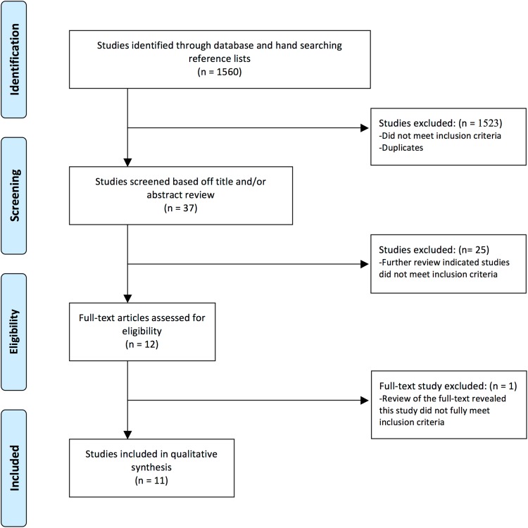
Methods: We searched the PubMed, Embase, and CINAHL databases for studies that conducted histological analyses of OCD lesions of the knee and identified 1560 articles. Of these, 11 met our inclusion criteria: a study of OCD lesions about the knee, published in the English language, and performed a histological analysis of subchondral bone and articular cartilage. These 11 studies were assessed for an etiology proposed in the study based on the study findings.

Results: Seven of 11 studies reported subchondral bone necrosis. Four studies reported normal articular cartilage, two studies reported degenerated or irregular articular cartilage, and five studies found a combination of normal and degenerated or irregular articular cartilage. Five studies proposed trauma or repetitive stress and two studies proposed poor blood supply as possible etiologies.

Conclusions: We found limited research on histological analysis of OCD lesions of the knee. Future studies with consistent methodology are necessary to draw major conclusions about the histology and progression of OCD lesions. Inconsistent histologic findings have resulted in a lack of consensus regarding the presence of osteonecrosis, whether the necrosis is primary or secondary, the association of cartilage degeneration, and the etiology of OCD. Such studies could use a standardized grading system to allow better comparison of findings.


Figures

Fig. 1
Fig. 1
PRISMA flow diagram for study selection. Moher D, Liberati A, Tetzlaff J, Altman DG, The PRISMA Group. Preferred Reporting Items for Systematic Reviews and Meta-Analyses: the PRISMA Statement. PLoS Med. 2009;6:e1000097. DOI: 10.1371/journal.pmed1000097.
Fig. 2
Fig. 2
Osteochondral plug harvested from the center of the lesion. A cleft separates the plug into two parts: a fragment side (F) and a basal side (B). Reprinted with permission from Uozumi H, Sugita T, Aizawa T, Takahashi A, Ohnuma M, Itoi E. Histologic findings and possible causes of osteochondritis dissecans of the knee. Am J Sports Med. 2009;37:2003–2008.
Fig. 3A–F
Fig. 3A–F
Safranin-O staining of cylindrical tissue sample with wide homogeneous cartilage (Patient 2). (A) Entire view of the slide (original magnification, ×40). Separation site (arrow). (B) CT image, sagittal view. (C) MRI T1-weighted image of the OCD lesion. Cylindrical tissue sample was obtained from central portion (dotted square). (D) Superficial layer of articular cartilage showing normal appearance of chondrocytes, no degenerative change, and no decrease of Safranin-O staining. (E) Cloning of chondrocytes (black arrow) and microfracture (arrowhead) were observed in the deep end of the cartilage layer just above the separation site. (F) Cartilage tissue with decreased Safranin-O staining was observed over the normal subchondral bone trabeculae. Fibrous tissue with blood vessels was observed within normal trabecular bone (DF, original magnification, ×100). Reproduced with permission from Yonetani Y, Nakamura N, Natsuume T, Shiozaki Y, Tanaka Y, Horibe S. Histological evaluation of juvenile osteochondritis dissecans of the knee: a case series. Knee Surg Sports Traumatol Arthrosc. 2010;18:723–730.
Fig. 4A–C
Fig. 4A–C
(A) Photomicrograph of the basal side of the osteochondral plug. The surface is covered with fibrillated cartilaginous tissue (asterisk). Beneath the surface, active bone remodeling (arrowheads) with proliferation of fibrovascular tissue, bone formation, and bone resorption is evident. The deep area (arrows) is composed of normal trabeculae and marrow tissue (Stain, hematoxylin and eosin; original magnification, ×20). (B) Photomicrograph of the deep area of the fragment side indicating necrotic subchondral trabeculae, because many of the lacunae are empty (arrowheads) (Stain, hematoxylin and eosin; original magnification, ×40). (C) Photomicrograph of the deep area of the fragment side showing viable subchondral trabeculae, because osteocytes are detected in the lacunae (arrowheads). The superficial part of this area consists of articular cartilage (asterisks) (Stain, hematoxylin and eosin; original magnification, ×40). Reproduced with permission from Uozumi H, Sugita T, Aizawa T, Takahashi A, Ohnuma M, Itoi E. Histologic findings and possible causes of osteochondritis dissecans of the knee. Am J Sports Med. 2009;37:2003–2008.

References

    1. Andrew TA, Spivey J, Lindebaum RH. Familial osteochondritis dissecans and dwarfism. Acta Orthop Scand. 1981;52:519–523. doi: 10.3109/17453678108992141. - DOI - PubMed
    1. Cahill BR. Osteochondritis dissecans of the knee: treatment of juvenile and adult forms. J Am Acad Orthop Surg. 1995;3:237–247. - PubMed
    1. Campbell CJ, Ranawat CS. Osteochondritis dissecans: the question of etiology. J Trauma. 1966;6:201–221. - PubMed
    1. Chiroff RT, Cooke CP., 3rd Osteochondritis dissecans: a histologic and microradiographic analysis of surgically excised lesions. J Trauma. 1975;15:689–696. doi: 10.1097/00005373-197508000-00011. - DOI - PubMed
    1. Enneking WF. Clinical Musculoskeletal Pathology. 1990. University Press of Florida. Available at: http://search.ebscohost.com/login.aspx?direct=true&db=nlebk&AN=20727&sit.... Accessed December 15, 2011.

Publication types