

doi: 10.1002/anie.201206749.
Epub 2012 Oct 10.
In Situ probing of newly synthesized peptidoglycan in live bacteria with fluorescent D-amino acids
Affiliations
- PMID: 23055266
- PMCID: PMC3589519
- DOI: 10.1002/anie.201206749
Item in Clipboard
In Situ probing of newly synthesized peptidoglycan in live bacteria with fluorescent D-amino acids
Angew Chem Int Ed Engl.
.
Abstract
Tracking a bug's life: Peptidoglycan (PG) of diverse bacteria is labeled by exploiting the tolerance of cells for incorporating different non-natural D-amino acids. These nontoxic D-amino acids preferably label the sites of active PG synthesis, thereby enabling fine spatiotemporal tracking of cell-wall dynamics in phylogenetically and morphologically diverse bacteria. HCC = 7-hydroxycoumarin, NBD = 7-nitrobenzofurazan, TAMRA = carboxytetramethylrhodamine.
Copyright © 2012 WILEY-VCH Verlag GmbH & Co. KGaA, Weinheim.
Figures
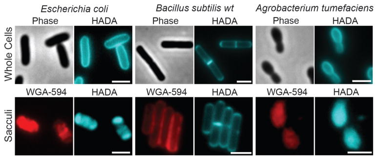
Long labeling pulses with HADA uniformly label PG in live cells of E. coli, B. subtilis and A. tumefaciens. The FDAA fluorescence is retained in isolated sacculi, which are also stained with the N-acetylglucosamine-specific WGA lectin conjugated to Alexa Fluor 594 (red). Scale bars, 2 μm.
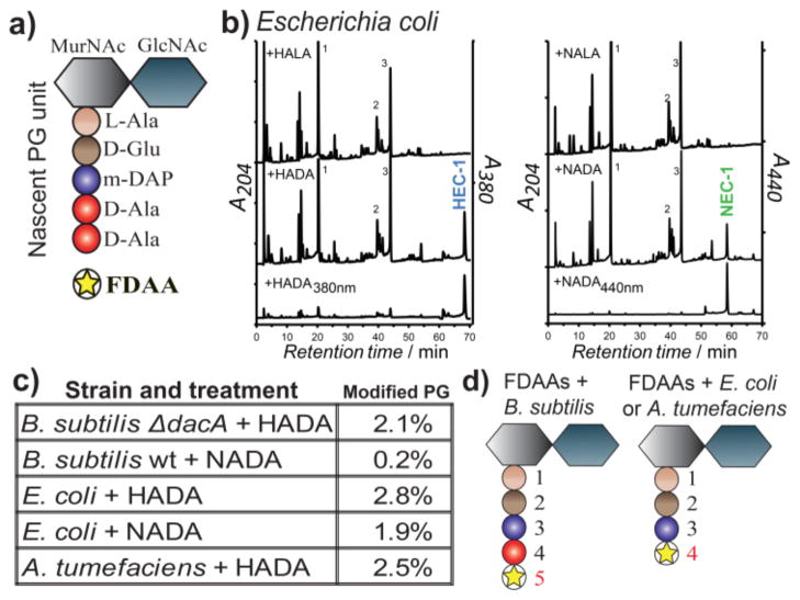
FDAA incorporation into the stem peptide of the PG subunit. a) Schematic representing the muramylpentapeptide precursor unit. b) HPLC detection of modified muropeptides in E. coli incubated with HADA or HALA and NADA or NALA. Samples were monitored using a dual wavelength UV monitor set for general muropeptide detection and for FDAA specific wavelengths. Peaks HEC-1 and NEC-1 correspond to the H ADA or N ADA modified muropeptides in E. c oli that were further characterized by electrospray ionization MS/MS (ESI-MS/MS). c) Percentage of FDAA incorporation into the total muropeptides varies among different bacteria as revealed by HPLC analysis. d) MS/MS analyses show that FDAAs exclusively incorporate into the 4th position of muropeptides in E. coli and A. tumefaciens and the 5th position in B. subtilis. See also Figures S6–7 and Table S1.
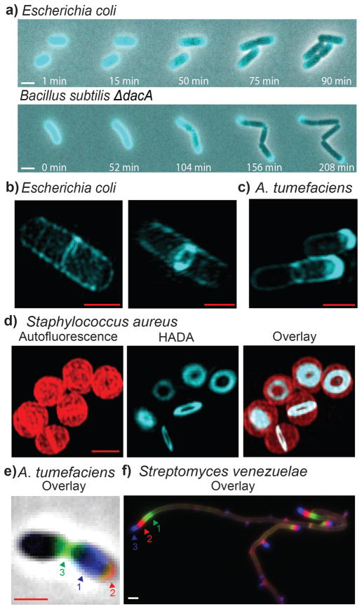
FDAAs label diverse bacterial growth patterns. a) Time-lapse microscopy of HADA labeled E. coli and B. subtilis ΔdacA cells imaged during growth on LB agarose pads. Superresolution microscopy of b) E. coli and c) A. tumefaciens after short pulses with HADA. d) Superresolution microscopy of S. aureus cells after a short pulse with HADA. Autofluorescence is shown in red. e) Triple labeling of A. tumefaciens with HADA (blue), EDA (clicked with red sulfo-Cy3-azide), and NADA (green). f) Triple labeling of S. venezuelae with NADA (green), TDL (red), and HADA (blue). Arrows in the triple labeling panels indicate the sequence of labeling. White scale bars, 2 μm; red scale bars, 1 μm.

Short pulses of HADA label distinct modes of growth in diverse bacteria. Strains were labeled for ~2–8% of the doubling time: E. coli (30 s), A. tumefaciens (2 min), B. subtilis ΔdacA (30 s), S. aureus (2 min), L. lactis (2 min), S. pneumoniae (4 min), C. crescentus (5 min), Synechocystis sp. PCC 6803 (1 h), S. venezuelae (2 min), B. conglomeratum (8 min), B. phytofirmans (20 min), V. Spinosum (10 min). Scale bars, 2 μm.

Modular structures of the fluorescent D-amino acids HADA, NADA, FDL and TDL and the ‘clickable’ D-amino acids EDA & ADA, color coded by the color of the fluorophores (blue, green and red).
References
-
- van Dam V, Olrichs N, Breukink E. Chembiochem. 2009;10:617–624. - PubMed
- Daniel RA, Errington J. Cell. 2003;113:767–776. - PubMed
- Tiyanont K, Doan T, Lazarus MB, Fang X, Rudner DZ, Walker S. Proc Natl Acad Sci U S A. 2006;103:11033–11038. - PMC - PubMed
- Olrichs NK, Aarsman MEG, Verheul J, Arnusch CJ, Martin NI, Herve M, Vollmer W, de Kruijff B, Breukink E, den Blaauwen T. Chembiochem. 2011;12:1124–1133. - PubMed
- Sadamoto R, Niikura K, Ueda T, Monde K, Fukuhara N, Nishimura SI. J Am Chem Soc. 2004;126:3755–3761. - PubMed
- de Pedro MA, Quintela JC, Höltje JV, Schwarz H. J Bacteriol. 1997;179:2823–2834. - PMC - PubMed
-
- Lam H, Oh DC, Cava F, Takacs CN, Clardy J, de Pedro MA, Waldor MK. Science. 2009;325:1552–1555. - PMC - PubMed
- Cava F, de Pedro MA, Lam H, Davis BM, Waldor MK. EMBO J. 2011;30:3442–3453. - PMC - PubMed
- Lupoli TJ, Tsukamoto H, Doud EH, Wang TSA, Walker S, Kahne D. J Am Chem Soc. 2011;133:10748–10751. - PMC - PubMed
Publication types
MeSH terms
Substances
Grants and funding
LinkOut - more resources
Full Text Sources
Other Literature Sources
Molecular Biology Databases
