. 2012 Oct 10;32(41):14265-70.
doi: 10.1523/JNEUROSCI.2693-12.2012.

Ectopic expression of select innexins in individual central neurons couples them to pre-existing neuronal or glial networks that express the same innexin

Affiliations

Ectopic expression of select innexins in individual central neurons couples them to pre-existing neuronal or glial networks that express the same innexin

Constantine P Firme 3rd et al. J Neurosci. .

Abstract

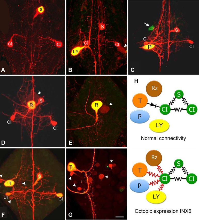
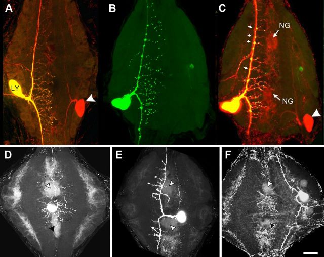
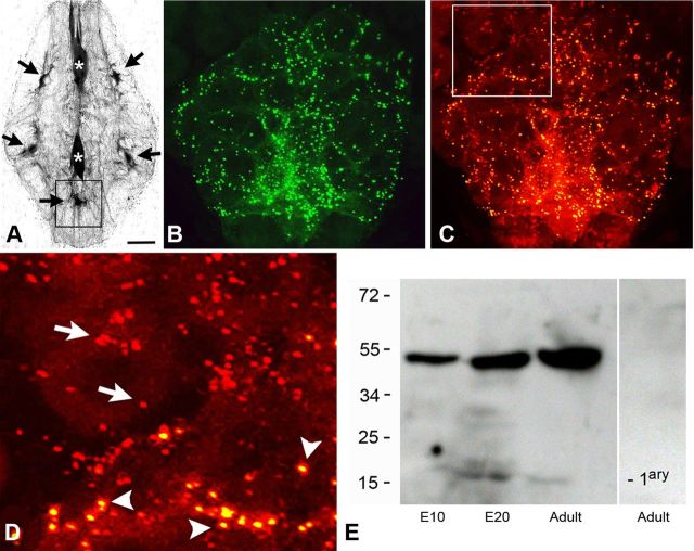
Fifteen of the 21 innexin (Inx) genes (Hve-inx) found in the genome of the medicinal leech, Hirudo verbana, are expressed in the CNS (Kandarian et al., 2012). Two are expressed pan-neuronally, while the others are restricted in their expression to small numbers of cells, in some cases reflecting the membership of known networks of electrically coupled and dye-coupled neurons or glial cells. We report here that when Hve-inx genes characteristic of discrete coupled networks were expressed ectopically in neurons known not to express them, the experimental cells were found to become dye coupled with the other cells in that network. Hve-inx6 is normally expressed by only three neurons in each ganglion, which form strongly dye-coupled electrical connections with each other [Shortening-Coupling interneuron (S-CI) network] (Muller and Scott, 1981; Dykes and Macagno, 2006). But when Hve-inx6 was ectopically expressed in a variety of central embryonic neurons, those cells became dye coupled with the S-CI network. Similarly, Hve-inx2 is normally uniquely expressed by the ganglion's large glial cells, but when it was ectopically expressed in different central neurons, they became dye coupled to the glial cells. In contrast, overexpression of the pan-neuronal Inx genes Hve-inx1 and Hve-inx14 did not yield any novel instances of dye coupling to pre-existent neuronal networks. These results reveal that expression of certain innexins is sufficient to couple individual neurons to pre-existing networks in the CNS. We propose that a primary determinant of selective neuronal connectivity and circuit formation in the leech is the surface expression of unique subsets of gap junctional proteins.


Figures

Figure 1.
Figure 1.
Ectopic expression of INX6 by select neurons leads to their inclusion into the S-CI circuit. A, Control Neurobiotin and Alexa Fluor 488 Dextran (molecular weight, 10,000) injection of a single S cell into a late embryonic ganglion (E20) shows tracer coupling only to the bilateral CIs, all of which normally express Hve-inx6 (Dykes and Macagno, 2006). B, Transgene expression of INX6 in a Leydig (LY) cell for 48 h, followed by Neurobiotin injection, shows strong tracer coupling to the S-CI circuit (E18). The Leydig cell's normal coupling with just its homolog is also detectable (arrowhead). C, Ectopic expression of INX6 in a P neuron in a late embryo (E26, 11 d after expression) revealing tracer coupling to the S-CI. Arrow indicates an unidentified neuron expressing INX6, which does not show tracer coupling with the network. D, Ganglion from a young juvenile animal 21 days after expressing INX6 in a Retzius (R) neuron reveals that tracer coupling with the S-CI network can be maintained for at least three weeks. Arrowhead points out the R homolog, showing normal tracer coupling. E, As a control, transgene expression of INX1 in the R cell does not alter its normal pattern of tracer coupling only to its contralateral homolog (arrowhead) (E19; 4 d of expression). F, G, Ectopic expression of INX6 in the T neuron (F; E24; 9 d of expression) also leads to tracer coupling with the S-CI, whereas, expression of INX1 does not (G; E23; 8 d of expression). Scale bar, 50 μm. H, Summary diagram showing the Hve-inx6 gene expressing S and CI cells (green), which are normally strongly electrically and dye coupled with one another (black resistors), while the R, P, and Leydig neuron, which do not normally express Hve-inx6, are not coupled to the S-CI (the T cell has a rectifying, non-dye-passing, synapse with the ipsilateral CI; black diode). However, upon ectopic expression of INX6, each of these cells now shows strong tracer coupling to the S-CI network (red resistors).
Figure 2.
Figure 2.
Embryonic transgene expression of INX2 in a Leydig (LY) neuron leads to tracer coupling with the glial Hve-inx2 network. A, Normal dye coupling between bilateral Leydig neurons. The left-side cell was injected with Neurobiotin and Alexa Fluor 488 Dextran (molecular weight, 10,000; yellow cell; arrowhead indicates Neurobiotin label in contralateral homolog (red). B, INX2-EGFP expression by the Leydig neuron shows a dispersed punctate distribution throughout the ganglionic neuropile. C, Overlay of Leydig cell in B with Neurobiotin staining (red) reveals outline of neuropile glia cells (NG) as well as contralateral homolog (white arrowhead). Note the larger puncta located at branch points along primary axon (small arrows). D–F, Examples of ectopic expression of INX2 in two unidentified interneurons (D, E) and a Touch sensory neuron (F) that lead to Neurobiotin tracer coupling with the neuropile glial cells (arrowheads). Scale bar, 50 μm.
Figure 3.
Figure 3.
The punctal distribution of the INX2-EGFP expression in ganglionic glial cells closely resembles the punctal distribution of the endogenous INX2 protein. A, The large glial cells in the leech ganglion are electrically coupled and tracer coupled. Neurobiotin injection into any one of them leads to labeling of all the cells. Fluorescent streptavidin labeling of a leech ganglion is shown here after the anterior neuropile glial cell (top asterisk) was injected with Neurobiotin. Strong streptavidin labeling can be seen in the posterior neuropile glial cell (bottom asterisk), as well as, five of the packet glial (PG) cells (black arrows). B, INX2-EGFP transgene expression in the posterior ventral medial PG cell (corresponding approximately with the location indicated by the box in A). A punctate pattern of staining can be seen distributed throughout the processes of the cell, which surround and outline many of the neuronal cell bodies. C, INX2 antibody staining (red) of the same cell and region as shown in B. D, Close-up view of the boxed area in C, showing the double labeling of the INX2 transgene (yellow spots, arrowheads) and the single labeling of the endogenous gap junctions (red, arrows) in adjacent glia, outside the boundary of the expressing cell. E, INX2 SDS-PAGE immunoblot analysis of leech lysates. From the left, the first lane shows lysate from an early embryo (E10); the second, a late embryo (E20); and the third, the adult CNS. The antiserum in each recognizes a band a little below 55 kDa. The lane on the right-hand side is the adult lysate with secondary antibody only. Scale bars: A, 75 μm; B, C, 15 μm; D, 7 μm.

References

    1. Altun ZF, Chen B, Wang ZW, Hall DH. High resolution map of Caenorhabditis elegans gap junction proteins. Dev Dyn. 2009;238:1936–1950. - PMC - PubMed
    1. Baccus SA, Burrell BD, Sahley CL, Muller KJ. Action potential reflection and failure at axon branch points cause stepwise changes in EPSPs in a neuron essential for learning. J Neurophysiol. 2000;83:1693–1700. - PubMed
    1. Baker MW, Macagno ER. Characterization of Hirudo medicinalis DNA promoters for targeted gene expression. J Neurosci Methods. 2006;156:145–153. - PubMed
    1. Chang M, Werner R, Dahl G. A role for an inhibitory connexin in testis? Dev Biol. 1996;175:50–56. - PubMed
    1. Chang Q, Gonzalez M, Pinter MJ, Balice-Gordon RJ. Gap junctional coupling and patterns of connexin expression among neonatal rat lumbar spinal motor neurons. J Neurosci. 1999;19:10813–10828. - PMC - PubMed

Publication types

Associated data

LinkOut - more resources