. 2012 Jul;35(3):557-66.
doi: 10.1590/S1415-47572012000400003. Epub 2012 Aug 17.

Clinical and molecular analysis of the enamelin gene ENAM in Colombian families with autosomal dominant amelogenesis imperfecta

Affiliations

Clinical and molecular analysis of the enamelin gene ENAM in Colombian families with autosomal dominant amelogenesis imperfecta

Sandra Gutiérrez et al. Genet Mol Biol. 2012 Jul.

Abstract

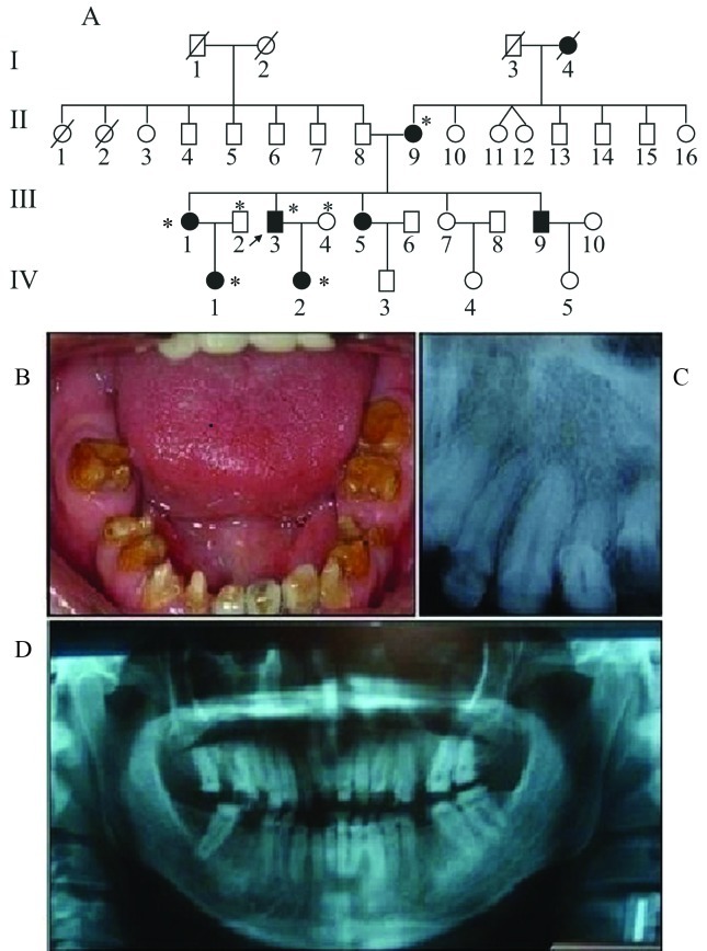
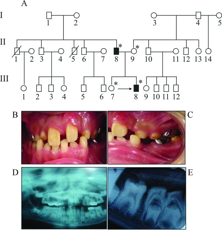
In this study, we analyzed the phenotype, clinical characteristics and presence of mutations in the enamelin gene ENAM in five Colombian families with autosomal dominant amelogenesis imperfecta (ADAI). 22 individuals (15 affected and seven unaffected) belonging to five Colombian families with ADAI and eight individuals (three affected and five unaffected) belonging to three Colombian families with autosomal recessive amelogenesis imperfecta (ARAI) that served as controls for molecular alterations and inheritance patterns were studied. Clinical, radiographic and genetic evaluations were done in all individuals. Eight exons and three intron-exon boundaries were sequenced for mutation analysis. Two of the five families with ADAI had the hypoplasic phenotype, two had the hypocalcified phenotype and one had the hypomaturative phenotype. Anterior open bite and mandibular retrognathism were the most frequent skeletal abnormalities in the families with ADAI. No mutations were found. These findings suggest that ADAI in these Colombian families was unrelated to previously described mutations in the ENAM gene. These results also indicate that other regions not included in this investigation, such as the promoter region, introns and other genes should be considered as potential ADAI candidates.

Keywords: ENAM gene; amelogenesis imperfecta; hypocalcified; hypoplasic; phenotype.


Figures

Figure 1
Figure 1
Family 1. A. The pedigree indicated autosomal dominant inheritance with eight affected members (black symbols). The asterisks indicate individuals who were analyzed clinically and whose ENAM gene was sequenced. Some individuals showed open anterior bite. The arrow indicates the proband III:3 patient. B. Photograph of proband III:3. The phenotype of this family was classified as generalized, rugged and severely hypoplasic with horizontal grooves. Many teeth showed caries and crown destruction while others were in the final stages of preparation for a prosthetic implant. C,D. Periapical and panorex radiographs of the same proband showing most of the teeth. Note the large areas of radiolucid enamel and the absence of several teeth.
Figure 2
Figure 2
Family 2. A. The pedigree indicated autosomal dominant inheritance with two affected members (black symbols). These two cases probably represent de novo mutations since none of the individuals in the first generation were affected. The arrow indicates the proband patient and the asterisks indicate individuals who were analyzed clinically and whose ENAM gene was sequenced. B, C. Photographs of proband III:8. The phenotype was classified as localized soft hypoplasic and was observed mainly in the anterior teeth. In this patient with mixed dentition, AI was present in deciduous and permanent dentition and was more severe in the latter. D,E. Panorex and periapical radiographs of the same proband showing premolars near eruption that were radiolucid in the enamel zone (similar to dentin). The periapical radiograph shows the marked contrast between dentin and enamel in the sixth permanent lower teeth.
Figure 3
Figure 3
Family 3. A. The pedigree indicated autosomal dominant inheritance with 22 affected members (black symbols). The arrows show the proband patients who were attended in this study and the asterisks indicate individuals who were analyzed clinically and whose ENAM gene was sequenced. B. Photograph of proband IV:8. The AI phenotype in this family was classified as hypomaturative (snow capped) with characteristics similar to fluorosis. Grooves with white stains that varied in location and intensity were observed among different members of the family. These alterations were observed mainly in the cuspids.
Figure 4
Figure 4
Family 4. A. The pedigree indicated autosomal dominant inheritance with eight affected members (black symbols). The arrows show the proband patients who were attended along with their mother. The asterisks indicate individuals who were analyzed clinically and whose ENAM gene was sequenced. B,C Photographs of proband III:1. The phenotype was classified as generalized hypocalcified. Note the characteristic indications of wear on the occlusal surfaces and therefore high tooth sensitivity. This patient also showed anterior open bite.
Figure 5
Figure 5
Family 5. A. The pedigree indicated autosomal dominant inheritance. The arrows indicate the proband patients and the asterisks indicate individuals who were analyzed clinically and whose ENAM gene was sequenced. B. Photograph of proband III:7. The AI phenotype in this family was classified as hypocalcified. However, AI was not generalized among members of this family but was restricted to hypocalcified areas. This individual showed no signs of wear on the occlusal surfaces, probably because of her young age.

References

    1. Atasu M, Eryilmaz A. Congenital hypodontia of lateral incisor in association with coloboma of the iris and hypomaduration type of amelogenesis imperfecta in a large kindred. J Clin Pediatr Dent. 1987;21:341–355. - PubMed
    1. Backman B, Holmgren G. Amelogenesis imperfecta: A genetic study. Hum Hered. 1988;38:189–206. - PubMed
    1. Backman B, Anneroth G, Horstedt P. Amelogenesis imperfecta: A scanning electron microscopic and micro-radiographic study. Oral Pathol. 1989;18:140–145. - PubMed
    1. Chan HC, Mai L, Oikonomopoulou A, Chan HL, Richardson AS, Wang SK, Simmer JP, Hu JC-C. Altered enamelin phosphorylation site causes amelogenesis imperfecta. J Dent Res. 2010;89:695–699. - PMC - PubMed
    1. Córdoba CA, Gutiérrez SJ, Gamboa LF. Anomalías dentarias presentes en Amelogenesis imperfecta. Rev Acad Col Odontoped. 2007;5:9–14.

LinkOut - more resources