. 2012;8(10):e1002930.
doi: 10.1371/journal.ppat.1002930. Epub 2012 Oct 4.

Structural basis of differential neutralization of DENV-1 genotypes by an antibody that recognizes a cryptic epitope

Affiliations

Structural basis of differential neutralization of DENV-1 genotypes by an antibody that recognizes a cryptic epitope

S Kyle Austin et al. PLoS Pathog. 2012.

Abstract

We previously developed a panel of neutralizing monoclonal antibodies against Dengue virus (DENV)-1, of which few exhibited inhibitory activity against all DENV-1 genotypes. This finding is consistent with reports observing variable neutralization of different DENV strains and genotypes using serum from individuals that experienced natural infection or immunization. Herein, we describe the crystal structures of DENV1-E111 bound to a novel CC' loop epitope on domain III (DIII) of the E protein from two different DENV-1 genotypes. Docking of our structure onto the available cryo-electron microscopy models of DENV virions revealed that the DENV1-E111 epitope was inaccessible, suggesting that this antibody recognizes an uncharacterized virus conformation. While the affinity of binding between DENV1-E111 and DIII varied by genotype, we observed limited correlation with inhibitory activity. Instead, our results support the conclusion that potent neutralization depends on genotype-dependent exposure of the CC' loop epitope. These findings establish new structural complexity of the DENV virion, which may be relevant for the choice of DENV strain for induction or analysis of neutralizing antibodies in the context of vaccine development.


Conflict of interest statement

MacroGenics has licensed some of the anti-DENV antibodies described in the work from Washington University. Syd Johnson is an employee of MacroGenics. Michael Diamond is a paid consultant of MacroGenics. None of the other authors have financial interests related to this work. This does not alter our adherence to all PLOS Pathogens policies on sharing data and materials.

Figures
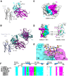
Figure 1
Figure 1. E111 in complex with DENV-1 DIII.
A ribbon diagram of the crystal structure of (A) E111 scFv in complex with DENV-1 strain 16007 DIII and (B) E111 Fab in complex with DENV-1 strain West Pac-74 DIII. The light chain is colored in cyan and the heavy chain in magenta. 16007 DIII is colored in light gray and West Pac-74 DIII is in gray. (C) A surface model showing the contact residues in the scFv complex. Contacts in the scFv are highlighted by light chain (cyan) or heavy chain (magenta) contacts. DIII contacts are highlighted by heavy chain (magenta), light chain (cyan), or both chains (green). (D) A ribbon diagram showing the residues of DIII contacted by the E111 scFv in the crystal structure. DIII residues that are contacted by the heavy and light chains of E111 (338, 344, and 345) are labeled. (E) A close-up of the contacts made by E111 in the CC′ loop of 16007 DIII (yellow) or West Pac-74 DIII (white). (F) Sequence of the four segments of 16007 DIII contacted by E111 aligned with the analogous residues of the other four DENV-1 genotypes and three serotypes. Residues are colored as in Figure 1D .
Figure 2
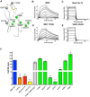
Figure 2. Kinetic analysis of E111 interaction with DENV genotypes and single variants.
(A) Ribbon diagram of DENV-1 16007 DIII with amino acids highlighted corresponding to tested mutants. Lateral ridge mutants and A-strand mutants are shown in gray. Amino acids representing genotypic variation of DENV-1 are shown in green. B–E. SPR traces are presented of E111 MAb interacting with (B) 16007 DIII, (C) West Pac-74 DIII, (D) 16007 DIII containing a T339S substitution or (E) 16007 DIII containing an A345V substitution. A single representative sensogram is shown for each DIII variant. The experimental curves (gray lines) were fit using a 1∶1 Langmuir analysis (black lines), after double referencing, to determine the kinetic parameters presented in panel F and Table 2 . Graphical representation of E111 binding to 16007 DIII mutants is presented according to the half-life (in seconds) of the interaction. The results are representative of a minimum of three independent experiments, with error bars showing standard deviation. Statistical significance was determined using a paired student t-test comparing the half-life of the E111 binding to 16007 DIII to that of another DIII variant.
Figure 3
Figure 3. E111 neutralization of different DENV-1 genotypes is only partially dependent on differences in epitope sequence.
(A) Plaque reduction and neutralization curves for five DENV-1 strains representing the five genotypes. The data is representative of three independent experiments performed in duplicate. PRNT50 values are shown in Table 3 . B–C. Serial six-fold dilutions of (B) DENV1-E103 or (C) E111 were incubated with wild type or V345A West Pac-74 RVPs for one hour at 37°C, and then added to Raji-DC-SIGNR cells. Infection was assessed by flow cytometry 48 hours later. One representative experiment of four is shown. The data is normalized relative to the infectivity of the RVPs in the absence of antibody. Error bars indicate standard error of the mean of replicate infections.
Figure 4
Figure 4. The structural basis of E111 neutralization.
(A) E111 Fab docked onto the DENV-2 dimer (PDB 1OAN). Transparent gray space filled-ribbon model of DENV-2 dimer with N-linked glycans colored green. The E111 Fab light (cyan) and heavy (magenta) chains are shown bound to the equivalent DIII of the three dimensional structure within the DENV-2 dimer. The WNV E16 Fab (DIII lateral ridge antibody), in green and gold, (PDB 1ZTX) is docked onto the analogous DENV-2 residues for comparison. (B) Surface representation of the DENV-1 post-fusion trimer structure. Upper panel: Each E protein in the trimer is colored independently (white, red and yellow), and the E111 epitope (colored as in Figure 1D ) is mapped onto DIII. The red surface of DI was made transparent to show the ribbon structure. Lower panel: The E111 scFv complex was superimposed onto a monomer of the post-fusion trimer (white); The E111 scFv light chain (cyan) bound to DIII clashes with DI of the neighboring E monomer (red) suggesting that E111 likely would inhibit formation of the E homotrimer required for virus fusion. Clashing beta strands from DI are labeled. (C) To determine whether E111 neutralizes infection before or after cellular attachment, BHK21-15 cells were pre-chilled to 4°C, and 102 PFU of DENV-1 (16007) was added to each well for 1 h at 4°C. After extensive washing at 4°C, increasing concentrations of E111 were added for 1 h at 4°C, and the PRNT protocol was then completed (dashed lines, Post). In comparison, a standard pre-incubation PRNT with all steps performed at 4°C is shown for reference. In this case, virus and MAb were incubated together for 1 h at 4°C, prior to addition to cells (solid lines, Pre). Data shown are representative from three experiments performed in duplicate.
Figure 5
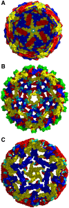
Figure 5. Mapping of the E111 epitope on the DENV virion.
The atomic structures of the (A) mature (PDB 1K4R), (B) immature (PDB 3C6D), and (C) 1A1D-2-bound DENV-2 (PDB 2R6P) are shown as determined by modeling of cryo electron-microscopy reconstructions. E proteins in each icosahedral symmetry axis are highlighted: yellow (5-fold), blue (3-fold), or red (2-fold). The E111 epitope is colored in cyan in each symmetry group. To visualize the localization of the E111 epitope (on the interior of the viral surface in certain axes) a cross-section of each viral particle is shown. prM is colored in green on the immature virus model. In panel C, the 1A1D-2 Fabs were removed from the deposited cryo-electron microscopy structure to show only the antibody-stabilized virus conformation.
Figure 6
Figure 6. Neutralization of DENV-1 by E111 varies with time and temperature in a genotype-dependent manner.
Serial dilutions of E111 were incubated with (A and E) DENV-1 16007, (B and F) West Pac-74, (C and G) V345A West Pac-74, or (D and H) A345V 16007 RVP for 1 hour at 37°C before the addition of Raji-DC-SIGNR cells to establish reference neutralization curves. Additional DENV-1 RVP-E111 complexes were incubated for 2, 4.5, 7, and 22 hours at 37°C (A–D) or 2 and 4.5 hours at 40°C (E–H) before addition to Raji-DC-SIGNR cells. Infection was carried out at 37°C and determined by flow cytometry 48 hours later. One representative experiment of three is shown. The data is normalized relative to the infectivity of the RVP in the absence of antibody at each time point for each temperature. Error bars indicate standard error of the mean of replicate infections.

References

    1. WHO. Dengue and severe dengue. Available: http://www.who.int/mediacentre/factsheets/fs117/en/index.html. Accessed 27 May 2012.
    1. Rico-Hesse R (1990) Molecular evolution and distribution of dengue viruses type 1 and 2 in nature. Virology 174: 479–493. - PubMed
    1. Holmes EC, Twiddy SS (2003) The origin, emergence and evolutionary genetics of dengue virus. Infect Genet Evol 3: 19–28. - PubMed
    1. Sabin AB (1952) Research on Dengue during World War II. Am J Trop Med Hyg 1: 30–50. - PubMed
    1. Halstead SB, Nimmannitya S, Yamarat C, Russell PK (1967) Hemorrhagic fever in Thailand; recent knowledge regarding etiology. Jpn J Med Sci Biol 20: 96–103. - PubMed

Publication types