. 2012;8(10):e1002966.
doi: 10.1371/journal.pgen.1002966. Epub 2012 Oct 4.

A mutation in the Srrm4 gene causes alternative splicing defects and deafness in the Bronx waltzer mouse

Affiliations

A mutation in the Srrm4 gene causes alternative splicing defects and deafness in the Bronx waltzer mouse

Yoko Nakano et al. PLoS Genet. 2012.

Abstract

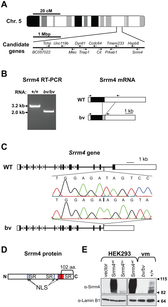
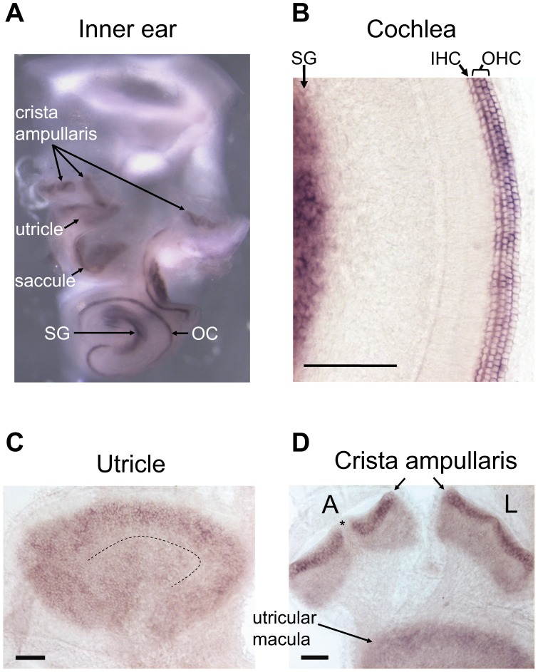
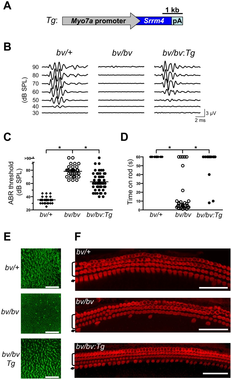
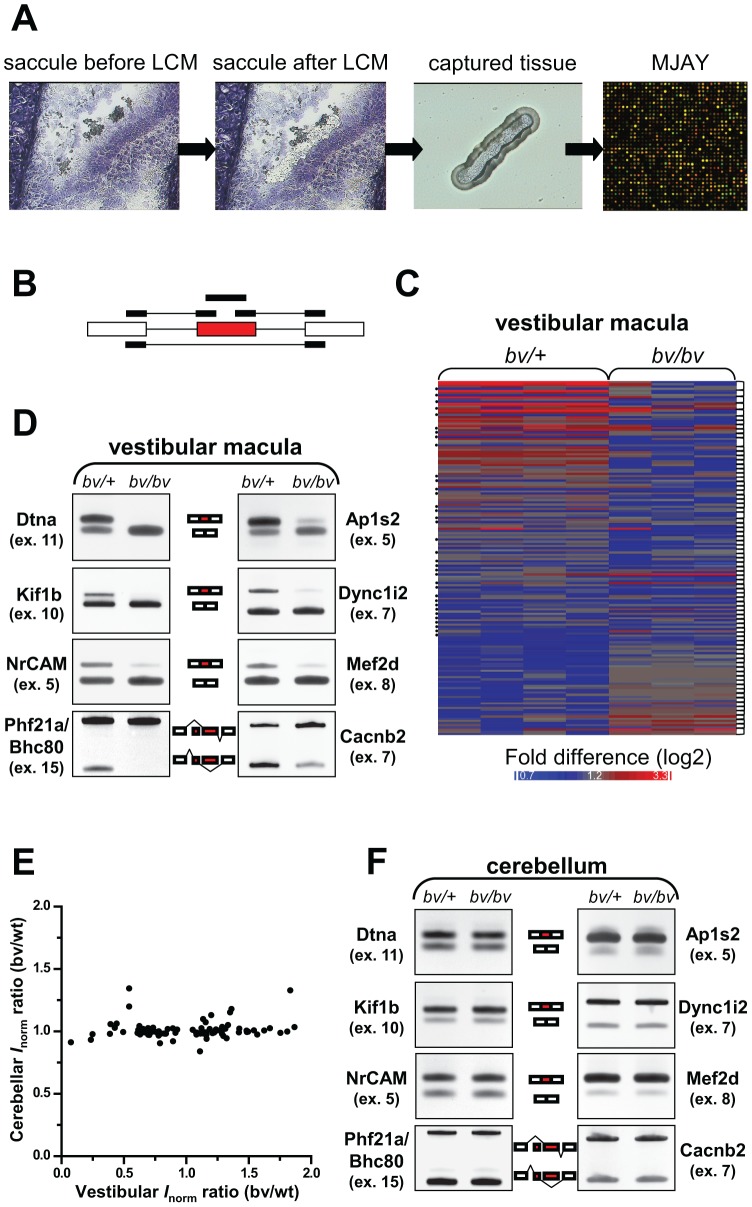
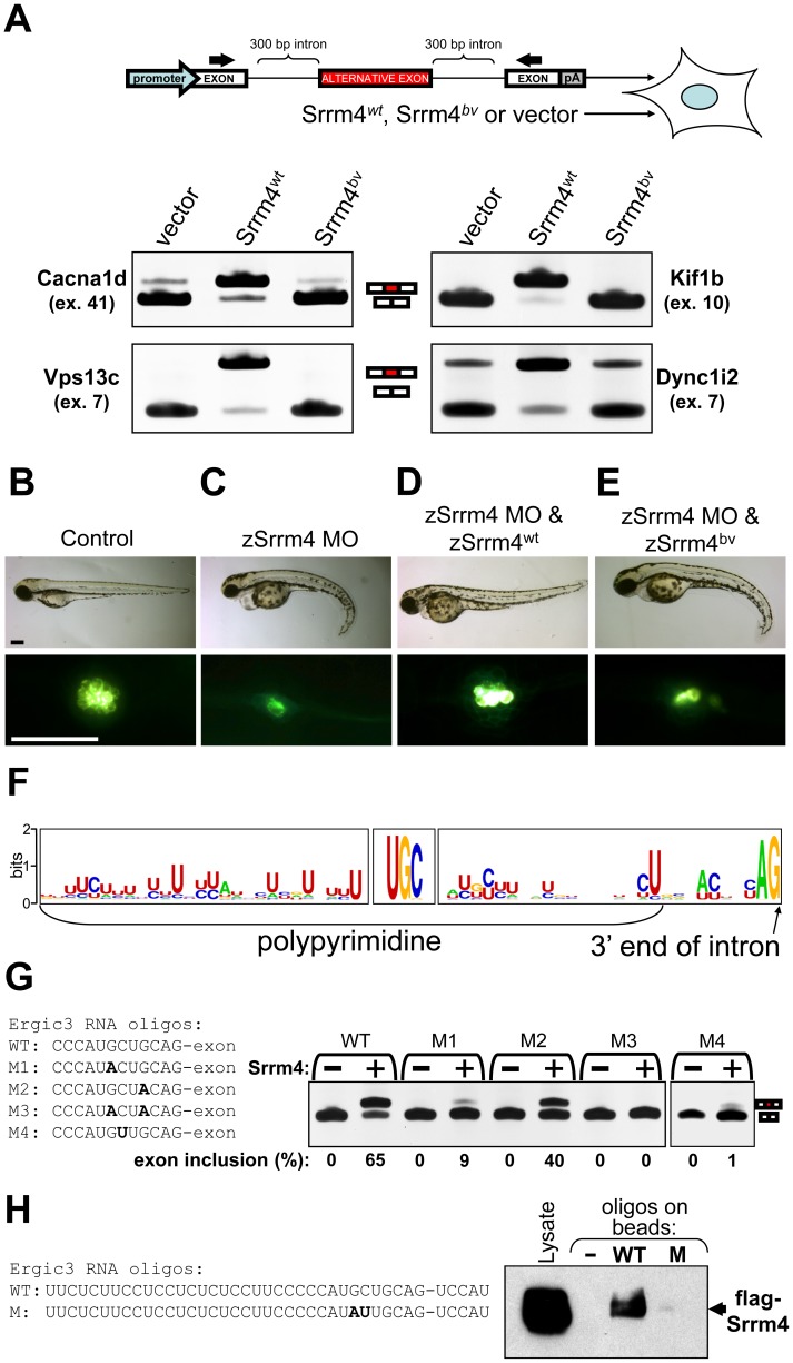
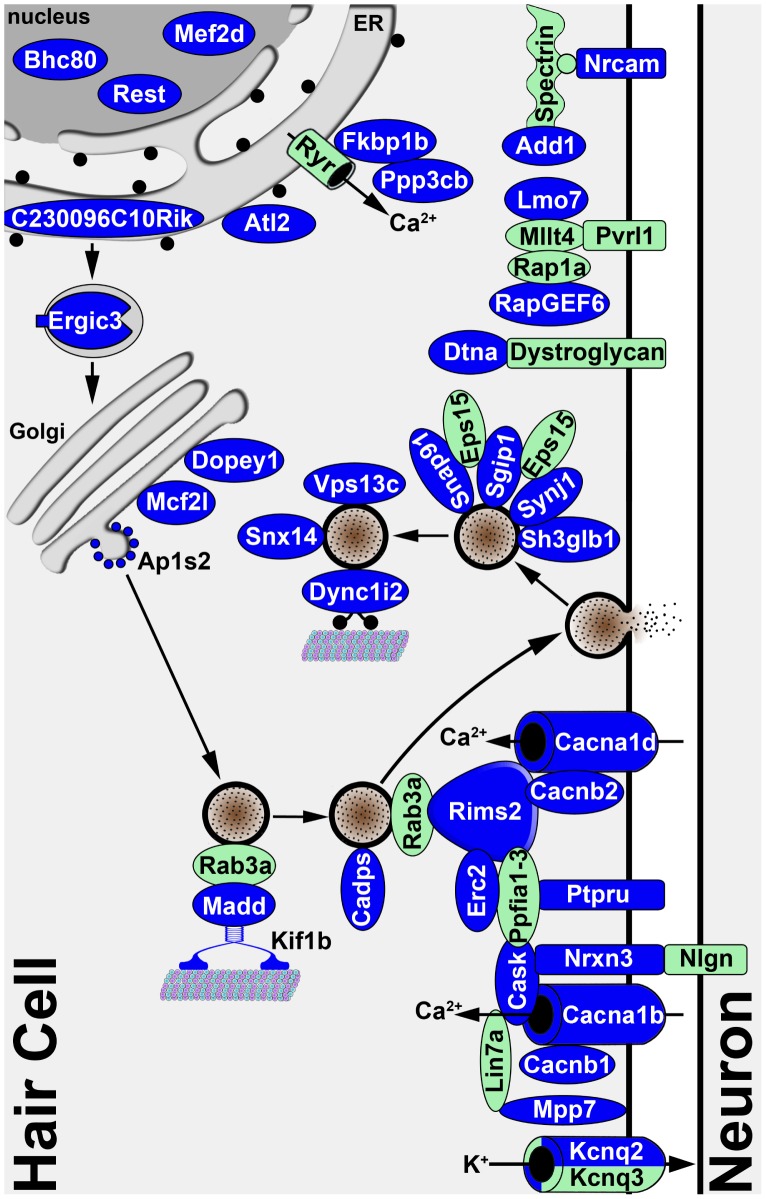
Sensory hair cells are essential for hearing and balance. Their development from epithelial precursors has been extensively characterized with respect to transcriptional regulation, but not in terms of posttranscriptional influences. Here we report on the identification and functional characterization of an alternative-splicing regulator whose inactivation is responsible for defective hair-cell development, deafness, and impaired balance in the spontaneous mutant Bronx waltzer (bv) mouse. We used positional cloning and transgenic rescue to locate the bv mutation to the splicing factor-encoding gene Ser/Arg repetitive matrix 4 (Srrm4). Transcriptome-wide analysis of pre-mRNA splicing in the sensory patches of embryonic inner ears revealed that specific alternative exons were skipped at abnormally high rates in the bv mice. Minigene experiments in a heterologous expression system confirmed that these skipped exons require Srrm4 for inclusion into the mature mRNA. Sequence analysis and mutagenesis experiments showed that the affected transcripts share a novel motif that is necessary for the Srrm4-dependent alternative splicing. Functional annotations and protein-protein interaction data indicated that the encoded proteins cluster in the secretion and neurotransmission pathways. In addition, the splicing of a few transcriptional regulators was found to be Srrm4 dependent, and several of the genes known to be targeted by these regulators were expressed at reduced levels in the bv mice. Although Srrm4 expression was detected in neural tissues as well as hair cells, analyses of the bv mouse cerebellum and neocortex failed to detect splicing defects. Our data suggest that Srrm4 function is critical in the hearing and balance organs, but not in all neural tissues. Srrm4 is the first alternative-splicing regulator to be associated with hearing, and the analysis of bv mice provides exon-level insights into hair-cell development.


Conflict of interest statement

The authors have declared that no competing interests exist.

Figures

Figure 1
Figure 1. Deletion mutation in the Srrm4 gene of bv mice.
(A) Schematic representation of genomic positions of the selected candidate genes in the 4-mega base pair (Mbp) interval to which the bv mutation has been mapped. (B) Left: RT-PCR-based detection of an abnormally short Srrm4 transcript in the inner ear of a bv/bv mouse. Right: schematic representation of wild-type (WT) and bv Srrm4 transcripts showing the positions of the RT-PCR primers (arrows), the translated and non-translated regions (black and white boxes, respectively), and a normally intronic sequence in the Srrm4 mRNA of the bv/bv mouse (gray box). (C) Comparison of the Srrm4 genes of wild-type and bv/bv mice. Horizontal lines represent introns, and black and white boxes represent the coding and non-coding regions of exons, respectively. The Srrm4 gene of bv/bv mice lacks parts of the last intron and exon. Chromatograms highlight differences between the WT and bv/bv mice with respect to the Srrm4 sequence near the deletion site (starting at vertical line in the lower chromatogram). (D) Schematic representation of the Srrm4 protein. The bracket indicates the portion of the protein that is encoded by the last Srrm4 exon in wild-type mice and is lacking in the bv/bv mice. The Ser/Arg (SR)-rich regions, putative nuclear localization signals (NLS), and a region that is highly conserved between Srrm4 and its closest paralogue, Srrm3 (amino acids 478–525 in Srrm4), are also indicated (red). (E) Upper panels: immunoblot analysis of Srrm4 expression in transfected HEK293 cells and in the vestibular macula (vm) of bv/bv and +/+ mice on E16.5. As indicated in the panel, the HEK293 cells were transfected with Srrm4wt, Srrm4bv, or an empty expression vector. Lower panels: comparable protein loading is demonstrated by the Lamin B1 signal present in all samples. Arrowheads and numbers (in kDa) indicate the positions of MW standards.
Figure 2
Figure 2. In situ hybridization of the mouse inner ear with an antisense Srrm4 probe.
(A) Whole-mount in situ hybridization of the inner ear revealing Srrm4 detection in each balance organ (i.e. crista ampullaris, saccule, and utricle), in the organ of Corti (OC), and in the spiral ganglion (SG). (B) In the cochlea, the antisense Srrm4 probe labeled all three rows of OHCs, the row of IHCs, and the spiral ganglion (SG). (C) In the utricular macula, the Srrm4 signal was present throughout, but weaker in the central (i.e. striolar) region than in the periphery. The dotted line indicates the estimated center of the striola. (D) In the crista amupllaris, Srrm4 signal was present in the sensory-cell layer. The anterior (A) and lateral (L) cristae are shown. Asterisk indicates the unstained, non-sensory septum cruciatum of the anterior crista. An adjacent segment of the utricular macula is also shown. Scale bars: 100 µm.
Figure 3
Figure 3. The bv phenotype is rescued by hair-cell targeted expression of an Srrm4 transgene.
(A) Schematic representation of the Srrm4 transgene (Tg) designed for rescue experiments. It consists of a mouse Myo7a promoter, the Srrm4 coding sequence, and a polyadenylation (pA) site. (B) Representative ABR waveforms for bv/+, bv/bv, and Srrm4-transgenic bv/bv mice (bv/bv:Tg). Broadband click stimuli were applied at the indicated sound pressure levels (SPL). (C) Statistical analysis of ABR thresholds for bv/+, bv/bv, and Srrm4-transgenic bv/bv mice; each symbol represents the value for a single mouse (one-way ANOVA, P<0.0001, post-hoc Tukey's test: *P<0.01). (D) Time spent on a fixed horizontal rod before falling, by bv/+, bv/bv, and Srrm4-transgenic bv/bv mice at P70–80. The maximal duration of the assay was 60 s (one-way ANOVA, P<0.0001, post-hoc Tukey's test: *P<0.01). (E) Vestibular macula preparations from bv/+, bv/bv, and Srrm4-transgenic bv/bv mice (P5) stained with phalloidin-Alexa Fluor 488 to visualize actin-rich structures, including stereocilia. (F) Mid-turn organ of Corti preparations from bv/+, bv/bv, and Srrm4-transgenic bv/bv mice (P5) stained with an anti-Myo7a antibody, which labels specifically the IHCs (arrows) and OHCs (brackets). Only the IHC row is affected by the bv mutation. Scale bars: 50 µm.
Figure 4
Figure 4. bv/bv mice are subject to splicing defects in the inner ear but not in the cerebellum.
(A) Schematic workflow for analyzing pre-mRNA splicing in the vestibular maculas of bv/bv and bv/+ mice. Vestibular maculas were isolated by laser capture microdissection (LCM), and RNA samples from the captured tissue were analyzed using mouse exon-junction microarrays (MJAYs). (B) The design of MJAY probe sets for cassette exons. Typically, 4 MJAY probe sets (black boxes) are used to measure the splicing of one cassette exon (red box); the probe sets anneal to the cassette exon itself, to the upstream and downstream exon-exon junctions, and to the skipping junction. (C) Microarray heat map of normalized probe-set signals. Probe-set signals are shown where at least two probe sets per alternative exon (connected by brackets) led to significantly different assessments of splicing rates in the vestibular maculas of bv/+ and bv/bv mice. Dots at the left margin represent the data generated by exon-skipping probe sets. (D) RT-PCR validation of 8 splicing differences between the vestibular maculas of bv/+ and bv/bv mice (additional RT-PCR data are shown in Figure S5). The RT-PCR primers were designed to anneal to constitutive exons (white boxes) flanking the tested alternative exons (red boxes). (E) The ratios of normalized probe-set intensities (I norm) from the vestibular maculas of bv/+ and bv/bv mice are plotted against those from the cerebellums of bv/+ and bv/bv mice. Only the probe sets that are indicative of splicing differences in the vestibular macula are included in the plot (Pearson's correlation test, r = 0.1, P = 0.3). (F) RT-PCR confirmation that the cerebellum of bv/bv mice lacks the splicing defects observed in the vestibular macula of the same mouse line.
Figure 5
Figure 5. Srrm4-dependent splicing requires the C-terminal region of Srrm4 and a novel motif in the pre–mRNA.
(A) RT-PCR-based testing of alternative splicing in HEK293 cells co-transfected with an Srrm4-encoding construct (Srrm4wt, Srrm4bv, or an empty expression vector) along with a minigene consisting of exons and introns (diagram). Each minigene contained an alternative exon (red box) and the adjacent intronic sequences, and these were situated between two constitutive exons (white boxes). The promoter and polyadenylation site (pA) of the minigene are indicated. Arrows indicate positions of the RT-PCR primers. Results obtained from the RT-PCR of 4 minigene-encoded mRNAs are shown. (B–E) Hair-cell survival in zebrafish injected with various combinations of the zSrrm4 MO, MO-insensitive zSrrm4wt mRNA, and MO-insensitive zSrrm4bv mRNA, as indicated. The upper panels show representative brightfield images of zebrafish (72 hpf) from each treatment group. The lower panels show representative fluorescence images of neuromasts from each treatment group following visualization of the hair cells with the FM1–43 dye (strong green signal). The faint green signal at cell-cell junctions is due to transgenic expression of membrane-anchored GFP in neuromasts of the zebrafish line. Scale bars: 200 µm. (F) A sequence-logo representation of the consensus sequence motifs found directly upstream of Srrm4-regulated exons. The detected consensus motifs include a polypyrimidine tract, a UGC motif, and an AG motif. (G) Results for RT-PCR testing of alternative splicing in HEK293 cells transfected with either the wild-type (WT) or a mutant (M1–4) version of an Ergic3 minigene, plus Srrm4wt (+) or an empty expression vector (−), as indicated. The WT and mutated sequences (M1–4) are shown. Mutated bases are shown in bold font. (H) RNA pull down of flag-tagged Srrm4 from the whole cell lysate (Lysate) of transfected HEK293 cells, using biotinylated RNA oligos or control empty streptavidin beads (−). The sequence of the wild-type RNA oligo (WT) contained 35 nucleotides from the intron preceding the Srrm4-regulated exon and 5 nucleotides from the exon. The boundary between the intron and exon is indicated by a hyphen. In the mutated RNA oligo (M), the GC motif was substituted with AU (bold characters). The amount of flag-Srrm4 in the cell lysate and on the washed beads was evaluated using an anti-flag antibody and Western blotting.
Figure 6
Figure 6. Diagram illustrating the known and predicted subcellular localizations of proteins encoded by validated Srrm4-regulated mRNAs.
The proteins encoded by the validated mRNA targets of Srrm4-dependent splicing are shown in blue. The references for the protein localization and interaction data are listed in Table S3. For 8 of the proteins encoded by Srrm4-regulated mRNAs, the subcellular localization either falls outside of the depicted area or is unknown. Proteins that are not known to be regulated by Srrm4 but link Srrm4-regulated proteins to each other or a membrane are shown in green. Arrows indicate the direction of each transport event.

References

    1. Dallos P (2008) Cochlear amplification, outer hair cells and prestin. Curr Opin Neurobiol 18: 370–376. - PMC - PubMed
    1. Géléoc GS, Holt JR (2003) Developmental acquisition of sensory transduction in hair cells of the mouse inner ear. Nat Neurosci 6: 1019–1020. - PMC - PubMed
    1. Lelli A, Asai Y, Forge A, Holt JR, Géléoc GS (2009) Tonotopic gradient in the developmental acquisition of sensory transduction in outer hair cells of the mouse cochlea. J Neurophysiol 101: 2961–2973. - PMC - PubMed
    1. Frolenkov GI, Belyantseva IA, Friedman TB, Griffith AJ (2004) Genetic insights into the morphogenesis of inner ear hair cells. Nat Rev Genet 5: 489–498. - PubMed
    1. Richardson GP, de Monvel JB, Petit C (2011) How the genetics of deafness illuminates auditory physiology. Annu Rev Physiol 73: 311–334. - PubMed

Publication types

MeSH terms