

Mesenchymal stem cells are short-lived and do not migrate beyond the lungs after intravenous infusion
- PMID: 23056000
- PMCID: PMC3458305
- DOI: 10.3389/fimmu.2012.00297
Mesenchymal stem cells are short-lived and do not migrate beyond the lungs after intravenous infusion
Abstract
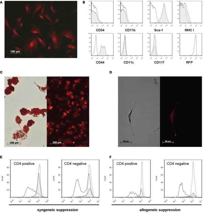
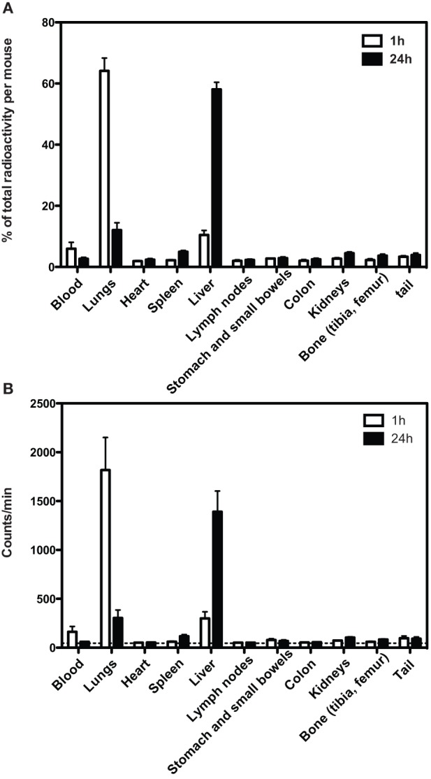
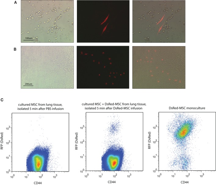
Mesenchymal stem cells (MSC) are under investigation as a therapy for a variety of disorders. Although animal models show long term regenerative and immunomodulatory effects of MSC, the fate of MSC after infusion remains to be elucidated. In the present study the localization and viability of MSC was examined by isolation and re-culture of intravenously infused MSC. C57BL/6 MSC (500,000) constitutively expressing DsRed-fluorescent protein and radioactively labeled with Cr-51 were infused via the tail vein in wild-type C57BL/6 mice. After 5 min, 1, 24, or 72 h, mice were sacrificed and blood, lungs, liver, spleen, kidneys, and bone marrow removed. One hour after MSC infusion the majority of Cr-51 was found in the lungs, whereas after 24 h Cr-51 was mainly found in the liver. Tissue cultures demonstrated that viable donor MSC were present in the lungs up to 24 h after infusion, after which they disappeared. No viable MSC were found in the other organs examined at any time. The induction of ischemia-reperfusion injury in the liver did not trigger the migration of viable MSC to the liver. These results demonstrate that MSC are short-lived after i.v. infusion and that viable MSC do not pass the lungs. Cell debris may be transported to the liver. Long term immunomodulatory and regenerative effects of infused MSC must therefore be mediated via other cell types.
Keywords: infusion; liver; localization; lung; mesenchymal stem cell; survival.
Figures
References
-
- Assis A. C., Carvalho J. L., Jacoby B. A., Ferreira R. L., Castanheira P., Diniz S. O., Cardoso V. N., Goes A. M., Ferreira A. J. (2010). Time-dependent migration of systemically delivered bone marrow mesenchymal stem cells to the infarcted heart. Cell Transplant. 19, 219–230 10.3727/096368909X479677 - DOI - PubMed
-
- Aurich H., Sgodda M., Kaltwasser P., Vetter M., Weise A., Liehr T., Brulport M., Hengstler J. G., Dollinger M. M., Fleig W. E., Christ B. (2009). Hepatocyte differentiation of mesenchymal stem cells from human adipose tissue in vitro promotes hepatic integration in vivo. Gut 58, 570–581 10.1136/gut.2008.154880 - DOI - PubMed
-
- Barbash I. M., Chouraqui P., Baron J., Feinberg M. S., Etzion S., Tessone A., Miller L., Guetta E., Zipori D., Kedes L. H., Kloner R. A., Leor J. (2003). Systemic delivery of bone marrow-derived mesenchymal stem cells to the infarcted myocardium: feasibility, cell migration, and body distribution. Circulation 108, 863–868 10.1161/01.CIR.0000084828.50310.6A - DOI - PubMed
-
- Boulland J. L., Leung D. S., Thuen M., Vik-Mo E., Joel M., Perreault M. C., Langmoen I. A., Haraldseth O., Glover J. C. (2012). Evaluation of intracellular labeling with micron-sized particles of iron oxide (MPIOs) as a general tool for in vitro and in vivo tracking of human stem and progenitor cells. Cell Transplant. [Epub ahead of print]. 10.3727/096368911X627598 - DOI - PubMed
LinkOut - more resources
Full Text Sources
Other Literature Sources