

Betulinic Acid inhibits growth of cultured vascular smooth muscle cells in vitro by inducing g(1) arrest and apoptosis
- PMID: 23056140
- PMCID: PMC3463985
- DOI: 10.1155/2012/251362
Betulinic Acid inhibits growth of cultured vascular smooth muscle cells in vitro by inducing g(1) arrest and apoptosis
Abstract
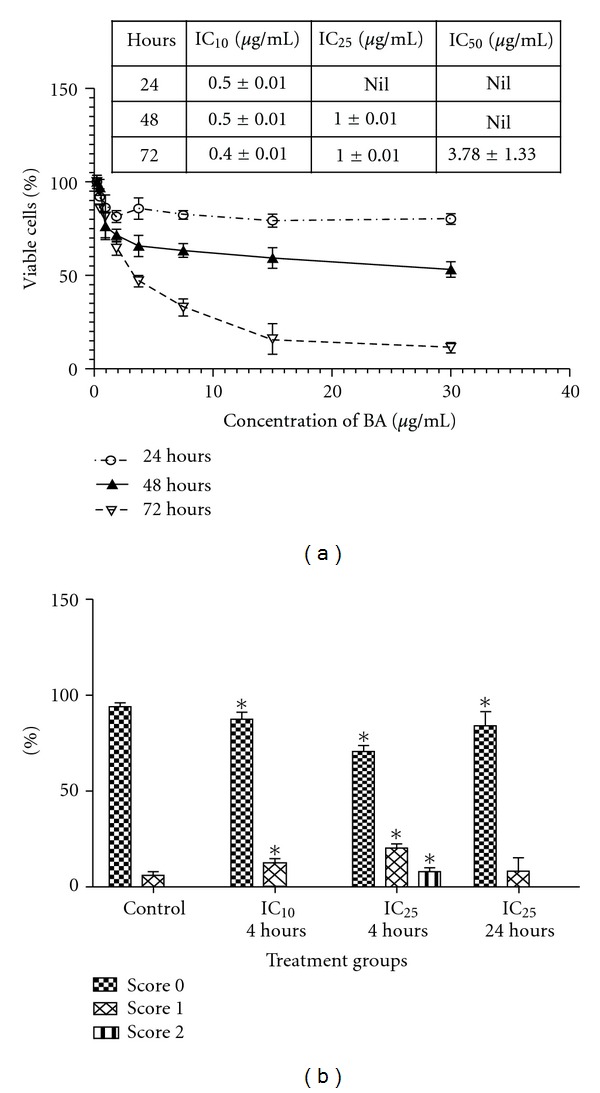
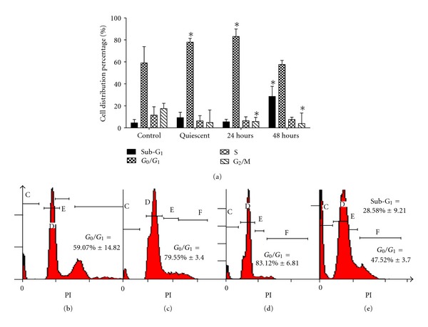
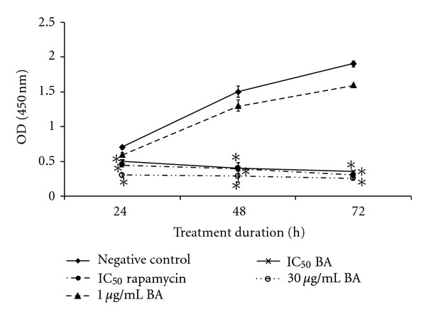
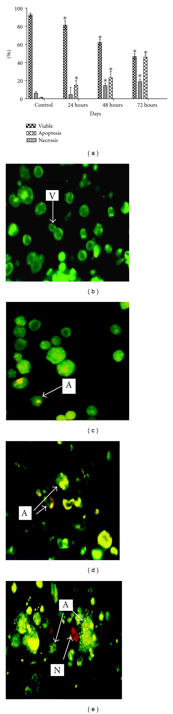
Betulinic acid is a widely available plant-derived triterpene which is reported to possess selective cytotoxic activity against cancer cells of neuroectodermal origin and leukemia. However, the potential of betulinic acid as an antiproliferative and cytotoxic agent on vascular smooth muscle (VSMC) is still unclear. This study was carried out to demonstrate the antiproliferative and cytotoxic effect of betulinic acid on VSMCs using 3-[4,5-dimethylthizol-2-yl]-2,5-diphenyltetrazolium bromide (MTT) assay, flow cytometry cell cycle assay, BrdU proliferation assay, acridine orange/propidium iodide staining, and comet assay. Result from MTT and BrdU assays indicated that betulinic acid was able to inhibit the growth and proliferation of VSMCs in a dose-dependent manner with IC(50) of 3.8 μg/mL significantly (P < 0.05). Nevertheless, betulinic acid exhibited G(1) cell cycle arrest in flow cytometry cell cycle profiling and low level of DNA damage against VSMC in acridine orange/propidium iodide and comet assay after 24 h of treatment. In conclusion, betulinic acid induced G(1) cell cycle arrest and dose-dependent DNA damage on VSMC.
Figures
References
-
- Dzau VJ, Braun-Dullaeus RC, Sedding DG. Vascular proliferation and atherosclerosis: new perspectives and therapeutic strategies. Nature Medicine. 2002;8(11):1249–1256. - PubMed
-
- Grube E, Gerckens U, Buellesfeld L. Drug-eluting stents: clinical experiences and perspectives. Minerva Cardioangiologica. 2002;50(5):469–473. - PubMed
-
- Rhee SJ, Yun KH, Lee SR, et al. Drug-eluting Stent thrombosis during perioperative period. International Heart Journal. 2008;49(2):135–142. - PubMed
-
- Shishehbor MH, Amini R, Raymond RE, et al. Safety and efficacy of overlapping sirolimus-eluting versus paclitaxel-eluting stents. American Heart Journal. 2008;155(6):1075–1080. - PubMed
LinkOut - more resources
Full Text Sources
