. 2012;7(10):e46216.
doi: 10.1371/journal.pone.0046216. Epub 2012 Oct 2.

The alternative Epac/cAMP pathway and the MAPK pathway mediate hCG induction of leptin in placental cells

Affiliations

The alternative Epac/cAMP pathway and the MAPK pathway mediate hCG induction of leptin in placental cells

Julieta Lorena Maymó et al. PLoS One. 2012.

Abstract

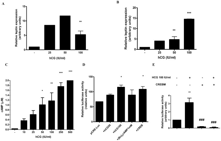
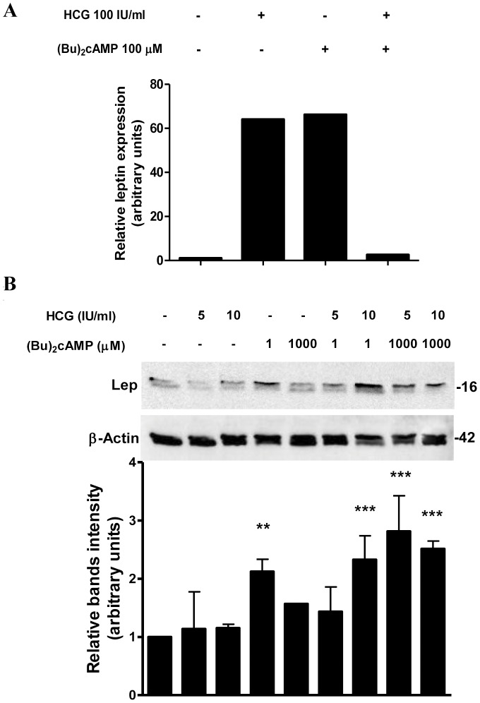
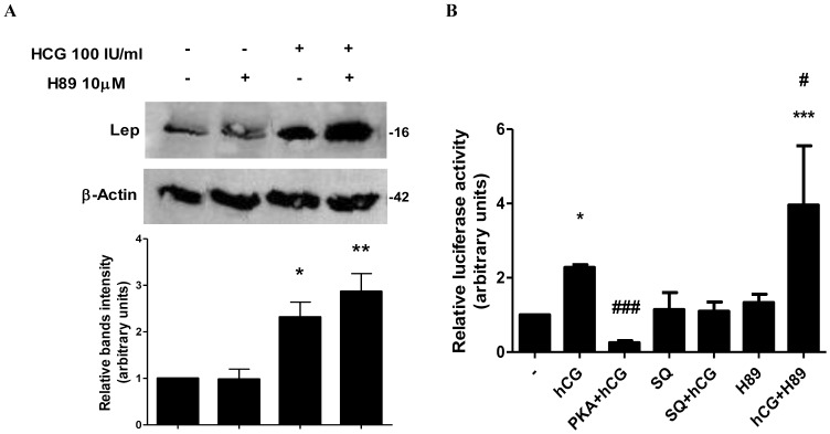
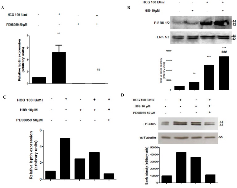
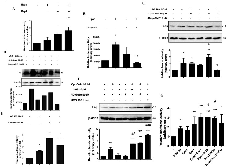
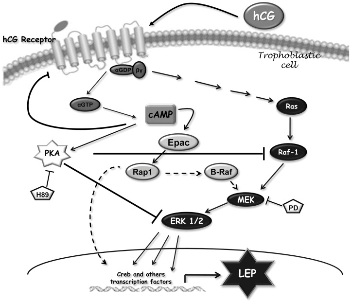
Pleiotropic effects of leptin have been identified in reproduction and pregnancy, particularly in the placenta, where it works as an autocrine hormone. In this work, we demonstrated that human chorionic gonadotropin (hCG) added to JEG-3 cell line or to placental explants induces endogenous leptin expression. We also found that hCG increased cAMP intracellular levels in BeWo cells in a dose-dependent manner, stimulated cAMP response element (CRE) activity and the cotransfection with an expression plasmid of a dominant negative mutant of CREB caused a significant inhibition of hCG stimulation of leptin promoter activity. These results demonstrate that hCG indeed activates cAMP/PKA pathway, and that this pathway is involved in leptin expression. Nevertheless, we found leptin induction by hCG is dependent on cAMP levels. Treatment with (Bu)(2)cAMP in combination with low and non stimulatory hCG concentrations led to an increase in leptin expression, whereas stimulatory concentrations showed the opposite effect. We found that specific PKA inhibition by H89 caused a significant increase of hCG leptin induction, suggesting that probably high cAMP levels might inhibit hCG effect. It was found that hCG enhancement of leptin mRNA expression involved the MAPK pathway. In this work, we demonstrated that hCG leptin induction through the MAPK signaling pathway is inhibited by PKA. We observed that ERK1/2 phosphorylation increased when hCG treatment was combined with H89. In view of these results, the involvement of the alternative cAMP/Epac signaling pathway was studied. We observed that a cAMP analogue that specifically activates Epac (CPT-OMe) stimulated leptin expression by hCG. In addition, the overexpression of Epac and Rap1 proteins increased leptin promoter activity and enhanced hCG. In conclusion, we provide evidence suggesting that hCG induction of leptin gene expression in placenta is mediated not only by activation of the MAPK signaling pathway but also by the alternative cAMP/Epac signaling pathway.


Conflict of interest statement

Competing Interests: The authors have declared that no competing interests exist.

Figures

Figure 1
Figure 1. hCG stimulates leptin mRNA expression and enhances cAMP levels in placenta.
(A) JEG-3 cells (1×10⁶ cells) were plated in complete DMEM-F12 media supplemented with 1% FBS and incubated during 3 days with different doses of hCG (IU/ml). (B) Placental explants were obtained as indicated in Materials and Methods and treated with increasing hCG concentrations. In (A) and (B), total RNA was extracted as described in Material and Methods. Leptin mRNA was quantified by real time RT-PCR. Cyclophilin was used as internal standard. (C) BeWo cells (1×10⁵) were seeded in 96-well plate and treated during 24 h with increasing concentrations of hCG, as indicated. cAMP-Glo assay kit was used to measure intracellular cAMP concentration. (D) Cells were transiently transfected with pCre-Luc plasmid construction and treated with hCG, (Bu)2cAMP or cotransfected with CREB, as indicated, during 72 h in DMEM-F12 media supplemented with 1% FBS. Luciferase activity was measured in cellular extracts and normalized to β-galactosidase activity. Activity obtained with empty vector (PGL-3 basic vector) was set as a control. (E) BeWo cells were transiently transfected with pL1951 and treated with 100 IU/ml hCG and/or cotransfected with CREBM plasmid. Cells were incubated during 72 h in DMEM-F12 1% FBS media. Luciferase activity was measured in cellular extracts and normalized to β-galactosidase activity. Activity obtained with empty vector (PGL-3 basic vector) was set as a control. Results shown are from a representative experiment and are expressed as means ± S.E.M. for three independent experiments performed in duplicates. *p<0.05, **p<0.01, ***p<0.001 vs control; ###p<0.001 vs hCG treatment.
Figure 2
Figure 2. cAMP induces leptin stimulation by hCG at low hormone concentrations.
(A) Placental explants were processed as previously described and treated with increasing hCG and/or (Bu)2cAMP concentrations during 4 h. Total RNA was extracted as described in Material and Methods. Leptin mRNA was quantified with real time RT-PCR. Cyclophilin was used as internal standard. (B) BeWo cells (1×10⁶ cells) were plated in complete DMEM-F12 media supplemented with 1% FBS and incubated during 3 days with different doses of hCG (IU/ml) and/or (Bu)2cAMP (µM), as indicated. Cell extracts were prepared as indicated in Materials and Methods. Proteins were separated on SDS-PAGE gels and leptin expression was determined by Western-blot. Molecular weights were estimated using standard protein markers. Loading controls were performed by immunoblotting the same membranes with anti-β-actin. Bands densitometry is shown in lower panels. Molecular weight (kDa) is indicated at the right of the blot. Representative results from three replicates are shown. **p<0.01, ***p<0.001.
Figure 3
Figure 3. PKA blocks hCG stimulation of leptin.
(A) BeWo cells were incubated during 3 days with hCG and/or H89, as indicated. Extracts from cells were prepared as previously described and loaded in a 12% SDS-PAGE. Leptin expression was determined by Western-blot. Loading controls were performed by immunoblotting the same membranes with anti-β-actin. Bands densitometry is shown in lower panels. Molecular weight (kDa) is indicated at the right of the blot. Representative results from three replicates are shown. (B) BeWo cells were transiently transfected with pL1951 and treated with 100 IU/ml hCG, 10 µM H89 and/or 100 µM SQ, or cotransfected with a plasmid expressing the catalytic subunit of PKA (PKA) (1 µg/ml) (C), or with a dominant negative mutant of the regulatory subunit of PKA (PKI) (1 µg/ml). Cells were incubated during 72 h in DMEM-F12 1% FBS media. Luciferase activity was measured in cellular extracts and normalized to β-galactosidase activity. Activity obtained with empty vector (PGL-3 basic vector) was set as a control. Results are expressed as mean ± S.E.M. for three independent experiments. *p<0.05, **p<0.01, ***p<0.001 vs. control; #p<0.05, ###p<0.001 vs. hCG treatment.
Figure 4
Figure 4. PKA inhibits ERK activation by hCG.
(A) JEG-3 cells (1×10⁶ cells) were plated in complete DMEM-F12 media supplemented with 1% FBS and incubated during 3 days with hCG (IU/ml) and/or PD98059. Total RNA was extracted as described in Material and Methods. Leptin mRNA was quantified by real time RT-PCR. Cyclophilin was used as internal standard. Results are expressed as mean ± S.E.M. for three independent experiments performed in triplicates. (B) BeWo cells were incubated for 15 min with hCG and/or H89 as indicated. Extracts from cells were prepared as previously described and loaded in a 12% SDS-PAGE. ERK 1/2 phosphorylation was determined by Western blot. Total ERK 1/2 protein level in extracts was determined as loading control. Molecular weights were estimated using standard protein markers. Bands densitometry is shown in lower panel. Results shown are from a representative experiment and are expressed as means ± S.E.M. for three independent experiments **p<0.01 ***p<0.001 vs control; ###p<0.001 vs hCG treatment. (C) Placental explants were processed as previously described pre-incubated during 30 min with 50 µM PD98059 and/or 10 µM H89 and treated with 100 IU/ml hCG during 4 h (C) or 15 min (D). (C) Total RNA was extracted as described in Material and Methods. Leptin mRNA was quantified by real time RT-PCR. Cyclophilin was used as internal standard. (D) Extracts were prepared as previously described and loaded in a 12% SDS-PAGE. ERK 1/2 phosphorylation was determined by Western blot. Total ERK 1/2 protein level in extracts was determined as loading control. Molecular weights were estimated using standard protein markers. Bands densitometry is shown in lower panel.
Figure 5
Figure 5. hCG induces leptin expression through the cAMP/Epac alternative signaling pathway.
(A) BeWo cells were transiently cotransfected with pL1951 and Epac (1 µg/ml) and/or Rap1b (1 µg/ml) proteins expression plasmids. (B) BeWo cells were transiently cotransfected with pL1951 Epac (1 µg/ml) and/or RapGAP (1 µg/ml) proteins expression plasmids. (C) and (E) BeWo cells (1×10⁶ cells) were plated in complete DMEM-F12 media supplemented with 1% FBS and incubated during 3 days with different doses of Cpt-OMe, hCG, (Bu)2cAMP, and H89, as indicated. Cell extracts were prepared as indicated in Materials and Methods. Proteins were separated on SDS-PAGE gels and leptin expression was determined by Western-blot. Molecular weights were estimated using standard protein markers. Loading controls were performed by immunoblotting the same membranes with anti-β-actin. Bands densitometry is shown in lower panels. Molecular weight (kDa) is indicated at the right of the blot. Representative results from three replicates are shown. (D) Cells were transfected with pL1951 plasmid construction and treated with hCG and/or Cpt-OMe, as indicated. (F) BeWo cells were cotransfected with pL1951 and/or Epac and Rap1b and treated with hCG (IU/ml). In (A), (B), (D) and (F) cells were incubated during 72 h in DMEM-F12 1% FBS media. Luciferase activity was measured in cellular extracts and normalized to β-galactosidase activity. Activity obtained with empty vector (PGL-3 basic vector) was set as a control. Results are expressed as mean ± S.E.M. for three independent experiments. *p<0.05, **p<0.01, ***p<0.001 vs. control; #p<0.05, ##p<0.01, ###p<0.001 vs. hCG treatment. (G) Placental explants were processed as previously described and treated with 100 IU/ml hCG and/or 10 µM Cpt-O-Me or 10 µM (Bu)2cAMP as indicated during 4 h. Proteins were separated on SDS-PAGE gels and leptin expression was determined by Western-blot. Molecular weights were estimated using standard protein markers. Loading controls were performed by immunoblotting the same membranes with anti-β-actin. Bands densitometry is shown in lower panels. Molecular weight (kDa) is indicated at the right of the blot.
Figure 6
Figure 6. The MAPK and the alternative cAMP/Epac signaling pathways participate in leptin stimulation by hCG in placenta.
Proposed model of the signaling pathways involved in hCG stimulation based on current data and its relation to leptin expression. Pointed arrow: Stimulation; Flat arrow: Inhibition. Dash arrow: possible pathways involved.

Similar articles

Cited by

References

    1. Fitzgerald JS, Busch S, Wengenmayer T, Foerster K, de la Motte T, et al. (2005) Signal transduction in trophoblast invasion. Chem Immunol Allergy 88: 181–199. - PubMed
    1. Zhang Y, Proenca R, Maffei M, Barone M, Leopold L, et al. (1994) Positional cloning of the mouse obese gene and its human homologue. Nature 372: 425–432. - PubMed
    1. Houseknecht KL, Portocarrero CP (1998) Leptin and its receptors: regulators of whole-body energy homeostasis. Domest Anim Endocrinol 15: 457–475. - PubMed
    1. Reitman ML, Bi S, Marcus-Samuels B, Gavrilova O (2001) Leptin and its role in pregnancy and fetal development–an overview. Biochem Soc Trans 29: 68–72. - PubMed
    1. Henson MC, Castracane VD (2005) Leptin in Pregnancy: An Update. Biol Reprod. - PubMed

Publication types