. 2012;7(10):e46412.
doi: 10.1371/journal.pone.0046412. Epub 2012 Oct 4.

Intron definition and a branch site adenosine at nt 385 control RNA splicing of HPV16 E6*I and E7 expression

Affiliations

Intron definition and a branch site adenosine at nt 385 control RNA splicing of HPV16 E6*I and E7 expression

Masahiko Ajiro et al. PLoS One. 2012.

Abstract

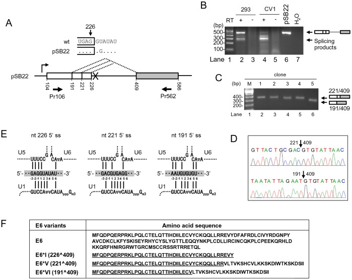
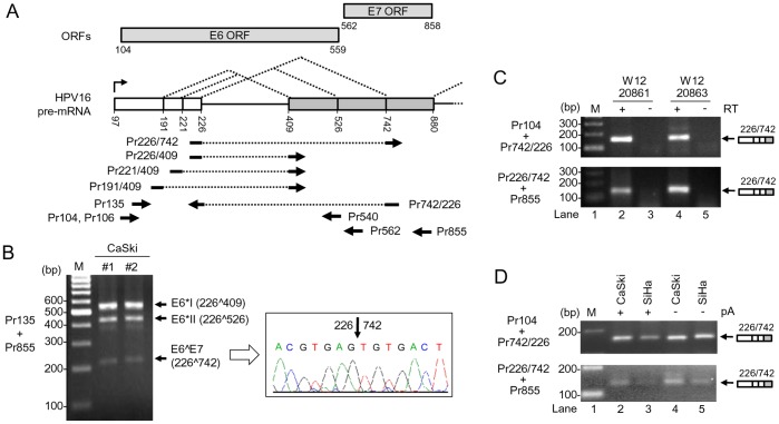
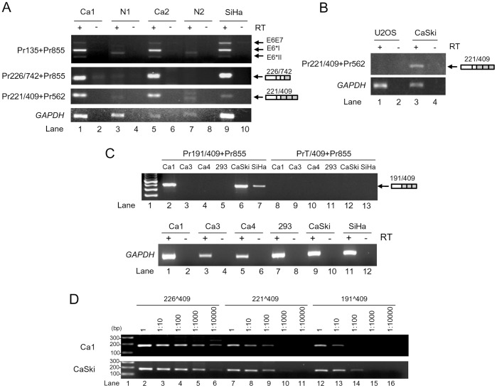
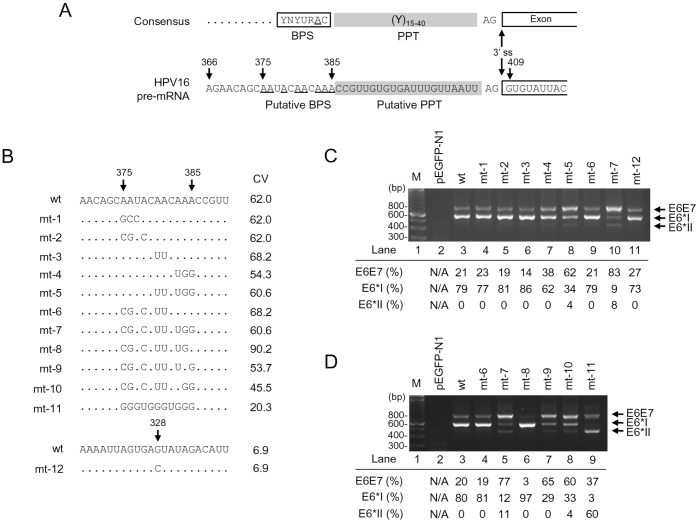
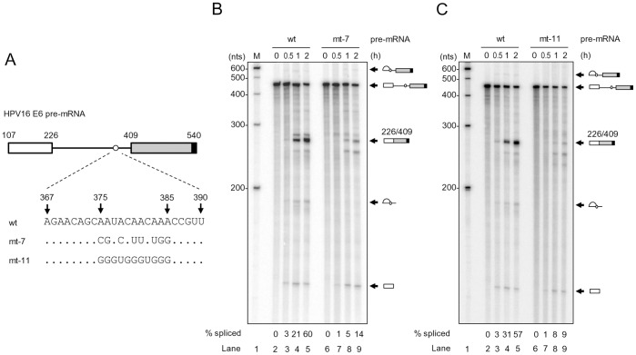
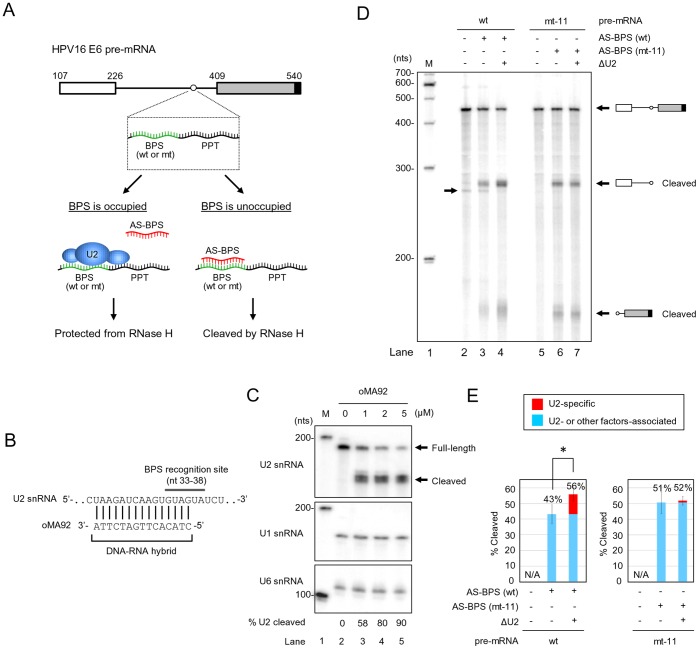
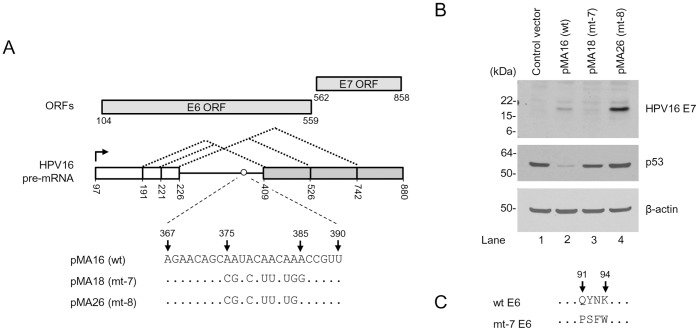
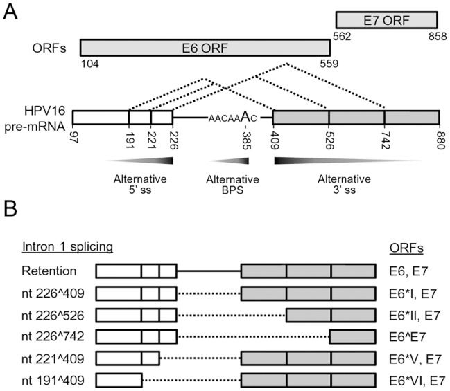
HPV16 E6 and E7, two viral oncogenes, are expressed from a single bicistronic pre-mRNA. In this report, we provide the evidence that the bicistronic pre-mRNA intron 1 contains three 5' splice sites (5' ss) and three 3' splice sites (3' ss) normally used in HPV16(+) cervical cancer and its derived cell lines. The choice of two novel alternative 5' ss (nt 221 5' ss and nt 191 5' ss) produces two novel isoforms of E6E7 mRNAs (E6*V and E6*VI). The nt 226 5' ss and nt 409 3' ss is preferentially selected over the other splice sites crossing over the intron to excise a minimal length of the intron in RNA splicing. We identified AACAAAC as the preferred branch point sequence (BPS) and an adenosine at nt 385 (underlined) in the BPS as a branch site to dictate the selection of the nt 409 3' ss for E6*I splicing and E7 expression. Introduction of point mutations into the mapped BPS led to reduced U2 binding to the BPS and thereby inhibition of the second step of E6E7 splicing at the nt 409 3' ss. Importantly, the E6E7 bicistronic RNA with a mutant BPS and inefficient splicing makes little or no E7 and the resulted E6 with mutations of (91)QYNK(94) to (91)PSFW(94) displays attenuate activity on p53 degradation. Together, our data provide structural basis of the E6E7 intron 1 for better understanding of how viral E6 and E7 expression is regulated by alternative RNA splicing. This study elucidates for the first time a mapped branch point in HPV16 genome involved in viral oncogene expression.


Conflict of interest statement

Competing Interests: The authors have declared that no competing interests exist.

Figures

Figure 1
Figure 1. Identification of two novel alternative 5′ ss in the HPV16 intron 1.
(A) Diagram of alternative 5′ ss identified in this study by using pSB22, an HPV16 E6 expression vector containing a mutated nt 226 5′ ss (GU to GG, U to G at nt 228). Numbers below the pre-mRNA indicate nucleotide positions in the HPV16 genome. Boxes, exons; lines, introns; dashed lines, splicing directions. Arrows below the exons are a pair of primers used for RT-PCR. (B) RT-PCR result of alternative RNA splicing of E6 pre-mRNA in HEK293 or CV1 cells transfected with pSB22. RT-PCR products with a size of ∼300 bp were gel-purified and cloned. (C) Plasmid DNAs extracted from five bacterial colonies were digested by EcoRI and analyzed by agarose gel electrophoresis, showing the insert size. (D) Sequence chromatograms showing splice junctions of colonies 1–4 (nt 221/409) and the colony 5 (nt 191/409). (E) Illustration of the base pairing of three alternative 5′ ss (shaded, middle) in HPV16 intron 1 with U1, U5 and U6 snRNAs. Exon sequences are labeled as −1, −2 and −3 from the 5′ end of the intron. Base pairing is indicated with solid lines. Ψ, pseudo-uridine; Gm3, 2,2,7-tri-methyl guanosine. (F) Coding potentials of spliced E6E7 transcripts by using the nt 221 (E6*V) or nt 191 (E6*VI) 5′ ss. Amino acid residues underlined in E6*I, E6*II, E6*V and E6*VI are identical to the N-terminus of wt E6.
Figure 2
Figure 2. Utilization of nt 742 3′ ss in multiple HPV16+ cervical cancer cell lines.
(A) Diagram of splicing directions of HPV16 E6E7 pre-mRNA from three alternative 5′ ss at nt 191, 221, and 226 to three alternative 3′ ss at nt 409, 526 and 742. Numbers represent nucleotide positions in the HPV16 genome. Arrows under the E6E7 pre-mRNA structure indicate primers used in RT-PCR in this and all other figures. Rectangles above the E6E7 pre-mRNA structure indicate ORFs of E6 and E7. (B) Two preps of total RNA extracted from CaSki cells were analyzed by RT-PCR with a primer pair of Pr135 and Pr855 (left). The 214 bp RT-PCR product was purified from the gel and sequenced as a splicing product from nt 226 to 742 (right). (C) E6∧E7 expression is detectable by RT-PCR from W12 subclone cell lines 20861 and 20863 which harbor integrated and episomal HPV16 genome, respectively. (D) E6∧E7 expressed in HPV16+ CaSki and HPV16+ SiHa cells are polyadenylated. Total RNA with or without polyadenylation (pA) was used for RT-PCR with the indicated splice junction primers on the left (C and D).
Figure 3
Figure 3. Usage of alternative 5′ ss and 3′ ss in HPV16+ cervical cancer tissues and cell lines.
(A) RT-PCR for total RNA from cervical tissues and SiHa cells with the indicated primer pairs (Fig. 2A) on the left, showing HPV16 splicing product from nt 221 5′ ss to nt 409 3′ ss in HPV16+ cervical cancer tissues (Ca) and SiHa cells. Expression of E6∧E7, E6*I, and E6*II served as positive controls. Normal cervical tissues (N) served as negative controls. A relative smaller or larger band in the normal tissue was a non-specific amplicon. (B) RT-PCR results for total RNA from HPV16+ CaSki cells and U2OS cells for splicing product from nt 221 to nt 409. HPV16-negative U2OS cells served as a negative control. (C) RT-PCR results of total RNA from HPV16+ cervical cancer tissues, CaSki, SiHa and HEK293 cells, showing the splicing product from nt 191 to nt 409. A 191∧409 splice junction 5′ primer and its 3′ half primer starting from nt 409 3′ ss were used in combination with a 3′ primer Pr855 for this comparison. HPV16-negative HEK293 RNA served as a negative control. GAPDH RNA served as a loading control. (D) Titration of the relative usage of nt 226, 221 and 191 5′ ss in cervical cancer tissues and CaSki cells by RT-PCR. The cDNAs of Ca1 and CaSki from panel C were serially diluted and amplified for the splicing products of 226∧409, 221∧409 and 191∧409 with each corresponding splice junction 5′ primer Pr226/409, Pr221/409, or Pr191/409 in combination of a common 3′ primer Pr562.
Figure 4
Figure 4. Mapping of the BPS in splicing of the HPV16 nt 409 3′ ss.
(A) Consensus splicing elements (3′ ss, BPS, polypyrimidine tract [PPT]) in a typical 3′ ss (upper) and their corresponding RNA sequences in the HPV16 nt 409 3′ ss (lower). (B) RNA sequences of putative wild type (wt) BPS and its derived mutants (mt 1–11) in HPV16 E6E7 constructs used in the study. Arrow heads indicate the nucleotide position in the HPV16 genome. Unchanged ribonucleotides in each mutant are indicated by dots. A mutant G328C BPS reported by another lab was included in this study for verification. The nucleotide G at nt 328 position in the HPV16 genome was substituted with a C in the mutant construct (mt-12). CV, consensus value of the putative BPS . (C and D) Identification of the nt 385A as a preferential branch site for splicing of the nt 409 3′ ss. RT-PCR results were obtained by using a primer pair of Pr106 and Pr855 (Fig. 2A) from total RNA extracted from HEK293 cells 24 h after transfection with a wt or mt construct as indicated above the agarose gel. Vector pEGFP-N1 served as a negative control (lane 2 in both C and D). M, size marker of 100-bp DNA ladders (lane 1 in both C and D). Identities of PCR products derived from unspliced E6E7 and spliced E6*I and E6*II RNA are shown on the right. Relative ratios (%) of the unspliced E6E7 (intron 1 retention) and spliced E6*I (nt 226∧409) and E6*II (nt 226∧526) RNA were calculated by the signal intensity of each product and shown below the gel.
Figure 5
Figure 5. Introduction of point mutations into the mapped BPS in HPV16 intron 1 inhibits RNA splicing in vitro.
(A) Diagram of HPV16 pre-mRNA used in in vitro splicing assays. First exon (nt 107–226) and second exon (nt 409–540), indicated by white and gray rectangles, respectively, are separated by its native intron 1 containing a wt, mt-7 or mt-11 BPS. An 11-nt U1 binding site (black rectangle) attached to each RNA 3′ end served as a 5′ ss or splicing enhancer to promote in vitro RNA splicing. (B and C) Reduction of HPV16 RNA splicing in vitro by introduction of point mutations into the mapped BPS. In vitro RNA splicing assay was performed with 32P-labeled HPV16 E6 pre-mRNA in the presence of HeLa nuclear extract at 30°C for the indicated splicing reaction time (h). Spliced products were resolved in a 6% denaturing PAGE gel and their identities are shown on the right. Splicing efficiency (% spliced) was calculated as described from each spliced gel and is shown at the bottom of the gel.
Figure 6
Figure 6. RNase H digestion to quantify U2 snRNP association with the wt or mt-11 BPS in the HPV16 intron 1.
(A) Flow chart of RNase H protection assays. A 32P-labeled HPV16 E6 RNA with a wt or mt-11 BPS was incubated with HeLa nuclear extract with or without U2 depletion in the presence of an antisense (AS) DNA oligo to wt or mt-11 BPS and RNase H. Association of U2 snRNA or other factors (unlabeled ovals) with the mapped BPS prevents the antisense DNA oligo to specifically access the correspondent region, thereby preventing RNase H cleavage of the testing pre-mRNA. Otherwise, binding of the antisense DNA oligo to the mapped BPS would form a DNA-RNA duplex and trigger RNase H digestion. (B) Diagram of DNA oligo-mediated U2 depletion from HeLa nuclear extract by RNase H digestion. A DNA oligo (oMA92) antisense to U2′s BPS recognition site was incubated at 30°C with HeLa nuclear extract for 30 min and the mixture was then digested by RNase H at 30°C for additional 10 min. (C) Determination of U2 depletion efficiency from HeLa nuclear extract by Northern blot. Total RNA from HeLa nuclear extract with or without U2 depletion by U2 DNA oligo-mediated RNase H digestion was quantified by Northern blot with a 32P-labeled DNA oligo specific for U2 (oKY50). U1 and U6 served as loading controls. U2 depletion efficiency was expressed as a ratio (%) of cleaved U2 vs remaining full-length U2 dividing by total signal intensity of U2 in each reaction condition of RNase H digestion. (D and E) U2 interaction with the mapped BPS prevents oligo-directed RNase H digestion. HeLa nuclear extracts with or without U2 depletion (ΔU2) were compared for DNA-oligo-mediated RNase H cleavage of HPV16 pre-mRNA with a wt or mt BPS (A). The reaction products were resolved in a 6% denaturing PAGE gel. Identities of RNase H cleavage products are shown on the right of the gel (D). An arrow on the left of the gel (D) indicates a spliced product from the wt pre-mRNA during 30 min incubation of RNase H digestion in the presence of HeLa nuclear extract. Cleavage efficiency (%) (E) of HPV16 E6 pre-mRNA with a wt or mt BPS from DNA oligo-mediated RNase H digestion in each reaction was calculated from four independent experiments by the sum of signal intensity of all cleavage products divided by the sum of signal intensity of both full-length pre-mRNA and cleavage products. *, P<0.05 by Student′s t-test.
Figure 7
Figure 7. Biological significance of the mapped BPS in the expression of E6 and E7 oncogenes.
(A) Diagrams of HPV16 E6E7 pre-mRNA expression vectors. Plasmid pMA16 has wild type sequence of E6E7 pre-mRNA, pMA18 expresses mt-7 pre-mRNA with a mt BPS, and pMA26 expresses mt-8 pre-mRNA similar to mt-7, except the 385A being restored as in wt. See RNA splicing profiles from each vector in Fig. 4D. (B) The expression of viral E6 and E7 oncoprotein from plasmid pMA16 (wt), pMA18 (mt-7), and pMA26 (mt-8). HCT116 cells were analyzed by Western blot 48 h after transfection with the indicated vectors above the gel for viral E7 and p53 (as an indicator of viral E6 expression and its activities). β-actin served as a loading control. (C) Amino acid residues in mt-7 E6 differs from wt E6 from aa 91 to 94 positions. Dots, unchanged aa residues.
Figure 8
Figure 8. Summary of the characterized features of HPV16 E6E7 intron 1.
(A) Preferential selection of a proximal splice site and branch site in splicing of the HPV16 E6E7 intron 1 which contains three suboptimal 5′ ss (nt 191, 221 and 226), three suboptimal 3′ ss (nt 409, 526 and 742), and a cluster of branch site As from nt 383 to nt 385. In this regard, a proximal nt 226 5′ ss is preferentially selected over the nt 221 and nt 191 5′ ss, a proximal nt 409 3′ ss is efficiently selected over the nt 526 and nt 742 3′ ss, and the branch site A at nt 385 is preferentially utilized over the nt 384 A or 383 A, to minimize splicing energy and length of the excising intron 1. The preferential selection of an alternative 5′ ss, 3′ ss and branch site is exercised by the principle of proximity and is indicated by graded grey arrows for more (dark grey) to less (light grey) splicing of the HPV16 E6E7 intron 1. (B) Alternative splicing of HPV16 E6 intron 1 leads to produce multiple E6 RNA species detectable in cervical cancer tissues and their derived cell lines. Dash lines indicate the spliced intron 1 from each isoform of E6E7 RNA and vertical lines in white (exon 1) and grey boxes (exon 2) stand for splice sites. ORFs for splicing variants of viral E6 and E7 are indicated on the right.

References

    1. Bernard HU, Burk RD, Chen Z, van Doorslaer K, Hausen H, et al. (2010) Classification of papillomaviruses (PVs) based on 189 PV types and proposal of taxonomic amendments. Virology 401: 70–79. - PMC - PubMed
    1. Howley PM, Lowy DR (2007) Papillomaviruses. In: Knipe DM, Howley PM, Griffin DE, Lamb RA, Martin MA, Roizman B, Straus SE, eds. Fields Virology. Fifth edition ed. Philadelphia: Lippincott Williams & Wilkins; 2299–2354.
    1. Walboomers JM, Jacobs MV, Manos MM, Bosch FX, Kummer JA, et al. (1999) Human papillomavirus is a necessary cause of invasive cervical cancer worldwide. J Pathol 189: 12–19. - PubMed
    1. Munoz N, Bosch FX, de Sanjose S, Herrero R, Castellsague X, et al. (2003) Epidemiologic classification of human papillomavirus types associated with cervical cancer. N Engl J Med 348: 518–527. - PubMed
    1. Munoz N, Castellsague X, de Gonzalez AB, Gissmann L (2006) Chapter 1: HPV in the etiology of human cancer. Vaccine 24 Suppl 3: S3–1–S310. - PubMed

Publication types

MeSH terms

Substances