. 2012;7(10):e46435.
doi: 10.1371/journal.pone.0046435. Epub 2012 Oct 4.

Solution structure, copper binding and backbone dynamics of recombinant Ber e 1-the major allergen from Brazil nut

Affiliations

Solution structure, copper binding and backbone dynamics of recombinant Ber e 1-the major allergen from Brazil nut

Louise Rundqvist et al. PLoS One. 2012.

Abstract

Background: The 2S albumin Ber e 1 is the major allergen in Brazil nuts. Previous findings indicated that the protein alone does not cause an allergenic response in mice, but the addition of components from a Brazil nut lipid fraction were required. Structural details of Ber e 1 may contribute to the understanding of the allergenic properties of the protein and its potential interaction partners.

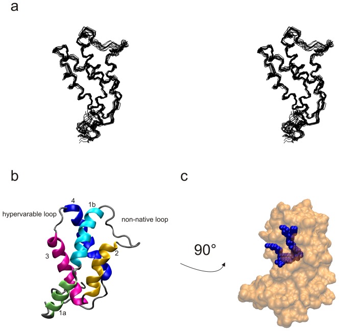
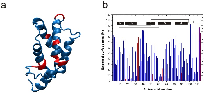
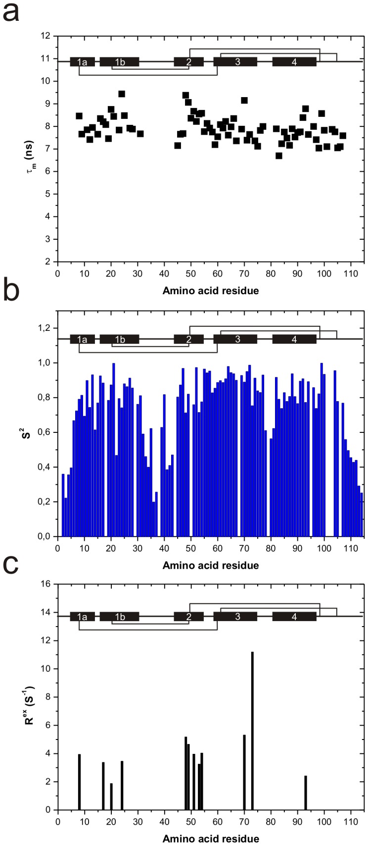
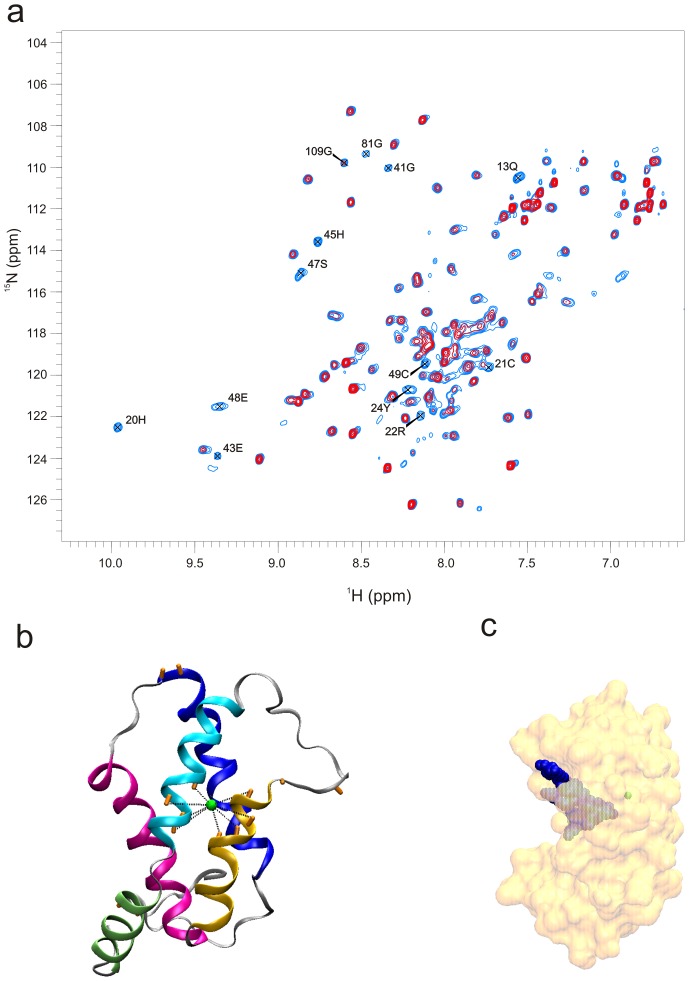
Methodology/principal findings: The solution structure of recombinant Ber e 1 was solved using NMR spectroscopy and measurements of the protein back bone dynamics at a residue-specific level were extracted using (15)N-spin relaxation. A hydrophobic cavity was identified in the structure of Ber e 1. Using the paramagnetic relaxation enhancement property of Cu(2+) in conjunction with NMR, it was shown that Ber e 1 is able to specifically interact with the divalent copper ion and the binding site was modeled into the structure. The IgE binding region as well as the copper binding site show increased dynamics on both fast ps-ns timescale as well as slower µs-ms timescale.

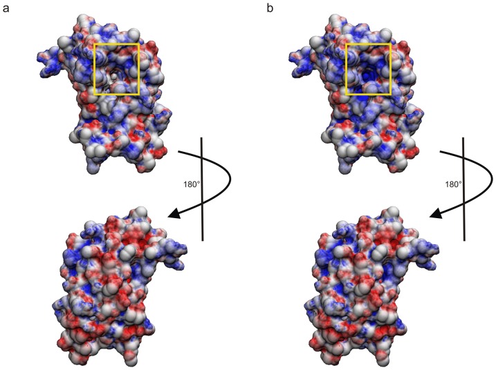
Conclusions/significance: The overall fold of Ber e 1 is similar to other 2S albumins, but the hydrophobic cavity resembles that of a homologous non-specific lipid transfer protein. Ber e 1 is the first 2S albumin shown to interact with Cu(2+) ions. This Cu(2+) binding has minimal effect on the electrostatic potential on the surface of the protein, but the charge distribution within the hydrophobic cavity is significantly altered. As the hydrophobic cavity is likely to be involved in a putative lipid interaction the Cu(2+) can in turn affect the interaction that is essential to provoke an allergenic response.


Conflict of interest statement

Competing Interests: The authors have declared that no competing interests exist.

Figures

Figure 1
Figure 1. Solution structure of Ber e 1.
a) Stereo view of the backbone of the 12 lowest energy structures after energy minimization. b) Cartoon representation of the Ber e 1 structure, with the different structure elements colored as follows: green = helix 1a, cyan = helix 1b, yellow = helix 2, pink = helix 3, blue = helix 4. c) Location of the hydrophobic cavity in Ber e 1. The structure has been rotated 90 degrees with respect to figure 1a and b.
Figure 2
Figure 2. Theroretical pepsin cleavage sites and solvent exposed residues.
a) Theoretical pepsin cleavage sites (red) mapped on the tertiary structure of Ber e 1. b) Exposed surface area of the N and HN atoms in the backbone of Ber e 1. Residues able to undergo pepsin cleavage are highlighted in red. The secondary structure elements, as well as the cysteine linkage are indicated in the top of the figure. Most of the theoretical pepsin cleavage sites are buried within the α-helices. The only surface exposed pepsin cleavage sites are located in the non-native loop and the C-terminal, and cleavage at these positions would not disrupt the integrity of the structure.
Figure 3
Figure 3. Ber e 1 backbone dynamics.
a) Residue-specific overall tumbling time (τm) values for 77 of 114 residues. The relatively uniform τm values indicate isotropic tumbling of Ber e 1. b) Order parameter, S2, of the N-H bond vector on a per residue basis. An S2 value of 1 equals a completely rigid N-H vector, whereas an S2 value of 0 implies complete rotational freedom of the N-H vector. Some flexibility is observed in the hypervariable loop, and the N- and C-terminal, as well as the non-native loop show high flexibility. c) Rex parameter, showing the residues where µs-ms dynamics could be identified. Slow dynamics is largely located to the interface of helix 1b and 2, as well as at the end of helix 3, leading into the hypervariable loop. The secondary structure elements, as well as the cysteine linkage are indicated in the top of each figure.
Figure 4
Figure 4. Ber e 1 copper interaction.
a) Paramagnetic copper relaxation enhancement experiment on Ber e 1. The spectrum shown in black is recorded in the absence of copper, whereas the spectrum shown in red has copper added in a 1∶1 (Cu2+:Ber e 1) stoichiometry. N-H groups in the backbone affected by paramagnetic relaxation enhancement by the addition of Cu2+ in a 1∶1 ration are HIS 20, CYS 21, ARG 22, TYR 24, GLU 43, HIS 45, SER 47, GLU 48, CYS 49, and GLN 52. In addition N-H groups form sidechains (s.c) of GLN 11, 13, 28 and 83 are also affected by Cu2+ at this stoichiometric ratio. b) A model of the copper atom positioning in Ber e 1, based on the nearby residues identified in (a). The N-H backbone groups that are bleached are indicated in the structure as orange rods. c) Due to the slow dynamics around the copper binding site, the copper atom is engulfed into the core of the protein. Interestingly, its position is very close to the bottom of the hydrophobic cavity.
Figure 5
Figure 5. Electrostatic surface potential of the Ber e 1 in absence and presence of Cu2+.
a) Electrostatic surface potential of the Ber e 1 at pH 7 in absence of Cu2+. b) Electrostatic surface potential of the Ber e 1 at pH 7 in presence of Cu2+. The entry of the hydrophobic cavity is highlighted with a yellow square. The protein shows an overall positive charge on the side of the protein that comprises the hypervariable loop and the entry to the hydrophobic cavity. In contrast, the other side of the protein, in particular helix 1a and most of helix 1b and 2, is negatively charged. The presence of histidines, taken together with the slow dynamics and the overall negative charge between helix 1b and 2, suggests that Cu2+ would enter the molecule from the negatively charged side of the protein. However, binding of Cu2+ only to a small degree changed the net surface charge on the helix 1b-2 side of the protein. The largest difference in surface potential is observed within the hydrophobic cavity; tuning the surface potential inside the cavity from neutral to more positive charge.

References

    1. Sampson HA (2004) Update on food allergy. J Allergy Clin Immunol 113: 805–819. - PubMed
    1. Radauer C, Bublin M, Wagner S, Mari A, Breiteneder H (2008) Allergens are distributed into few protein families and possess a restricted number of biochemical functions. J Allergy Clin Immunol 121: 847–852 e847. - PubMed
    1. Ruiter B, Shreffler WG (2012) The role of dendritic cells in food allergy. Journal of Allergy and Clinical Immunology 129: 921–928. - PubMed
    1. Radauer C, Breiteneder H (2007) Evolutionary biology of plant food allergens. J Allergy Clin Immunol 120: 518–525. - PubMed
    1. Kreis M, Forde BG, Rahman S, Miflin BJ, Shewry PR (1985) Molecular evolution of the seed storage proteins of barley, rye and wheat. J Mol Biol 183: 499–502. - PubMed

Publication types

MeSH terms